“Screening of Bioaffinity Ligands for Human Soluble
COMT Purification”
Sara Rute Afonso Costa
Dissertation for purpose of Master Degree in Biochemistry
Supervisors: Luís António Paulino Passarinha, PhD
João António de Sampaio Rodrigues Queiroz, PhD
Covilhã, 2010
Universidade da Beira Interior
Departamento de Química
“By three methods we may learn wisdom: First, by reflection, which is noblest; Second, by imitation, which is easiest; and third by experience, which is the bitterest.”
Acknowledgements
First and foremost, I would like to thank you to my supervisors Prof. Luís Passarinha and Prof. João Queiroz for their valuable scientific support and continuous help.
To all members of Health Sciences Research Centre (CICS) of University of Beira Interior, tanks all help in the development of this work.
Aos meus exemplos de vida, pai e mãe, dedico-vos tudo o que consegui. Ao meu irmão…por seres tu! Não é possível agradecer-vos tudo o que fazem por mim!
Agradeço a minha familia, em especial aos meus padrinhos e avó…e familia de coração pela força que sempre me deram sem voces nada disto seria possivel.
To all my friends, especially Joana, Bruno, Francisca, Ricardo, Inês, Ana, Carina e Irina by being unique and reliable friendship! For all the moments in this year, thank you for cry and laugh with me…this year would not have been the same without you!
My lab colleagues for the teachings about the science life, because now we are prepared for good and not so good things…
Tiago, for your unconditional patience and love, despite the distance you being still my shelter seaport. Thank you for looking me that way…
Contents
1.1 Enzymatic reaction and physiological role ... 3
1.2 Gene and Protein characterization ... 4
1.2.1 The COMT gene: localization and structure ... 4
1.2.2 The COMT protein: localization and characterization ... 5
1.3 Expression of recombinant COMT proteins ... 9
1.4 Purification procedures of COMT proteins ... 10
1.4.1 COMT stability ... 11
1.5 Genetic polymorphism of COMT and diseases association ... 11
1.5.1 COMT role in Parkinson disease ... 14
1.6 Analytical methods in COMT assays ... 15
3.1 Material ... 20
3.2 Methods ... 20
3.2.1 Recombinant hSCOMT production and recuperation... 20
3.2.2 Total protein quantification: Bradford micro-assay ... 21
3.2.3 Analytical chromatography: hSCOMT specific activity assay ... 22
Figures Index ... iv
Tables Index ... viii
Abbreviations list ... ix
Resumo ... x
Palavras-Chave ... xii
Abstract ... xiii
Keywords ... xiv
Chapter I. Catechol-O-methyltransferase: An overview ... 1
Chapter II. Aims and Outline ... 17
3.2.4 SDS-PAGE and Western blot ... 23
4.1 General considerations ... 26
4.2 Procedure ... 26
4.3 Octyl-sepharose support and destabilizing elution condition using L-arginine .... 27
4.3.1 Results and discussion ... 28
4.4 Epoxy-sepharose support and dual salt system ... 31
4.4.1 Results and discussion ... 32
5.1 General considerations ... 34
5.2 Procedures ... 35
5.2.1 Econo-Pac® disposable Chromatography Columns system ... 35
5.2.2 FPLC system ... 35
5.3 Results and discussion ... 36
5.3.1 Retention using salt manipulation ... 36
5.3.2 Retention using pH manipulation ... 38
6.1 Concluding remarks ... 51
6.2 Future work ... 52
I. Solutions composition ... 66
II. Abstract and poster presentation: “6º Encontro Nacional de Cromatografia in Madeira, Portugal - 2009” ... 67
Chapter IV. Hydrophobic Interaction Chromatography: Octyl and Epoxy supports ... 25
Chapter V. Pseudo-Bioaffinity Chromatography: Amino Acids as Immobilized Ligands ... 33
Chapter VI. Concluding remarks and Future work ... 50
Chapter VII. References ... 53
III. Abstract: “8th
European Symposium on Biochemical Engineering Science (ESBES) in Bologna, Italy - 2010”... 68 IV. Manuscript ... 69
iv
Figures Index
Figure 1. Reaction mechanism of COMT. AdoMet and S-adenosyl-L-homocysteine.
(Adapted from (Lundstrom et al., 1995)).
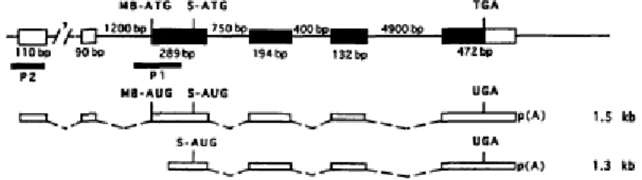
Figure 2. The structure of human COMT gene. Thin line represents introns and the
boxes the exons. The black boxes indicate the protein coding regions. Two identified promoters, P1 and P2, are shown by black bars. COMT mRNA species expressed from the genes (1.3 and 1.5 kb) are presented as white bars. The positions of translation initiation codons for MB-COMT polypeptide (MB-ATG, MB-AUG), S-COMT polypeptide (S-ATG, S-AUG) and for translation stop codons (TGA, UGA), and the sizes of exons and introns are also shown (Lundstrom et al., 1995).
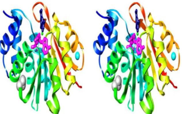
Figure 3. Crystal structure of hSCOMT. Ribbon diagrams of human 108V S-COMT
colored from blue (N-terminus) to red (C-terminus) are shown in stereo. AdoMet (magenta) and dinitrocatechol (dark blue) are shown in stick representation. K+ (cyan), Mg2+ (green), and the side chain of residue V108 (gray) are shown in space-filling representation (Rutherford et al., 2008b).
Figure 4. A22S, A52T, and V108M COMT polymorphisms. Ribbon diagram of
wild-type COMT colored from blue (N-terminus) to red (C-terminus). AdoMet and 3,5-dinitrocatechol are shown in stick representation and colored by atom type. Polymorphic residues 22, 52, and 108 are shown in space-filling representation and colored blue, cyan, and green, respectively (Rutherford and Daggett, 2009).
Figure 5. Dopamine replacement in PD therapy with dopamine precursor (L-DOPA).
Scheme of the triple combination therapy in PD. (European Parkinson’s Disease Association, 2007).
Figure 6. Analytical methods applied to COMT activity analysis (Adapted from
(Pihlavisto and Reenila, 2002)).
v
Figure 8. hSCOMT specific activity at L-arginine concentrations range from 0.5 to 2 M
in comparison with the positive control achieved with 10 mM Tris-HCl at pH 7.8. The experiments were performed at 4ºC during 3 h.
Figure 9. Chromatographic profile of an E. coli lysate extract onto the octyl-sepharose
support with a dual salt system and stepwise gradients from buffer A, B to C. (A) buffer A: NH2SO4 and Na3C6H5O7 at 25 mM in 10 mM Tris–HCl, pH 7.8; buffer B: NH2SO4 and Na3C6H5O7 at 25 mM, L-arginine 1 M in 10 mM Tris–HCl, pH 7.8 and buffer C: 10
mM Tris–HCl, pH 7.8. (B) buffer A: NH2SO4 and Na3C6H5O7 at 25 mM, L-arginine
0,5 M in 10 mM Tris–HCl, pH 7.8; buffer B: NH2SO4 and Na3C6H5O7 at 25 mM,
L-arginine 1 M in 10 mM Tris–HCl, pH 7.8 and buffer C: 10 mM Tris–HCl, pH 7.8.
Figure 10. Typical chromatographic profile of an E. coli lysate extract onto an octyl
support with a dual salt system and stepwise gradient from buffer A, B to C. Buffer A:
NH2SO4 and Na3C6H5O7 at 25 mM in 10 mM Tris–HCl, pH 7.8; buffer B: NH2SO4 and Na3C6H5O7 at 1 mM, L-arginine 1M in 10 mM Tris–HCl, pH 7.8 and buffer C: 10 mM
Tris–HCl, pH 7.8.
Figure 11. Chromatographic profiles of an E. coli lysate extract with recombinant
hSCOMT onto the six AAIL’s tested with a stepwise gradient from 100 to 0% buffer A (buffer A: 1.5 M NH2SO4 in 10 mM Tris–HCl pH 7.8; buffer B: 10 mM Tris–HCl, pH 7.8).
Figure 12. (A) SDS-PAGE analysis of E. coli recombinant hSCOMT lysates, loaded
onto the several AAIL`s supports: (lanes 1, 2) L-arginine, (lanes 3, 4) L-aspartate, (lanes 5, 6) glutamine, (lanes 7, 8) histidine, (lanes 9, 10) leucine, (lanes 11, 12) L-methionine. The representative first and second lanes corresponded respectivelly to fractions collected at 100% buffer A and 0% buffer B. (B) Western blot from the same fractions obtained from AAIL’s chromatographic trials.
Figure 13. Activity (%) of hSCOMT at pH ranges from 1 to 13 in comparison to
vi
Figure 14. Comparative elution profiles on two AAIL’s, methionine (A, C, E) and
L-histidine (B, D, F), supports. (A, B) binding buffer 10 mM Tris-HCl at pH 5 and elution
steps without salt (10 mM Tris-HCl at pH 6, pH 7, pH 7.8). (C, D) binding buffer 10 mM Tris-HCl at pH 5 and elution steps with 0.5 M NaCl and 1 M in 10 mM Tris-HCl at pH 7.8. (E, F) binding buffer 10 mM Tris-HCl at pH 5 and elution steps with 1 M NaCl in 10 mM Tris-HCl at pH 7,8.
Figure 15. (A, C) SDS-PAGE analysis of E. coli recombinant hSCOMT lysates, loaded
onto the L-methionine (A) and L-histidine (C) supports. The representative first lanes corresponded to fractions collected at 10 mM Tris-HCl at pH 5 (A, C). (A) second lanes corresponded to fractions collected at 1 M NaCl in 10 mM Tris-HCl at pH 7.8 and (C) second and third lanes corresponded to fractions collected at 0.5 and 1 M NaCl in 10 mM Tris-HCl at pH 7.8, respectively. (B, D) Western blot of fractions obtained from AAIL’s chromatographic trials visualized in (A) and (C).
Figure 16. Elution profiles on L-methionine and L-histidine supports. The binding
buffer is 10 mM Tris-HCl at pH 5 and the elution steps in L-methionine and L-histidine resins is respectively 65 mM/250 mM NaCl and 50 mM/250 mM NaCl. All these solutions were prepared in 10 mM Tris-HCl at pH 7.8.
Figure 17. (A, C) SDS-PAGE analysis of E. coli recombinant hSCOMT lysates, loaded
onto the L-methionine (A) and L-histidine (C) supports. The representative first lanes corresponded to fractions collected at 10 mM Tris-HCl at pH 5, second and third lanes corresponded to fractions collected at 0.5 and 1M NaCl in 10 mM Tris-HCl at pH 7.8, respectively. (B, D) Western blot of the same fractions obtained from the AAIL’s chromatographic trials.
Figure 18. Chromatographic profiles of an E. coli lysate extract with recombinant
hSCOMT onto the L-methionine resin. The binding buffer was 10 mM Tris-HCl at pH 5. The first elution step with 65 mM in 10 mM Tris-HCl at pH 7.8 and the second elution stage was performed with (A) DTT 50 mM and (B) cysteine 250 mM, both in 10 mM Tris-HCl at pH 7.8.
vii
Figure 19. Control of hSCOMT specific activity in a chromatographic process on the
L-methionine support. (A, B) E. coli lysates was in contact with solution II (250 mM NaCl in 10 mM Tris-HCl, pH 7.8) during 2 h at 4ºC and RT. (C, D) E. coli lysates was in contact with solution I (10 mM Tris-HCl, pH 5) during 4 h at 4ºC and RT. (E) E. coli lysates was in solution A during 2 h and next in the presence of solution B during 4 h at RT.
Figure 20. Chromatographic profiles of an E. coli lysate extract with recombinant
hSCOMT onto the six AAIL’s tested with a stepwise gradient from 0 to 100% buffer A (buffer A: 10 mM Tris–HCl, pH 7.8 (L-arginine), 4 (L-aspartate), 6,5 (L-glutamine), 5 (L-methionine and L-histidine), 5,7 (L-leucine); buffer B: NaCl 1M in 10 mM Tris– HCl, pH 7.8).
Figure 21. (A) SDS-PAGE analysis of E. coli recombinant hSCOMT lysates, loaded
onto the several AAIL`s supports: (lanes 1, 2) L-arginine, (lanes 3, 4) L-aspartate, (lanes 5, 6) glutamine, (lanes 7, 8) histidine, (lanes 9, 10) leucine, (lanes 11, 12) L-methionine. The representative lanes corresponded to the respective binding pH: 7.8 (1), 4 (3), 6.5 (5), 5 (7), 5.7 (9), 5 (11) and elution at 1 M of NaCl (lanes 2, 4, 6, 8, 10, 12). (B) Western blot from the same fractions obtained from AAIL’s chromatographic trials.
viii
Tables Index
Table I. Quantization of S- and MB-COMT polypeptides in human tissues expressed as
% of total COMT (Lundstrom et al., 1995; Tenhunen and Ulmanen, 1993).
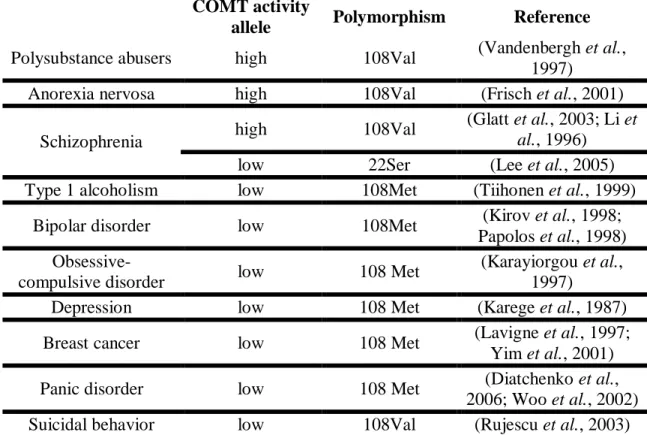
Table II. Polymorphisms of COMT and diseases association.
Table III. Dual salt system concentrations used in hSCOMT-Epoxy Sepharose
retention assays.
Table IV. Screening of binding buffer pH in six AAIL’s supports.
Table V. Typical pKa’s and pI’s of amino acids immobilized in the chromatographic
supports study.
Table VI. Summary of elution steps used for hSCOMT L-methionine/histidine
ix
Abbreviations list
AADC - Aromatic amino acid decarboxylase AAIL’s - Amino acids immobilized ligands AdoMet - S-adenosyl-L-methionine BSA - Bovine serum albumine
COMT – Cathecol-O-methyltransferase DTT - Dithiothreitol
FPLC - Fast performance liquid chromatography HPLC - High pressure liquid chromatography
hSCOMT - human soluble Cathecol-O-methyltransferase IPTG - isopropylthiogalactosidase
MB-COMT – Membrane-bound Cathecol-O-methyltransferase OD - Optical density
PD - Parkinson’s disease pI - Isoeletric point
PVDF - Polyvinylidene difluoride RT - Room temperature
S-COMT - Soluble Cathecol-O-methyltransferase SDS - Sodium dodecyl sulfate
SDS-PAGE - Sodium dodecyl sulfate -polyacrylamide gel electrophoresis TBS-T - Tris buffer saline-Tween 20
x
Resumo
Nos finais de 1950, Alxelrod e co-autores descreveram a enzima responsável pela O-metilação de catecolaminas e outros catecóis, e no mesmo ano a Catecol-O-metiltransferase (COMT) foi parcialmente purificada e caracterizada. Desde então, foram efectuados vários estudos para caracterizar os três polimorfismos mais frequentes da forma humana da COMT, responsáveis por lhe conferir susceptibilidade térmica e oxidativa. Estes polimorfismos podem ter implicações clínicas, e são o factor genético preponderante em várias doenças neurodegenerativas que envolvam os sistemas catecolaminergicos. Nos últimos 40 anos, foram descritas várias estratégias de purificação, utilizando vários extractos biológicos, e consequentemente vários métodos bioquímicos de separação. Apesar de todos os desenvolvimentos tornam-se necessários métodos de purificação mais simples, rápidos e eficazes que confiram um grau de pureza mais elevado sem comprometer a actividade enzimática da Catecol-O-metiltransferase humana na forma solúvel (hSCOMT).
O objectivo global deste trabalho foi desenvolver/melhorar os processos de purificação da hSCOMT, aumentando o grau de purificação e de recuperação da actividade enzimática para aplicação em domínios farmacológicos, cinéticos e estruturais. Deste modo o trabalho desenvolvido foi assente em três objectivos; os dois primeiros utilizando Cromatografia de Interacção Hidrofóbica (HIC), sendo que no suporte Octil-sepharose o objectivo foi analisar o efeito de agentes destabilizadores, a L-arginina; e no suporte Epoxi-sefarose foi analisar o efeito do sistema de dois sais nomeadamente, citrato de sódio (Na3C6H5O7) e sulfato de amónio (NH2SO4). Por
último, utilizar cromatografia de pseudo-bioafinidade em suportes com aminoácidos imobilizados, com o objectivo de analisar a selectividade destes suportes utilizando estratégias de eluição não específicas, sal e alteração de pH.
Estudos realizados pelo nosso grupo de investigação, demonstraram que a estratégia do sistema de dois sais apresenta uma selectividade adequada e consequentemente um factor de purificação incrementado, reduzindo-se assim os efeitos de desnaturação da proteína hSCOMT na presença de elevadas concentrações de sal. Deste modo, tornou-se conveniente analisar a aplicação de uma estratégia de dois sais no suporte Epoxi-sefarose com o objectivo de melhorar o processo de purificação da enzima hSCOMT. Especialmente os resultados demonstraram que, neste suporte, as concentrações de sal necessárias para promover a retenção da hSCOMT são superiores a
xi 0,7 M NH2SO4/0,15 M Na3C6H5O7, tornando assim esta estratégia inadequada em
comparação com estratégias já descritas. Em geral, devido à forte ligação hidrofóbica entre proteínas e suportes cromatográficos, a eluição poderá ser comprometida, não permitindo eluições com a simples diminuição da concentração de sal. Este facto é relevante na interacção entre hSCOMT-Octil, em que a utilização dos sistemas de dois sais necessita da diminuição da temperatura de modo a promover a eluição completa da proteína alvo.
Em geral, os amino ácidos, têm sido amplamente utilizados, para melhorar estratégias de purificação ou para estabilizar proteínas durante o processo cromatográfico. Especificamente, a L-arginina é altamente eficiente melhorando o desempenho de colunas cromatográficas. Neste caso, torna-se conveniente a optimização dos métodos de fraccionamento no suporte Octyl, testando condições de eluição destabilizadoras pela incorporação da L-arginina na fase móvel. No trabalho desenvolvido, estudos preliminares mostram que este aminoácido poderá interferir na estrutura nativa da COMT, promovendo uma diminuição da actividade específica. Adicionalmente, o grau de purificação alcançado denota que a estratégia apresentada não cumpre os requisitos necessários para a purificação da proteína alvo.
Os suportes de aminoácidos têm sido utilizados na separação de várias biomoléculas, no entanto, estes suportes nunca foram utilizados na purificação da proteína hSCOMT. Deste modo, o último objectivo é analisar a performance de ligandos típicos de pseudo-bioafinidade (arginina, metionina, histidina, L-aspartato, L- glutamina and L-leucina) como etapa central no isolamento da COMT, de modo a compreender a selectividade alcançada. Especialmente, foram utilizados perfis de eluição não específicos baseados na manipulação do pH e na concentração de sal. As condições utilizadas de forma a promover a retenção da hSCOMT mostraram que: 1) são necessárias altas concentrações para promover a retenção da proteína, que favorecem o decréscimo da actividade da proteína; 2) na manipulação de pH, a necessidade de pH’s ácidos para promover a retenção da hSCOMT, levam a uma alteração do peso molecular e consequente perda de actividade enzimática. Em síntese conclui-se, comparando as duas estratégias utilizadas, que o factor crítico para este facto é independente dos suportes utilizados, e altamente influenciável pelas condições estabelecidas para promover a ligação da hSCOMT.
Como conclusão, a comparação entre as três abordagens descritas na dissertação demonstram as limitações existentes no delineamento das estratégias de purificação
xii desta proteína a partir de misturas biológicas complexas. No entanto, comparando entre as três partes exploradas, e apesar de serem necessários estudos estruturais de modo a caracterizar e compreender o mecanismo de ligação, os suportes de aminoácidos tornam-se mais vantajosos devido a simplicidade e eficiência nos processos cromatográficos.
Palavras-Chave
Catecol-O-metiltransferase humana, Cromatografia de Interacção Hidrofobica (HIC), Cromatografia de Pseudo-Bioafinidade, Purificação.
xiii
Abstract
Catechol-O-methyltransferase (COMT), play an important role in the metabolism of catecholamines, catecholestrogens and catechol drugs and consequently has a closer relationship with several mental disorders. As a result, while the development of pharmaceutical Human Soluble COMT (hSCOMT) trials for a rational drug design depends on the availability of high purified samples, more suitable purification strategies must be developed and emerged in order to fulfil the requirements of pharmaceutical industry.
In this context, the global aim of this work was the improvement of recovery yields and activity protein results in a hSCOMT purification process. Therefore, the work was being oriented according to three intermediate goals. Two first’s concerning Hidrophobic Interaction Chromatography; (1) Octyl-sepharose support in order to analyse new fractionation methods by testing destabilizing elution conditions with the incorporation of L-arginine in the mobile phase; (2) the application of a dual salt system in Epoxy-sepharose resin; (3) the pseudo-bioaffinity chromatography using six commercial resins with immobilized amino acids, examining the ligands performance, in order to understand the selectivity’s achieved based on non-specific desorption profiles supported on pH and ionic strength manipulation.
In general for Epoxy-sepharose support, the concentrations of dual salt system required to allow hSCOMT retention was above 0,7 M NH2SO4/0,15 M Na3C6H5O7.
Indeed, this strategy is needless, considering previous purification strategies described that allow satisfatory protein selectivity and purification factor with less salt concentration; and consequently with a reduction in hSCOMT denaturation effects.
The L-arginine is highly effective in improving the performance of various chromatographic columns. However, is this study preliminary stability assays showed that this amino acid decreases hSCOMT specific activity; showing that this strategy do not comprise hSCOMT purification strategy requests.
This is the first report using amino acids as immobilized ligands (AAIL’s) as pseudo-bioaffinity supports in hSCOMT isolation. Based on non-specific desorption profiles the results show that; (1) the high concentrations necessary to promote hSCOMT retention, decreasing product activity recovery; (2) for pH approach, the requirement of acidic pH’s to allow hSCOMT retention leads to a molecular weight alteration and consequently loss of enzymatic activity. The comparison between these
xiv two strategies, reveal that the hSCOMT molecular weight discrepancy are not AAIL
dependent, but due to binding conditions.
In conclusion, the comparison of these three approaches in this dissertation demonstrated the complexity of hSCOMT purification processes. In spite of, structural studies are need to understand the hSCOMT-AAIL binding mechanism, these supports have furthermost advantages over the earlier methods published due of its simplicity and efficiency for hSCOMT purification.
Keywords
Human Cathecol-O-methyltransferase, Hydrophobic Interaction Chromatography (HIC), Pseudo-bioaffinity chromatography, Purification.
1
2
Chapter I. Catechol-O-methyltransferase: An overview
In the late of 1950s, Axelrod and coworkers described the enzyme catalyzed-O-methylation of catecholamines and other catechols (Axelrod et al., 1958), in the same year Cathecol-O-methyltransferase (COMT) was partly purified and characterized (Axelrod and Tomchick, 1958). The subsequent basic research occured since 1958 as far as 1975, and the protein purification has revealed some heterogeneity in COMT activity, stability and molecular weights (Guldberg and Marsden, 1975). Also multiple forms of COMT have been demonstrated in a variety of tissues, supporting the hypothesis of isozymes (Assicot and Bohuon, 1971; Huh and Friedhoff, 1979). The enzyme has been found in organisms at various phylogenic levels, from bacteria and yeast to man (Guldberg and Marsden, 1975; Männisto, 1994), Afterwards two distinct forms of COMT have been identified, based on subcellular fractionation studies, a soluble (S-COMT) and a membrane-bound form (M-COMT) (Roth, 1992).
Although, the interest in COMT was rekindled in the late 1980s, when the potent and selective second-generation COMT inhibitors were developed (Mannisto and Kaakkola, 1989, 1990) as adjuncts in L-DOPA therapy for Parkinson’s diseases (PD). Since that, much work on the biochemical characterization of COMT was done with purified enzyme from rat tissues and human placenta. However, with the disclosure of mammalian COMT cDNAs (Bertocci et al., 1991; Lundstrom et al., 1991) and genes it was possible to express both rat and human S- and MB-COMT in heterologous systems, eukaryotic (Tilgmann et al., 1992; Ulmanen et al., 1997) and prokaryotic (Bonifacio et
al., 2001; Lundstrom et al., 1992; Malherbe et al., 1992; Passarinha et al., 2007),
allowing a more detailed characterization of the functional properties of the enzyme, its subcellular localization, and its three-dimensional structure (Vidgren et al., 1994).
In spite of the crystal structures of rat COMT have provided a useful basis for development of COMT inhibitors used in the primary treatment of PD (Bonifacio et al., 2002; Kaakkola et al., 1994; Learmonth et al., 2005; Learmonth et al., 2004; Lerner et
al., 2003; Lerner et al., 2001; Masjost et al., 2000; Palma et al., 2003; Palma et al.,
2006), and Entacapone and Tolcapone (first COMT inhibitor commercialized in 2003) are currently being used clinically in treatment of Parkinson disease, structures of the human protein are desirable. However, was only in 2009 the first succeeded crystallization of soluble form of Human COMT (hSCOMT) (Rutherford et al., 2008b).
In the last years, several studies have been performed in order to characterize the polymorphism of human COMTs. Three common polymorphisms were described (A22S, A52T, and V108M), two of which (A22S and V108M) render the protein
3
Chapter I. Catechol-O-methyltransferase: An overview
susceptible to deactivation by temperature or oxidation (Rutherford and Daggett, 2009). Tipically, polymorphisms could have clinical implications, and are a candidate gene in many neurologic disorders involving catecholaminergic systems.
1.1 Enzymatic reaction and physiological role
COMT enzyme catalyzes the transfer of a methyl group from the coenzyme S-adenosyl-L-methionine (AdoMet) to one of the hydroxyls (preferentially 3-hydroxyl) in a variety of endogenous and exogenous catechol or substituted catechols substrates in the presence of an Mg+2 ion (Figure 1). The chemical step of the reaction was revealed to be an SN2-like process (Woodard et al., 1980), and likely proceeds by sequential
order kinetics mechanism with AdoMet-binding first, followed by an Mg2+ ion, and then by the catechol substrate. S-adenosyl-L-homocysteine the last ligand, wich is released in the catalytic cycle (Lotta et al., 1995).
Figure 1. Reaction mechanism of COMT. AdoMet and S-adenosyl-L-homocysteine. (Adapted from (Lundstrom et al., 1995)).
This enzymatic O-methylation plays an important physiological role in the inactivation of biologically active and toxic catechols. These substrates include: a wide variety of catechols, namely catecholamines with hormonal and neurotransmission activities (dopamine, norepinephrine, epinephrine, catecholestrogens) and their hydroxylated metabolites, ascorbic acid (Guldberg and Marsden, 1975; Kopin, 1985; Mannisto et al., 1992), dietary phytochemicals, indolic intermediates of melanin metabolism (Smit et al., 1990), xenobiotic catechols like carcinogenic catechol-containing flavonoids (Zhu et al., 1994) and a multitude of drugs with a catechol structure by methylation and consequently inactivation (Guldberg and Marsden, 1975).
4
Chapter I. Catechol-O-methyltransferase: An overview
1.2 Gene and Protein characterization
1.2.1 The COMT gene: localization and structure
In humans, COMT gene was localized in locus 22q11.2 (Grossman et al., 1992a; Winqvist et al., 1992). The regulation of the COMT gene expression seems to occur at several levels, (i) transcription initiation, (ii) translation initiation and (iii) mRNA splicing. The gene organization together with the complex regulation enables the expression of two different COMT proteins, S- and MB-COMT (Lundstrom et al., 1995).
The overall structure of the COMT gene, showed in Figure 2, is composed of six exons. At 5’ region, the first two exons are noncoding and the translation initiation codons for the membrane bound (MB-ATG for MB-COMT polypeptide) and soluble (S-ATG for S-COMT form) isoforms are located on the third exon.
The size of the 3' untranslated region is 274 bp and the larger COMT mRNA also has several 5' ends (Lundstrom et al., 1995). The shorter transcript initiates at multiple sites in the region between the two translation initiation ATG codons, and these two alternative splicing products have different capacity for the translation of MB- and S-COMT polypeptides (Tenhunen and Ulmanen, 1993).
The expression of the COMT gene is controlled by two distinct promoters located in exon 3 (Lundstrom et al., 1991). The upper promoter (P2) is constitutively expressed. In contrast, the lower promoter (P1) is regulated in a tissue-specific manner and so, the amount of the shorter transcript varies from one tissue to another (Lundstrom et al., 1995).
5
Chapter I. Catechol-O-methyltransferase: An overview
Figure 2. The structure of human COMT gene. Thin line represents introns and the boxes the
exons. The black boxes indicate the protein coding regions. Two identified promoters, P1 and P2, are shown by black bars. COMT mRNA species expressed from the genes (1.3 and 1.5 kb) are presented as white bars. The positions of translation initiation codons for MB-COMT polypeptide (MB-ATG, MB-AUG), S-COMT polypeptide (S-ATG, S-AUG) and for translation stop codons (TGA, UGA), and the sizes of exons and introns are also shown (Lundstrom et al., 1995).
1.2.2 The COMT protein: localization and characterization
The COMT protein is a monomeric (Tilgmann and Kalkkinen, 1991) and nonglycosylated enzyme (Tilgmann and Ulmanen, 1996). This protein exists as two isozymes: a ubiquitous 221-residue soluble form and a 271-residue membrane-bound form (Bertocci et al., 1991; Lotta et al., 1995), and their molecular weight are 24.4 and 29 kDa, respectively (Bertocci et al., 1991; Lundstrom et al., 1995). In addition, chromatofocusing revealed that isoelectric points of S-COMT is 5.5 (White and Wu, 1975).
The membrane isoform of COMT is an integral membrane protein with the catalytic portion of the enzyme oriented toward the cytoplasmic side of the membrane (Ulmanen and Lundstrom, 1991). Their 50 additional amino acids can owning a stretch of 17 hydrophobic amino-acid residues (Bertocci et al., 1991; Lundstrom et al., 1991). The arrangement of positively charged amino acids spanning the putative anchor sequence, suggests that MB-COMT may be oriented towards the cytoplasmic side of the membrane, reminding the membrane proteins of the class Ib type (Singer, 1990; von Heijne and Gavel, 1988). Nevertheless, the protein appears mostly in a soluble form (S-COMT), and only a minor fraction is in the particular form (MB-COMT) (Guldberg and Marsden, 1975; Roth, 1992).
6
Chapter I. Catechol-O-methyltransferase: An overview
1.2.2.1 Subcellular localization
In general, the indication of COMT subcellular distribution was obtained through differential centrifugation. The major S-COMT activity is in the non-sedimenting and cytoplasmic fractions (Guldberg and Marsden, 1975; Roth, 1992; Tilgmann et al., 1992), while MB-COMT was previously assigned to the outer mitochondrial and plasma membrane (Lundstrom et al., 1992; Tilgmann et al., 1992). Indeed, currently MB-COMT subcellular localization was described in the rough endoplasmic reticulum (Tilgmann et al., 1992; Ulmanen et al., 1997).
1.2.2.2 Tissues distribution
In mammals, COMT is widely distributed throughout the organs. The S-COMT form represents approximately 70-80% of the total COMT proteins whereas in brain only 30% of COMT proteins is in S-form (Table 1) (Lundstrom et al., 1995; Tenhunen
et al., 1994). The highest activity is found in the liver, followed by the kidneys and
gastrointestinal tract (both stomach and intestine) (Lundstrom et al., 1995; Tenhunen and Ulmanen, 1993).
As observed in table I, the difference in the expressed protein levels can be explained by different translation efficiency of COMT polypeptides from the larger COMT mRNA (Mannisto et al., 1992; Nissinen et al., 1988). In most tissues the level of the shorter COMT mRNA, capable of expressing only the S-COMT polypeptide, exceeds the longer transcript. Tipically, the long transcript has been found in all tissues analyzed, with higher levels in human liver, brain, kidneys, adrenals, and lungs. On the other hand, the short transcript, is particularly abundant in liver, kidneys, and mammary glands and it is found in very small amounts in the human brain (Hong et al., 1998; Tenhunen et al., 1994).
Table I.Quantization of S- and MB-COMT polypeptides in human tissues expressed as % of
total COMT (Lundstrom et al., 1995; Tenhunen and Ulmanen, 1993).
S-COMT MB-COMT Liver 85 15 Kidney 77 23 Adrenal medulla 74 26 Duodenum 89 11 Brain 30 70
7
Chapter I. Catechol-O-methyltransferase: An overview
1.2.2.3 Differences of S- and MB-COMT
Total In spite of different molecular weight, and subcellular localization, S-COMT and MB-S-COMT have a similar kinetic mechanism (Mannisto et al., 1992), similar affinities for AdoMet (Jeffery and Roth, 1987; Lotta et al., 1995), magnesium, inhibition by calcium, and optimal pH for the activity (Mannisto and Kaakkola, 1999). However, the most distinct difference between S- and MB-COMT is the different substrate specificities, and the regioselectivity of methylation (Gordonsmith et al., 1982; Lau and Bruice, 1998; Lotta et al., 1995; Malherbe et al., 1992; Mannisto and Kaakkola, 1999). As S-COMT and MB-COMT seem to exhibit different functions, and is the membrane isoform that is more relevant in the inactivation of xenobiotic catechols and the latter playing an important role in the termination of catecholaminergic neurotransmission (Lotta et al., 1995; Roth, 1992).
1.2.2.3.1 Substrate specificities and regioselectivity
MB-COMT has a higher affinity for dopamine and others catechols (Gordonsmith et al., 1982; Malherbe et al., 1992). The adjacent membrane with a charged structure or an additional structural part in the amino end of MB-COMT contributes significantly to the higher binding affinity of the substrates (Lotta et al., 1995). Indeed, for an extended charged side chain of the substrate, the membrane anchor region of MB-COMT (a possible helix) or the membrane itself causes a more favorable binding interactions (Lotta et al., 1995). Also, physiological substrate concentrations and possible differences in substrate selectivity have to be considered when the relative importance of either enzyme subtype is assessed (Mannisto and Kaakkola, 1999).
It is well known that, both enzyme isoforms promote 3-0-methylation, and MB-COMT is even more regioselective than S-MB-COMT. The meta/para ratio is higher, 22 to 88 (depending on the substrate) in MB-COMT than in S-COMT, 4 to 15 (depending on the substrate) (Gordonsmith et al., 1982; Lotta et al., 1995). The reason may be that as the p-hydroxyl group (i.e., 4-O-hydroxyl) approaches the AdoMet; this forces the side chain to become orientated in an unfavorable position with the hydrophobic protein residues of the catalytic site (Gordonsmith et al., 1982; Lau and Bruice, 1998).
8
Chapter I. Catechol-O-methyltransferase: An overview
1.2.2.4 COMT structure: Crystallographic studies
The first COMT crystallization was obtained in 1994 (Vidgren et al., 1994), of rat soluble Cathecol-O-methyltransferase. In XXI century, crystallization of rSCOMT structure, complexed with various inhibitors, had provided considerable insight into the recognition of substrates (Bonifacio et al., 2002; Lerner et al., 2001; Palma et al., 2006). Rat and human COMT share 81% sequence identity and both belong to the highly structurally conserved AdoMet-dependent methyltransferase fold family (class I) (Cheng and Roberts, 2001; Martin and McMillan, 2002). In spite of, rat and human COMT similarities the structure of the human COMT are desirable because the proteins of two species differ in their kinetic properties (Km values generally are higher in the rat enzyme than in human COMT, and the relative specificities for specific catechols can differ). However, only in 2008 hSCOMT was crystallized. (Rutherford et al., 2008b).
Specifically, the structure of hSCOMT is composed of a seven-stranded β-sheet core (3↑2↑1↑4↑5↑7↓6↑) and wiched between two sets of α-helices (Figure 3). The active site of COMT consists of the AdoMet binding domain and the actual catalytic site (Veerapandian, 1997). The results of crystallographic studies indicated that the catalytic site of S-COMT was formed by the Mg2+ ion and relevant amino acids for substrate-binding and methylation catalysis. The substrate-binding motif of the AdoMet site is similar to the Rossman fold, which is a common feature of many nucleotide binding proteins (Mannisto and Kaakkola, 1999).
Figure 3. Crystal structure of hSCOMT. Ribbon diagrams of human 108V S-COMT colored
from blue (N-terminus) to red (C-terminus) are shown in stereo. AdoMet (magenta) and 3,5-dinitrocatechol (dark blue) are shown in stick representation. K+ (cyan), Mg2+ (green), and the side chain of residue V108 (gray) are shown in space-filling representation (Rutherford et al., 2008b).
9
Chapter I. Catechol-O-methyltransferase: An overview
In summary, the Lys144 and Glu199 residues participated in the methylation reaction (Veerapandian, 1997; Woodard et al., 1980), and Trp38 and Pro174 residues were positioned at the surface of the enzyme and sandwiched the planar catechol ring system (Bonifacio et al., 2002; Lerner et al., 2001; Vidgren et al., 1994), in order to maintain the proper positioning for catalytic reaction. The Leu198, Met201, and Trp38 residues made a hydrophobic wall around the ligand binding site, while Met201 residue has variable conformations depending on the bound ligand and adjusts the size of the ligand-binding site (Bonifacio et al., 2002). The Pro174 and Leu198 residues are known to contribute significantly to the stabilization of the complex (Learmonth et al., 2004). Furthermore, lipophilic Leu198 residue influences the regioselectivity of ortho- and meta-nitrated inhibitors (Palma et al., 2006). Indeed, positively charged Lys144 and S-Met group of AdoS-Met influence the electrostatic effects of catechol ring substitution and the selectivity of several inhibitors.
1.3 Expression of recombinant COMT proteins
Recombinant production of proteins is a major step forward in the development of biotechnology products that became possible with the advent of recombinant DNA technologies.
In the past decades, with the disclosure of mammalian COMT gene and cDNA synthesis (Bertocci et al., 1991; Lundstrom et al., 1991) it was possible to express recombinant COMT proteins in heterologous system such as eukaryotic (insect (Tilgmann et al., 1992) and mammalian cells (Malherbe et al., 1992; Tilgmann et al., 1992; Ulmanen et al., 1997)) and prokaryotic (Bonifacio et al., 2001; Lundstrom et al., 1992; Malherbe et al., 1992; Passarinha et al., 2007; Xu et al., 1999) systems. The last one used different strains of Escherichia coli (E. coli). In spite of all the afore-mentioned systems having produced functional forms of the protein, the improvement of recombinant COMT, in large scale expression E. coli is a promising host. Since it is completely lacks endogenous COMT enzyme (Tilgmann and Ulmanen, 1996) and in order to improve the volumetric and mass productivity for several biopharmaceutical and neurological domains (Bonifacio et al., 2001). Indeed, this expression system allows the highest levels of expressed soluble, non-glycosilated and moderate size proteins (Ibdah et al., 2003; Vilbois et al., 1994), such as COMT.
10
Chapter I. Catechol-O-methyltransferase: An overview
1.4 Purification procedures of COMT proteins
The development of techniques and methods for the separation and purification of proteins have been essential for several advancements in biotechnology research. The purity of a protein is a pre-requisite that has to be established for structure and function studies or its potential application.
During the last 40 years, several purification procedures were been described to COMT from different biological extracts. The enzyme has been enriched by various biochemical separation methods and strategies, such as differential centrifugation, ammonium sulfate (NH2SO4) fractionation (Ball et al., 1971), size exclusion
(Lundstrom et al., 1992) and anion (Ball et al., 1971; Lundstrom et al., 1992) or cation exchange chromatography (Tilgmann and Ulmanen, 1996; White and Wu, 1975). Recently, affinity chromatographic methods have been described for the purification of recombinant rat and human COMT as a fusion protein (Bonifacio et al., 2001; Cotton et
al., 2004). In spite of this chromatographic procedure leads to low percentage recovery,
it allows the purification of the recombinant enzyme in sufficient amounts for structure– function studies (Bonifacio et al., 2002; Palma et al., 2003; Rodrigues et al., 2005). Also, preparative reversed-phase chromatography is described for the purification of human COMT (Lundstrom et al., 1992), but the denaturing conditions made it impossible to use the enzyme for biochemical and functional analysis. Only a few authors described hydrophobic interaction chromatography (HIC) as part of the downstream purification process for this recombinant protein (Nunes et al., 2009; Passarinha et al., 2006; Passarinha et al., 2008). In fact, this strategy could offer great advantages in terms of increased yields and reduced the number of chromatographic steps.
Accordingly, most of the formerly published purification protocols for COMT have presented multi-step procedures requiring application of multiple chromatography types and chemical and physical manipulations in order to achieve the desired level of purity. Therefore, the demands of more simple, reliable and rapid purification methods are needed to recover highly pure, homogeneous and active COMT protein.
11
Chapter I. Catechol-O-methyltransferase: An overview
1.4.1 COMT stability
The global aim of a protein purification process is not only the removal of contaminants, but also the concentration of the desired protein and their transfer to an environment where it is stable and in a formulation for the intended application. In general, proteins for pharmaceutical applications must be stable over 2 years or longer, against several stress factors encountered during storage, shipping and handling. Various additives (designed as excipients) are used to enhance stability and reduce aggregation of the proteins against these effects (Arakawa et al., 2007a).
Several purification data suggested that COMT is fairly labile and loses rapidly its activity during the isolation process and storage (Tilgmann and Kalkkinen, 1990), probably due to the oxidation of the free cysteine-SH groups. Indeed, S-COMT highly purified fractions show a more rapid decrease in activity (Cotton et al., 2004). Experimental observations reveal that EDTA and MgCl2 in equimolar concentrations
(Ball et al., 1971) conjugated with the reducing agent DTT (Cotton et al., 2004) into buffers, have a stabilizing effect on all enzyme preparations. Similarly, the use of reducing agents as mercaptoethanol, DTT or L-cysteine by other investigators had stabilized the enzyme and allowed the purification and partial characterization of human COMT (Assicot and Bohuon, 1970). In this context, it can be concluded that since reducing agents can restore COMT activity, the rapid inactivation observed in purified S-COMT samples can be a result of the oxidation of sulphydryl group(s) (Assicot and Bohuon, 1970).
1.5 Genetic polymorphism of COMT and diseases association
The human COMT gene contains three common coding polymorphisms: A22S, A52T, and V108M (Cargill et al., 1999; Lee et al., 2005; Saito et al., 2001; Shield et
al., 2004), leaving the protein susceptible to deactivation by temperature or oxidation.
Although, the structural and epidemiological effects of the V108M mutation are the best characterized. The side chain of residue 108 is buried within a hydrophobic pocket in a loop between R5 and β3 from the protein’s active site. The mutation distorted the overall structure of COMT, increasing the solvent exposure of both the AdoMet- and catechol-binding sites, as showed in Figure 4 (Rutherford et al., 2006). While the wild-type and V108M proteins display similar kinetic properties in vitro (Chen et al., 2004; Goodman et al., 2002; Lotta et al., 1995), the V108M polymorphism destabilizes the
12
Chapter I. Catechol-O-methyltransferase: An overview
protein, increasing its susceptibility to thermal (Rutherford et al., 2008a; Shield et al., 2004) and chemical denaturation (Rutherford et al., 2008a) as well as oxidative stress (Cotton et al., 2004; Li et al., 2005; Li et al., 2004). This destabilization results in the protein levels decrease (Chen et al., 2004; Doyle et al., 2004; Doyle and Yager, 2008), and therefore activity (Boudikova et al., 1990; Grossman et al., 1992b; Scanlon et al., 1979; Spielman and Weinshilboum, 1981; Weinshilboum and Dunnette, 1981) in vivo relative to the wild-type protein.
In the A22S mutation, residue 22 is positioned in a surface loop between R1 and R2, 13 Å from the AdoMet-binding site and ∼20 Å from the catechol-binding site. This polymorphism significantly increases COMT’s affinity for AdoMet but decreases COMT activity by 30% relative to the wild-type protein (Li et al., 2005; Rutherford and Daggett, 2009).
In addition, while the A52T mutation slightly decreases COMT’s affinity for AdoMet, it has no significant effect on COMT activity (Li et al., 2005). Typically, residue 52 is positioned on the protein surface in helix R3 which contains critical residues for the binding of AdoMet and catechol substrates. The 52T protein have a T50 (temperature resulting in 50% inactivation) value more low to wild-type (Shield et al., 2004).
Figure 4. A22S, A52T, and V108M COMT polymorphisms. Ribbon diagram of wild-type
COMT colored from blue (N-terminus) to red (C-terminus). AdoMet and 3,5-dinitrocatechol are shown in stick representation and colored by atom type. Polymorphic residues 22, 52, and 108 are shown in space-filling representation and colored blue, cyan, and green, respectively (Rutherford and Daggett, 2009).
In spite of both the 22S and 108M alleles have been associated with neuropsychiatric dysfunction (Frisch et al., 2001; Glatt et al., 2003; Karayiorgou et al., 1997; Karege et al., 1987; Kirov et al., 1998; Lee et al., 2005; Li et al., 1996; Papolos et
13
Chapter I. Catechol-O-methyltransferase: An overview
al., 1998; Rujescu et al., 2003; Woo et al., 2002), only the allele 108M is known to be
related to an increased risk of cancer (Lavigne et al., 1997; Yim et al., 2001). Interestingly, recent studies have linked the 108M allele with increased sensitivity to pain (Woo et al., 2002) and with improved prefrontal cognition (Sheldrick et al., 2008), as described in table II. No data associating the A52T polymorphism with disease have been published.
Table II. Polymorphisms of COMT and diseases association
COMT activity
allele Polymorphism Reference
Polysubstance abusers high 108Val (Vandenbergh et al., 1997)
Anorexia nervosa high 108Val (Frisch et al., 2001) Schizophrenia high 108Val
(Glatt et al., 2003; Li et
al., 1996)
low 22Ser (Lee et al., 2005) Type 1 alcoholism low 108Met (Tiihonen et al., 1999)
Bipolar disorder low 108Met (Kirov et al., 1998; Papolos et al., 1998)
Obsessive-compulsive disorder low 108 Met
(Karayiorgou et al., 1997)
Depression low 108 Met (Karege et al., 1987) Breast cancer low 108 Met (Lavigne et al., 1997;
Yim et al., 2001) Panic disorder low 108 Met (Diatchenko et al.,
2006; Woo et al., 2002) Suicidal behavior low 108Val (Rujescu et al., 2003)
In general, the level of COMT enzyme activity is genetically polymorphic in human tissues with a trimodal distribution of low (COMTLL), intermediate (COMTLH), and high (COMTHH) activity (Boudikova et al., 1990; Weinshilboum and Raymond, 1977). The enzyme activity is ubiquitous and these levels vary not only among different species (Ellingson et al., 1999; Schultz et al., 1989), but also in individuals of the same species (Palmatier et al., 1999) as well as in tissues from the same individuals (Ellingson et al., 1999; Guldberg and Marsden, 1975; Mannisto and Kaakkola, 1999).
14
Chapter I. Catechol-O-methyltransferase: An overview
1.5.1 COMT role in Parkinson disease
Parkinson’s disease is a progressive neurodegenerative disorder caused by the loss of dopaminergic nigrostriatal neurons, leading to characteristic motor symptoms. Until now, the most effective treatment for this disease is the dopaminereplacement therapy with L-DOPA (dopamine precursor) together with an inhibitor of aromatic amino acid decarboxylase (AADC), as showed in Figure 5. The efficacy of this therapy, however, decreases with time and most patients develop fluctuating responses and dyskinesias (Bene et al., 2009). The last decade showed that the use of COMT inhibitors as adjuvants to the L-DOPA/AADC inhibitor therapy, in order to increase the bioavailability of L-DOPA, improve the clinical benefits of this therapy (Figure 5) (Bene et al., 2009). A historical overview of the discovery and development of COMT inhibitors reveal a special emphasis on nebicapone, presently under clinical development, as well as entacapone and tolcapone, which are already approved as adjuncts in the therapy of PD (Bonifacio et al., 2007). In spite of the association of the COMT alleles with PD has been extensively studied, no association has been found (Hoda et al., 1996; Syvanen et al., 1997; Xie et al., 1997). Nevertheless, some results demonstrated that some Japanese individuals with the low COMT activity allele may have an increased risk for PD (Kunugi et al., 1997; Yoritaka et al., 1997).
Figure 5. Dopamine replacement in PD therapy with dopamine precursor (L-DOPA). Scheme
15
Chapter I. Catechol-O-methyltransferase: An overview
1.6 Analytical methods in COMT assays
Typically, COMT activity analysis has been applied clinically since COMT inhibitors have been introduced as adjuvant drugs in the treatment of PD (Pihlavisto and Reenila, 2002). The discovery of COMT as a drug target and the identification of polymorphic COMT forms (soluble and membrane-bound) have increased the demand for reliable, sensitive and fast analytical activity assays for COMT activity. For example, for the:
-Measurement of the recombinant protein activity; -Testing in vitro the efficacy of new inhibitor candidates; -Determination of structure–activity relationships;
-Activity measurement in various physiological and pathophysiological states. In general, there is a great variation in analytical methods of COMT assays. These strategies consist in the sample handling and incubation followed by separation and detection of the reaction products. The major combinations methods are shown in Figure 4, such as separation of the COMT reaction products by solvent extraction and detection by fluorometry (Axelrod et al., 1958) or radiochemical techniques (Bates et
al., 1979; Hong et al., 1998; Zurcher and Da Prada, 1982). However, the introduction of
High Pressure Liquid Chromatography (HPLC) techniques coupled with UV detection (Pennings and Van Kempen, 1979), fluorometric (Nohta et al., 1984; Smit et al., 1990), radiochemical (Nissinen, 1985) and electrochemical detection (Nissinen and Mannisto, 1984; Passarinha et al., 2006; Schultz et al., 1989) have improved the sensitivity and specificity of the analysis. However, activities between different COMT enzyme sources are not necessarily comparable and the distinct protein assay methods in samples should also be considered (Pihlavisto and Reenila, 2002), in order to access COMT activity.
16
Figure 6. Analytical methods applied to COMT activity analysis (Adapted from (Pihlavisto
17
18
Chapter II. Aims and Outline
The global aim of this work was the improvement of recovery yields and activity protein results in hSCOMT purification process. Therefore, the work will be oriented according to the following intermediate goals:
Chapter IV. Hydrophobic Interaction Chromatography: Octyl and Epoxy supports
Octyl-sepharose support and destabilizing elution conditions using L-arginine
- To optimize new fractionation methods in Octyl-sepharose sorbents by testing destabilizing elution conditions with the incorporation of L-arginine in the mobile phase.
Epoxy-sepharose support and dual salt system application
- To analyze the application of dual salt system effects in epoxy-sepharose matrice, in order to improve hSCOMT downstream processes.
Chapter V. Pseudo-Bioaffinity Chromatography: Amino Acids as Immobilized Ligands
- To examine the performance of pseudo-bioaffinity ligands, based on amino acids matrices in the isolation of hSCOMT from E. coli lysates. In this point, the intermediate aims will be understanding the selectivity’s achieved by these new supports and consequently their influence in the purity and kinetic properties of the target protein. Typical elution methods will be developed based on non-specific desorption profiles supported on pH and ionic strength manipulation.
19
20
Chapter III. General methodologies
3.1 Material
Ultrapure reagent-grade water was obtained with a Milli-Q system (Milipore/Waters). Carbenicillin (disodium salt), isopropylthiogalactosidase (IPTG), tryptone, bact-yeast extract, lysozyme, dithiothreitol (DTT), ammonium sulphate (NH2SO4), Tris(hydroxymethyl)aminomethane (Tris), sodium citrate (Na3C6H5O7),
L-cysteine, L-arginine, AdoMet, CAPS, DNase, RNase, epinephrine (bitartrate salt), disodium ethylenediaminetetraacetic (EDTA), citric acid monohydrate, sodium octyl sulfate, dibutylamine, Bovine serum albumin (BSA) and arginine, methionine, L-histidine, L-aspartate, L-glutamine and L-leucine agarose supports were obtained from sigma were obtained from Sigma Chemical Co (St Louis, MO, USA). Potassium chloride (KCl), sodium acetate (anhydrous). Sodium chloride (NaCl) were supplied by Fluka (Buchs, Switzerland). Bis-acrylamide 30% and Bio-Rad protein assay reagent was purchased from (Bio-Rad, Hercules, CA).The full range rainbow protein standards used for estimation of subunit molecular weight, anti-rabbit IgG alkaline phosphatase secondary antibody and Octyl-Sepharose 6FF support were purchased by GE Healthcare Biosciences (Uppsalla, Sweden). Polyclonal rabbit anti-COMT antibody was produced in BIAL using purified recombinant rat COMT (Bonifacio et al., 2001). The epoxy-Sepharose CL-6B was prepared by covalent immobilization of 1,4-butanediol diglycidyl ether on Sepharose CL-6B according to the protocol described elsewhere (Sundberg and Porath, 1974). All other reagents were of analytical grade and used without further purification.
3.2 Methods
3.2.1 Recombinant hSCOMT production and recuperation 3.2.1.1 Plasmid and bacterial strain
The Champion pET101 Directional TOPO expression kit (Invitrogen Corporation, Carlsbad, CA, U.S.A.) was used for the expression of hSCOMT in its native form on E. coli BL21-Star (Invitrogene, USA) strain. The overexpression of hSCOMT protein is under the control of the IPTG inducible promoter and employing carbenicillin supplementation as a selection marker.
21
Chapter III. General methodologies
3.2.1.2 Recombinant hSCOMT protein production
The recombinant plasmids pET101, with construct hSCOMT, were transformed into E. coli BL21-star cells and grown overnight at 37 ºC in agar plates with LB medium containing carbenicilin disodium salt (50 µg/ml). Next, a single colony was inoculated in 62,5 mL of SOB medium in 250 mL shake flasks and grown at 37 ºC. When they reach an optical density at 600 nm (OD600nm) of 2.6 units, an aliquot was
added in 250mL of SOB medium in 1 L shake flasks, since the inoculation volume was fixed to achieve an initial OD600nm of 0.2–0.3 units. As the OD600nm reached 0.6 units, was induced the recombinant hSCOMT production by the addition of IPTG (final concentration of 1 mM). After induction, cells were grown at 37 ºC during 4 h and collected by centrifugation at 4600 rpm for 20 min at 4 ºC and the pellet was stored at -20 ºC.
3.2.1.3 Cell lysis
The bacterial cell pellet (250 mL) was resuspended in 10 mL of Bial buffer, and disrupted by lysozyme treatment (0.5 mg/mL) during 15 minutes at room temperature (RT). After this, six consecutive freeze (-196ºC) / thaw (42ºC) cycles were performed. After, DNase (250 µg/mL) and RNase (500 µg/mL) are added and the lysate was incubated for 10 minutes at 37ºC. Next, the lysate obtained was centrifuged at 16000 g for 20 min at 4 ºC to remove the cell debris. The supernatant was then applied to chromatographic columns and used in stability assays.
3.2.2 Total protein quantification: Bradford micro-assay
The Bio-Rad Protein Assay, based on the Bradford method, is a dye-binding assay in which a differential color change of a dye occurs in response to various proteins concentrations. The maximum absorbance for an acidic solution of Coomassie® Brilliant Blue G-250 dye shifts from 465 nm to 595 nm when the binding to a protein occurs. The Coomassie blue dye binds essentially to basic and aromatic amino acid residues, especially arginine.
22
Chapter III. General methodologies
3.2.2.1 Procedure
The protein content in samples was measured by the Bio-Rad protein assay reagent (Bio-Rad, Hercules, CA), with BSA as standard samples (1.2 to 10.0 μg/ml), according to manufacturer’s indications.
y = 0,0709x + 0,0102 R² = 0,9951 0 0,2 0,4 0,6 0,8 0 2 4 6 8 10 A b so rv an ce (5 9 5 n m ) BSA concentration (μg/ml)
Figure 7. BSA bial buffer calibration curve.
3.2.3 Analytical chromatography: hSCOMT specific activity assay
The activity assay was designed to evaluate the methylation efficiency of recombinant hSCOMT by measuring the amount of O-methylated reaction products with electrochemical detectors. These detectors have excellent limits of detection and application of a wide range of analytes, suitable for the assay of catecholamines and phenolic hydroxyls of the O-methylated products in an oxidative/reductive mode at inert electrodes. These methods are commonly reported because of the high sensitivity and selectivity achieved (Saxer et al., 2004).
3.2.3.1 Procedure
In hSCOMT activity assay an aliquot of 150 µg/mL (500 µL) of the soluble extract was incubated in reaction mixture, at 37 ºC during 5 min. After, was added 0.1 mL of Epinefrine (1 mM) and the reaction was stopped by adding 200 µL of percloric acid (0.4 M) 10 minutes later. The samples were centrifuged at 6000 rpm for 10 min at 4ºC and after the supernatants were filtered through a 0.22 µm pore size filter to remove precipitated debris. Finally, the injection was performed into the HPLC system (Waters) with an amperometric electrochemical detection.
Chromatographic separation was achieved using a 5 µm particle size XTerra MS C18 ODS reversed-phase analytical column (waters, 250 × 4.6 mm i.d.) connected to a precolumn (Waters, 5 µm, 10 × 4.6 mm i.d.). All buffers pumped in HPLC system were
23
Chapter III. General methodologies
filtered through a 0.22 µm pore size membrane (Schleicher Schuell, Dassel, Germany) and degassed ultrasonically. The mobile fase was pumped by an isocratic mode through the chromatographic system at 1.0 mL/min. All the injections were made with a rheodyne valve, equipped with a 20 µL sample loop. The electrochemical oxidation of metanephrine to their respective O-quinones was performed in a flow single-cell, equipped with a 3 mm diameter glassy carbon working electrode set at +750 mV over an ISAAC reference electrode. The eluent, analytical column and flow single-cell were maintained with a constant temperature (33 ºC) using a preheated module installed in the electrochemical amperometric detector. The method sensitivity was set at 100 nA.
3.2.4 SDS-PAGE and Western blot
Typically, the SDS-PAGE (Sodium Dodecyl Sulfate – Polyacrylamide Gel Electrophoresis) method is useful for molecular weight and purity analysis of proteins. In the presence of the negatively charged detergent, Sodium Dodecyl Sulfate (SDS) binds to the polypeptides in order to form complexes with constant charge to mass ratios. This results in a means of differentiation based on the molecular weight of the molecules. After the electrophoresis, proteins are immobilized on membrane, usually a
polyvinylidene difluoride (PVDF) membrane, and the blot is then probed for a specific
protein using primary antibodies directed against the protein of interest. Subsequently, secondary antibody recognizes the primary antibody. The secondary antibody has been conjugated to an enzyme wich in addition of a specific substract produces a colorimetric reaction in membrane site corresponding to protein.
3.2.4.1 Procedure
Were boiled in a loading buffer for 10 min at 100 ºC and then deposit and run, respectively on stacking (4%) and resolving (12,5%) SDS-PAGE, with a running buffer at 150 V for approximately 90 min. Then, one gel was stained by Comassie brilliant blue R-250 and the second was used to perform Western blot, for the analysis of purity and immunoreactivity, respectively.
For comassie brilliant blue staining procedure, the gel was placed in the staining solution for 30 min on agitation. After this, for excess staining, the gel was placed in the
24
distaining solution with the same procedure. The gel could be stored indefinitely in fixing solution.
The second gel was transferred to a PVDF membrane (GE Healthcare Biosciences, 0.45 µm) in blotting buffer, over a 10 min at 750 mA at 4°C. The PVDF membrane were blocked with Tris buffer saline – Tween 20 (TBS-T) containing 5% (w/v) skimmed milk powder for 1 h and incubated overnight at 4 ºC with rabbit anti-ratSCOMT polyclonal antibody (diluted 1:2000 in TBS-T), that cross reacts with the human protein. After washing three times (15 min) with TBS-T, PVDF membrane was incubated with anti-rabbit IgG alkaline phosphatase secondary antibody (diluted 1:10,000 in TBS-T) for 1 h at RT. Posteriorly, the membranes were washed three times again with TBS-T. Revelation reaction were developed using 200 µL of ECF substrate (GE Healthcare Biosciences) and images of blots were captured with the Molecular Imager FX Pro Plus MultiImager system by chemiluminescence’s detection
25
C
hapter IV
.
Hydrophobic Interaction Chromatography:
26
Chapter IV. Hydrophobic Interaction Chromatography: Octyl and Epoxy supports
4.1 General considerations
Hydrophobic interaction chromatography, a form of multivalent interaction chromatography, complementary to other separation techniques, is a powerful technique in modern biotechnology for the downstream processing of several biomolecules (Mahn
et al., 2005). In general, it is a method that can be applied to all proteins, at
laboratory-scale applications and industrial processes, since HIC can be adapted to the special hydrophobic needs of each protein. Typically, HIC involves the separation of protein molecules owing to the differential hydrophobic interactions between immobilized hydrophobic ligands on the gel and non polar regions on the surface of proteins (Queiroz et al., 2001). The adsorption (and retention) of proteins is carried out by moderate to high concentrations of anti-chaotropic salts followed by a linear or stepwise decrease in the ionic strength of the eluent. Recovery and resolution levels of HIC are often satisfactory (Lienqueo et al., 2003) if a comprehension of certain parameters governing HIC, such as the stationary phase and the mobile phase (Queiroz et al., 2001), could offer a rational optimization scheme for the purification process as a whole. Four parameters govern the adsorption of a protein to a homologous series of hydrophobic stationary phases: (1) the type of ligand, (2) the chain-length, (3) the surface concentration of immobilized ligand and (4) the type of the matrix or support (Tomaz et al., 2002). The most widely used ligands for HIC are aromatic groups (e.g. phenyl) due to the presence of hydrophobic and aromatic (Π–Π) interactions and linear chains alkanes, such as Octyl (Queiroz et al., 2001). Furthermore, ligands with intermediate hydrophobic character, such as Epoxy-Sepharose, have been useful in HIC processes, since they promote a mild binding strength and one elution by simple decreasing the ionic strength of the eluent (Tomaz et al., 2002).
4.2 Procedure
Chromatographic separations were performed on a Fast Performance Liquid Chromatography (FPLC) system (GE Healthcare Biosciences, Uppsalla, Sweden). All buffers pumped in FLPC system were filtered through a 0.22 µm pore size membrane (Schleicher Schuell, Dassel, Germany) and degassed ultrasonically.
The media were packed according to company guidelines (20 mL of gel volume) into a C16 glass column (GE Healthcare Biosciences). The column was equilibrated