Clínica Universitária de Otorrinolaringologia
Mucopolysaccharidoses: Otorhinolaryngologic
manifestations and management
Ana Rita Duarte Serejo Alcobia Portugal
Clínica Universitária de Otorrinolaringologia
Mucopolysaccharidoses: Otorhinolaryngologic
manifestations and management
Ana Rita Duarte Serejo Alcobia Portugal
Orientado por: Dr. Marco Simão
3
Agradecimentos
A realização desta dissertação de mestrado contou com importantes apoios e incentivos sem os quais não se teria tornado uma realidade e aos quais estarei eternamente grata.
Ao Professor Doutor Óscar Dias, pela sua orientação, total apoio, disponibilidade, pelo saber que transmitiu, pelas opiniões e criticas, total colaboração no solucionar de dúvidas e problemas que foram surgindo ao longo da realização deste trabalho e por todas as suas palavras de incentivo.
Ao Dr. Marco Simão, por me ter orientado e pela sua disponibilidade, pela sua participação e colaboração, uma vez que sem ele não seria possível a realização deste trabalho.
À Doutora Sónia Albuquerque, da Biomarin, pela total disponibilidade em fornecer bibliografia necessária e importante para o desenvolvimento da revisão.
Por último, tendo consciência que sozinha, nada disto teria sido possível, dirijo um agradecimento especial aos meus pais, pelo seu apoio incondicional, incentivo, amizade e paciência demonstrados e total ajuda na superação dos obstáculos que ao longo desta caminhada foram surgindo
n Abstract
The mucopolysaccharidoses (MPS) belong to a group of rare lysosomal storage diseases and are caused by a deficiency in enzymes involved in the sequential degradation of glycosaminoglycans (GAGs) leading to a substrate accumulation in various tissues and organs, which can cause multisystemic manifestations. The accumulation of partially degraded GAGs in tissues leads to clinical symptoms that characterize particular MPS subtypes, including growth retardation and progressive damage to respiratory, cardiovascular, musculoskeletal, nervous, gastrointestinal, auditory and visual systems. People with this disorder can be born already with symptoms but are generally born without these alterations and develop them progressively, with potentially life-threatening consequences.
The otorhinolaryngologic abnormalities of these patients encompass otologic alterations, adenotonsilar hypertrophy and airway alterations, to name but a few. Hearing loss is initially mechanical due to otitis media with effusion and thickening of the outer ear structures and, later, with progressive deterioration, with sensorineural component.
Being a progressive disease it is essential to be aware to worsening symptoms and ENT (ear, nose and throat manifestations) evaluation should be done periodically. Thus, otolaryngologists have an opportunity to play an increasingly integral role in the multidisciplinary approach to the diagnosis and management of many children with MPS. Clinical suspicion, early recognition, and prompt diagnosis of these challenging disorders is crucial, as outcomes of treatment in many cases appear time-sensitive, with better results being achieved when intervention is initiated at a younger age or prior to progression of the disease.
Keywords: Otolaryngology; Mucopolysaccharidoses
As MPS pertencem a um grupo de doenças de depósito lisossomal e têm uma incidência global entre 1 em150,000 e os 1 em 10,000 nascimentos vivos.
São causadas por deficiência das enzimas envolvidas na degradação sequencial de glicosaminoglicanos, presentes em todos os tecidos conjuntivos, levando a uma acumulação de substrato em vários tecidos e órgãos, podendo causar manifestações multissistémicas, nomeadamente falência multiorgânica progressiva . Estão descritos sete subtipos de MPS, cada um com uma deficiência congénita de enzimas lisossomais diferente. No entanto, uma
5
seguem uma padrão autossómico recessivo, com exceção da MPS II, que tem um padrão ligado ao X.
Nos doentes afectados, esta doença pode estar presente desde o nascimento, ou mais tarde à medida que a mesma vai progredindo.
A acumulação de GAGs parcialmente degradados nos tecidos causa uma grande variedade de sintomas clínicos, com desenvolvimento crónico e progressivo, incluindo atraso de crescimento e danos progressivos nos sistemas respiratório, cardiovascular, músculo-esquelético (dismorfismo facial e displasia das articulações e esqueleto, incluindo contracturas e mãos caracteristicamente grandes), nervoso (atraso no desenvolvimento ou atraso mental), gastrointestinal, auditivo e visual. Complicações respiratórias recorrentes também são prevalentes e correspondem a uma porção significativa de morbilidade e mortalidade associadas a estas condições.
As anomalias otorrinolaringológicas destes doentes estão entre as manifestações mais frequentes e abrangem, entre outras, obstrução da via aérea (causa da apneia do sono nestes doentes), infecções recorrentes (otites, sinusites e rinites) e défices auditivos. De facto, a avaliação por otorrinolaringologia precede o diagnóstico em 30% dos casos.
Vários factores podem contribuir para o estreitamento da via área nos doentes com MPS e este pode ocorrer apenas ao nível superior ou em vários níveis simultaneamente. A acumulação de GAGs tanto nas amígdalas ,causando hipertrofia das mesmas, como nas paredes da faringe e laringe, causando macroglossia e espessamento dos tecidos moles da laringofaringe é o principal fator, levando a estreitamento da via área superior. Para esta contribuem também a presença de deformidades no crâneo ou coluna, tal como pescoço curto, ponte nasal achatada, laringe anterior alta, cavidade nasofaríngea funda e estreita, deformações mandibulares ou vértebras cervicais com alterações, nomeadamente instabilidade da articulação atlantoaxial. Se estes fatores (que definem a presença de obstrução da via superior) forem acompanhados de traqueobroncomalácia ou acumulação de GAG na mucosa da traqueia, distorção da traqueia em combinação com laxidez do tecido da mesma ou secreções excessivamente espessas podemos estar perante uma obstrução da via aérea em múltiplos níveis.
É importante avaliar o grau de obstrução durante o sono, uma vez que é um factor importante na apresentação clinica. Apneia obstrutiva do sono ocorre em mais de 80% dos doentes (AOS nos adultos é definida como IAH>5, AOS pediátrico aceite como IAH>ou=1,5).
Infecções recorrentes como traqueítes e laringites são frequentes e relacionam-se com a presença de muco viscoso e clearance mucociliar reduzida devido à acumulação de GAG. Para além disto, os depósitos de GAG nas aéreas retronasais, trompas de eustáquio e ouvido médio aumentam o risco de otite media com efusão e otite media aguda.
A Perda auditiva verificada é inicialmente mecânica, devido a otite media com efusão e outras infecções recorrentes ou espessamento das estruturas do ouvido externo ou deformações na cadeia ossicular e, mais tarde, com deterioração progressiva, com componente sensorineural. No passado poucas pessoas com fenótipos graves de MPS atingiam o estado adulto. No entanto, avanços ao nível de métodos de diagnóstico, tratamento multidisciplinar e novas descobertas terapêuticas que aumentam a sobrevida levaram a um aumento do número de indivíduos que sobrevivem para além da infância. Em adultos a diminuição da acuidade auditiva e as alterações mecânicas na laringe são responsáveis por provocar alterações no discurso e dificuldades na comunicação, que por sua vez podem levar a depressão, isolamento e menor compromisso nos cuidados de saúde.
A abordagem diagnóstica destes casos é baseada na suspeita clínica (baseada nas alterações mencionadas anteriormente), exames imagiológicos e avaliação laboratorial, incluindo resultados de analise de GAG urinários e ensaios da actividade enzimática. Análise de mutações ou testes moleculares têm sido utilizados para confirmar o diagnóstico de um tipo particular de mucopolissacaridoses ou para avaliar membros da família quando o tipo de MPS e a mutação familiar já é conhecida; testes moleculares podem ter valor prognóstico se as mutações identificadas estiverem bem caracterizadas.
Como o diagnóstico destas patologias pode ser complexo, é recomendado que os doentes com suspeita de diagnóstico de mucopolissacaridoses sejam referenciados a um geneticista ou especialista metabólico para avaliação.
Nas MPS é recomendado avaliação audiológica sistemática (anualmente), polissonografia e exames fibróticos da via aérea superior e estudos imagiológicos da traqueia e laringe, nomeadamente tomografia computorizada e ressonância magnética). A Polissonografia é a primeira linha de investigação para qualquer individuo com suspeita de MPS e AOS. As técnicas imagiológicas da via aérea superior têm um papel importante na avaliação não invasiva da obstrução da via aérea uma vez que informam sobre diversos locais de possível obstrução e, para
7
além disso, a Tomografia computorizada foi selecionada como uma medida de outcome fiável para a eficácia de intervenção médica e não-médica da AOS.
Tradicionalmente, este grupo de doenças era considerado incurável, com tratamento apenas sintomático e paliativo. Mais recentemente , têm sido feitos esforços no sentido de controlar os processos patológicos com um todo, numa tentativa de bloquear ou pelo menos atrasar a evolução da doença.
Como as mucopolissacaridoses afetam múltiplos sistemas é necessário controlar um diverso espectro de manifestações como parte dos cuidados integrados. A gestão destes sintomas pode incluir utilização de aparelhos adaptativos ou de suporte, terapia física e ocupacional, terapêutica sintomática, intervenções cirúrgicas e terapêuticas para fornecer a enzima em défice. Neste contexto, a terapêutica de reposição enzimática e transplantes de células hematopoiéticas têm tido sucesso em casos selecionados.
Vários estudos revelam que o transplante de células hematopoiéticas pode alterar o curso natural da doença, aumentando a esperança media de vida e melhorando algumas anomalias sistémicas. No entanto, este é um procedimento de alto risco com elevada morbilidade e mortalidade. Assim, a sua indicação depende do tipo de MPS, situação clinica do doente, idade e presença ou não de défices neurológicos.
Mais recentemente, a terapia de reposição enzimática (TRE) tem sido desenvolvida e usada no tratamento de diferentes tipos de mucopolissacaridoses. Actualmente há quatro fármacos aprovados para o tratamento da MPS I (Laronidase; AldurazymeTM – Biomarin Pharmaceutical/Genzyme Co.), MPS II (Idursulfase; ElapraseTM – Shire Human Genetic Therapies Inc.), MPS IV (GALNS-elosulfase alfa; VimzymTM – Biomarin Pharmaceutical) e MPS VI (Galsulfase; NaglazymeTM – Biomarin Pharmaceuticals) e consistem na administração endovenosa periódica da enzima especifica em falta, permitindo uma maior degradação de GAG nos órgãos e tecidos e promovendo uma melhoria significativa em algumas manifestações clinicas. Esta terapêutica tem também as suas limitações: uma vez que é necessária uma administração semanal, pode cursar com risco de reações alérgicas e pode necessitar de um acesso venoso central com risco inerente de infecção e risco subsequente de endocardite. O elevado custo económico e baixa penetração no sistema nervosa central são outras das limitações desta terapêutica.
Apesar dos ensaios clínicos parecerem promissores, ainda não há extensa experiência com estas terapêuticas, e ainda não estão disponíveis dados de longa duração quanto à sua eficácia.
O tratamento cirúrgico das manifestações otorrinolaringológicas melhora significativamente a qualidade de vida destes indivíduos. O tratamento cirúrgico actual não é curativo e é limitado aos doentes sintomáticos, mas pode ser usado como uma abordagem preventiva para evitar futuras complicações e melhorar consideravelmente a qualidade de vida destes doentes, reduzindo a rinorreia persistente, reduzindo a frequência e gravidade das infecções otológicas e aliviando os sintomas da obstrução da via aérea superior.
Como se trata uma doença progressiva é essencial ter atenção ao agravamento de sintomas e uma avaliação de otorrinolaringologia deve ser feita periodicamente. Os otorrinolaringologistas têm a oportunidade de desempenhar um papel integrante na abordagem multidisciplinar ao diagnostico e tratamento das crianças com MPS. Uma vez que estas são referenciadas para o tratamento de otites médias recorrentes, perda de capacidade auditiva e obstrução da via aérea superior. Suspeita clinica, reconhecimento precoce e diagnóstico imediato destas doenças é crucial, uma vez que os resultados do tratamento são muitas vezes sensíveis ao tempo, com melhores resultados quando a intervenção é iniciada numa idade mais precoce ou anteriormente à progressão da doença
Palavras chave: Otorrinolaringologia, Mucopolissacaridoses
9
x
Abbreviations
MPS Mucopolysaccharidoses GAGs Glycosaminoglycans ENT Ear, nose and throat CHL Conductive Hearing loss SHL Sensorineural hearing loss OSA Obstructive sleep apneia
CPAP Continuos positive airway pressure BiPAP Bilevel positive airway pressure CT Computed Tomography
MRI Magnetic resonance imaging ERT Enzyme replacement therapy BMT Bone marrow transplantation
Index
Introduction... 12
Clinical presentation... 13
Diagnosis... 18
Laboratory testing for an MPS disorder………. 18
Urinary GAG analysis……….. 19
Enzyme activity………..…………... 20 Mutation analysis...20 Biomarkers... 21 Follow up... 21 Treatment/Management ... 23 Surgical treatment ... 24 Anaesthesic considerations………... 25 Conclusion ... 26 References ... 27
11
List of Tables
Table 1. Overview of the mucopolysaccharidoses... 12
Table 2. Clinical manifestations and medical issues of the mucopolysaccharidoses... 13
Table 3. Summary of general and ENT features of the MPS syndromes ... 17
List of Figures Figure 1 Clinical manifestations of MPS ……… 14
Figure 2 Excess arytenoid mucosa, distended by MPS deposits ... 15
Figure 3 Diagnostic algorith for MPS ... 19
Figure 4. Trachea in a patient with MPS ... 22
Introduction
This review aims to raise awareness and inform clinicians about these disorders, provide the reader with an overview of the variety and scope of otorhinolaryngologic problems in patients with storage disorders and how an ENT specialist can have an important role in diagnosis and follow up of these patients. Since mucopolysaccharidoses is a progressive systemic disorder, one of the many alert signs are ENT manifestations, which will worsen over time. Early diagnosis is of main importance before irreversible complications appear.
In this review are included selected advances in the diagnostic and treatment aspects of MPS and a systematic review of the otorhinolaryngologic findings of this disorder.
The mucopolysaccharidoses represent a clinically diverse group of metabolic disorders within a larger family of genetically inherited lysosomal storage diseases. MPS disorders have an overall incidence reported as anywhere from 1 in 150,000 to as high as 1 in 10,000 live births. They are caused by deficiencies in enzymes involved in the sequential degradation of glycosaminoglycans, which are present in all connective tissues. The resulting impaired degradation of GAGs in cells and tissues leads to substrate accumulation causing progressive multi-organ dysfunction. As illustrated in Table 1, seven types of MPS disorders have been described, each with a different congenital deficiency of lysosomal enzymes, however a comprehensive account of each syndrome is beyond the scope of this review.
All MPS disorders follow an autosomal recessive inheritance pattern, with the exception of MPS Table 1: Overview of the mucopolysaccharidoses. Adapted from Mitchell J et al 20161
13
Clinical Presentation
Presentation of the disorder in these patients may be at birth or later as the condition proceeds. Patients with MPS have a wide range of multisystemic symptoms, with progressive and chronic course, affecting predominantly bone (facial dysmorphism, skeletal or joint dysplasia, including contractures and characteristic broad hands) and cardiopulmonary systems, cornea, skin, liver, spleen, brain and cerebral membranes (developmental delay or mental retardation). Recurrent respiratory complications are also prevalent and likely account for a significant portion of the severe morbidity and mortality associated with these conditions.1 A brief synopsis of general features of each syndrome is shown in Table 2.
ENT manifestations are among the most frequent, in fact, in individuals affected by MPS disorders, the structures of the head and neck are nearly always involved and often at an early age. As a result, the otorhinolaryngologist is in a prime position to initiate diagnostic workup and referral for definitive testing.
The more common otorhinolaryngologic manifestations are obstruction of the airways (causing sleep apnea), recurrent infections (otitis, sinusitis, rhinitis) and hearing impairment. ENT evaluation can precede diagnosis in 30% of the cases. 1,2,3,4
In a study, Bicalho et al. found that apneia was reported in 31% of the patients and in physical exam, macroglossia (Fig.1) was the main alteration (41%) in oropharingoscopy. Among the changes seen in otoscopy, tymphanic membrane retraction was present in 33% of the individuals and hipoacusia in 45%.3
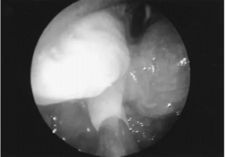
The upper airway in MPS patients can be narrowed due to accumulation of GAGs in adenotonsilas, causing adenotonsilar hypertophy, walls of pharynx and larynx, causing macroglossia, and thickened soft tissues in the laryngopharynx (Fig.1). Simmons et al. refer having seen cases where there are festoons of excessive tissue associated with substance deposition over the arytenoid cartilages and the laryngoepiglottic folds (Fig1). In extreme cases these may prolapse into the laryngeal inlet and cause stridor and severe airways compromise. There may also be narrowing of the tracheal lumen due to GAG deposition in the wall (Fig1).4,7 In some cases tracheostomy has been necessary to secure a safe airway. Simmons et al describe cases where they have been successful performing per oral laser excision of the excess tissue using a microscope and micromanipulator or laser bronchoscope. 4
Moreover, progressive upper airway obstruction can be compounded by deformities of the skull or spine, such as short neck, flattened nasal bridge, high anterior larynx, deep and narrow
Figure 1. Clinical manifestations of MPS. A: Oropharyngeal image showing macroglossia and gingival hyperplasia; B: normal lyaryngoscopic image; C-D: laryngoscopic images showing a thick laryngeal wall, tonsillar hyperplasia, and redundant arytenoid mucosa prolapse into laryngeal inlet; E-F: nasal endoscopic images showing inflammation and abnormal mucous secretion; G: normal bronchoscopic image (Canani et al 2003)5; H: bronchoscopic image showing tracheal deformity and narrowing; (images from Berger KI et al. 2012)6
15
cervical vertebrae such as atlantoaxial joint instability. 4,8,9,10 Multilevel airway obstruction may occur if upper airway obstruction is accompanied by tracheobrochomalacia or accumulation of GAG in the tracheal mucosa. 9,12,13,14,15 Airway obstruction can also occur due to tracheal distortion in combination with laxity of tracheal tissue. 13,16 Excessively thick secretions through the upper and lower airways can worsen airway obstruction.
The degree of snoring or airway obstruction while sleeping is a very important part of the clinical presentation. Obstructive sleep apnea occurs in >80% of patients. 9,17,18 In adults OSA is defined as AHI>5, whereas pediatric OSA is accepted as AHI> ou =1,5.
Although patients with MPS may improve airway obstruction with more conservative treatment approaches including positive airway pressure devices (CPAP/BIPAP), management often requires early adenotonsilectomy and in extreme cases tracheostomy to ensure a patent airway in the short-or long-term.4
Recurrent infections like tracheitis and laryngitis are frequent in MPS patients. The reason for this is the presence of viscous mucous and reduced mucociliary clearance due to GAG accumulation. Moreover, the GAG deposits in retronasal areas, eustachian tubes and in the middle ear increase the risk of otitis media with effusion and acute otitis media. 4
In patients with MPS, hearing loss is initially mechanic/conductive (Conductive hearing loss) and latter, with progressive deterioration, with sensorineural component (Sensorineural hearing
Figure 2. Excess arytenoid mucosa, distended by MPS deposits, that was prolapsing into the airway with each inspiration(image from Simmons M.A et al, 2005)4
loss) and is observed due to various reasons. CHL may develop due to recurrent upper respiratory tract infections and serous otitis media or thickening of external ear components or bone chain deformities. SHL is thought to be caused by the accumulation of GAG in the cochlea, auditory nerve, and brain stem. In many patients, mixed-type hearing loss, along with the symptoms of conductive and sensorineural hearing loss, can also be seen. A degree of sensorineural loss is common in MPS types I and II. WOLD et al. while evaluating patients with MPSI, II and VI showed 78% of hearing loss with audiometric evaluation. From these 71% presented mixed-type hearing loss, 14% SHL and 14 %CHL 2. In CHL related to middle ear effusion, while the ventilation tube/pressure equalization tubes implementation is frequently used as treatment, although hearing loss is usually not resolved, hearing aids are advised in SHL. Nerve compression from GAG storage at the internal auditory canal can reduce the efficacy of cochlear implants. 1,19
As described by Wold et al., in cases with MPS, the initiation of treatment in early stages carries great importance in terms of the prognosis of the disease. Nonetheless, due to the late appearance of the symptoms, the definite range is 3-4 years of age. 20
In addition to the findings previously mentioned, common characteristic facial features may include frontal bossing, hypertelorism, depressed nasal bridge, large nares, thickened lips, gingival hyperplasia, gapped teeth, coarse or grotesque facies, hirsutism, and corneal clouding (characteristically absent in MPS II).
17
In the past, few people with severe phenotypic mucopolysaccharidoses reached adulthood. However better methods of diagnosis, multidisciplinary care, and new therapies have extended lifespan, leading to an increasing number of patients surviving beyond childhood. In adults with MPS hearing impairment and mechanical alterations to the larynx commonly cause speech problems and poor communication due to hearing, and speech problems can lead to depression, isolation, and less participation in care.1
x
Diagnosis
The diagnostic approach in MPS is based on clinical suspicion, radiological examination and laboratory testing.
We should suspect of MPS when is present 1) recurrent Upper Respiratory Tract Infections (URTIs) in children with dysmorphism and/or hepatomegaly, and/or cardiac abnormalities, and/or skeletal abnormalities, and/or hernias; 2) Obstructive Sleep Apnea and/or macroglossia and coarse facial features; 3) obstruction of the airways, restrictive lung disease; 4) Hearing loss3.
As diagnosis of these disorders can be complex, it is recommended that patients suspected of having MPS be referred to a geneticist or metabolic specialist for laboratory testing. 21
n Laboratory testing for an MPS disorder
An MPS diagnosis is based on laboratory results from urinary GAG analyses and enzyme activity assays. Enzyme activity assays measure enzyme activity in tissue (blood or fibroblasts). Quantitative GAG assays measure overall elevation of GAG as compared with GAG levels expected in age-matched normal subjects. Qualitative GAG assays detect the type of GAG excreted. Determining the correct MPS type is essential to ensure appropriate therapeutic management. The type of MPS cannot be reliably determined on the basis of clinical presentation or test results alone. High clinical suspicion and thorough testing help ensure an accurate diagnosis of MPS (Fig.3). 21
19 Urinary GAG analysis
In normal subjects, urinary GAG excretion varies with age, higher values being found during the first years of life, followed by a slow and constant decrease thereafter. Qualitatively, 90% of the GAG content in normal urine consists of chondroitin-4 and -6 sulphate, with the remaining. being heparan sulphate. Most MPS patients have higher GAG excretion in urine compared with age-matched normal subjects; however, as not all MPS patients have a clear elevation of total GAG excretion, an accurate diagnosis requires a full GAG profile including both quantitative and qualitative analysis done in tandem.23,24 In fact, patients with different types of MPS differ not only in the total amount of GAG excreted in urine, but also in the relative proportion of various types of GAG.25 As a consequence, the demonstration of an abnormal pattern is diagnostic for an MPS disorder; moreover, the presence of specific GAGs can suggest the MPS subtype and may direct the appropriate enzyme analyses. For example, high amounts of heparan sulphate or keratan sulphate essentially characterize MPS III and IV, respectively. Within a
single subtype of MPS, GAG excretion can also vary depending on the severity of the disease phenotype.26 Thus an MPS diagnosis should neither be confirmed nor ruled out on the basis of a single GAG test, although GAG testing usually is the first step in the diagnostic pathway. An elevated level of urinary glycosaminoglycan suggests a MPS disorder but does not provide a specific diagnosis.21,27
Enzyme activity
The diagnosis of MPS should be confirmed by enzyme activity testing.21 Enzyme activity typically is measured in leucocytes or cultured fibroblasts (skin biopsy or punch). For many types of MPS, enzyme activity can be measured from a dried blood spot (DBS). DBS-based assays offer considerable practical advantages with respect to sample collection, storage and transport, and multiple enzyme activity tests can be performed on a single DBS. Recent technological advances have made these assays sensitive and specific if appropriate controls are performed. It is recommended that a positive result from a DBS be confirmed by a tissue-based assay. Due to the complexity of these assays, referral to a geneticist or metabolic specialist is essential.21
Mutation analysis
Mutation analysis or molecular testing includes looking for the known disease-causing mutations in addition to looking for abnormal sequences in the gene coding for a particular enzyme. Molecular testing has limited utility in initial screening because of the extreme genetic heterogeneity that characterizes all types of MPS. Although several MPS disorders are associated with specific commonly occurring mutations, most families have private mutations.28,29,30 Thus the primary use of molecular testing is to confirm a diagnosis of a particular type of MPS or to evaluate family members when the type of MPS and the family mutation is known. Molecular testing can have prognostic value if the mutations that are identified have been well characterized.31,32 When a patient has been diagnosed with a specific type of MPS and his/her mutation is known, the information can be used for carrier testing and prenatal testing of siblings. With regard to carrier testing, it is important to note that except for MPS II, the MPSs are inherited in an autosomal recessive fashion, so carriers have a very small likelihood of having children affected with MPS unless the union is consanguineous.21
21 Biomarkers
Currently there are no established biomarkers for any of the MPS types. Urinary GAG levels usually decrease with treatment 33,34,35, but are not an ideal biomarker. Other biomarkers under investigation include heparin cofactor II-thrombin complex measured in serum 36,37, the ratio of dermatan to chondroitin sulphate in urine 37,38, and dipeptidyl peptidase IV in plasma 39. An ideal biomarker would be specific to a particular type or types of MPS, would help to differentiate more severe from less severe disease phenotypes, would respond to treatment and would be easily detected and quantified.
n Follow-up
In MPS disorders it is recommended systematic audiological evaluation (annually), polysomnography and upper airway fiberoptic examinations and imaging studies of the trachea and larynx.4
Polysomnography is first line of investigation for any individual with suspicion of MPS and OSA. Polissomnographic studies in children with MPS show a bigger prevalence of OSA secondary to adenotonsilar hypertrophy and GAG deposition on oropharynx.40, 41
Upper airway imaging tecniques including MRI and CT play an importante role in noninavsive evaluation of OSAS.41,42 CT findings from a study show that in children with MPS the retropalatine e retroglossal spaces are substantially smaller, resulting in a disorderly breathing during sleep .41
Figure 4. Trachea in a patient with MPS 4
Figure 5. Imagiologic studies in MPS. I -CT scan showing tracheal deformity and narrowing; K-L typical frontal and lateral plain X-rays of the chest showing skeletal abnormalities and the presence of a tracheostomy and possibly infective changes in the lungs (hazy shadow in the right lower zone of the frontal image). Images from Berger KI et al 20126
In addition to this, CT also has been selected as a reliable outcome measure of efficacy of medical or non-medical interventions in OSA. CT avoids the use of sedation but provides high radiation doses, and this issue is relevant to children.41,43 On the other hand, MRI allows multiple scanning planes, provides high contrast resolution and does not require radiation exposure 44, but as long as times for each imaging slice are needed, patients may need sedation. Sedation, with the consequent loss of muscle tone, may be a better model of the situation of upper airways during sleep. However the high anesthetic risk in MPS is universally recognized (will be discussed later in this review) and anesthesia has been recommended only for life-threatening procedures.4 There is growing awareness that CT can be used an imaging biomarker to establish the presence and/or severity of disease. 45
Data from a study also confirmed that adenoid hypertrophy secondary to GAG deposition is usually a frequent complication of MPS and that adenoid volume can be evaluated with fibroscopy .40,41
In MPS patients with OSA there are multiple sites of obstruction and upper airway imaging techniques including MRI and CT and nasofibroscopy offer information about different locations of possible obstruction. Conversely, CT explores some aspects of upper airway disease that are not revealed on Polysomnography or nasal endoscopy. Data from each of the three techniques are useful to gain a thorough evaluation of MPS upper airway obstructive disease. In MPS, CT plus Polysomnography findings and nasal obstruction endoscopic degree might be transformed into quantitative data to track the clinical course and to evaluate the efficacy of novel therapies. Pediatric otorhinolaryngologic specialists have a crucial role in the diagnosis of patients with MPS since these individuals are referred for treatment of recurrent otitis media, loss of hearing and upper airway obstruction. Early clinical suspicion, identification and diagnosis are crucial because in many cases treatment outcomes are sensible to passage of time, best outcomes are reached with early intervention or in younger patients.
Treatment/Management:
Traditionally, this group of conditions was regarded as incurable, with only symptomatic and palliative treatment, with a multidisciplinary team. More recently however, further to the management of the symptoms and individual manifestations, efforts have been made to control
23
disease. Because MPS affects multiple body systems, management of a diverse spectrum of disease manifestations is an important part of providing integrated care. Management may include use of adaptive or supportive devices, physical and occupational therapy, symptom based medications, surgical interventions, and therapies to provide the deficient enzyme.
In this context, enzyme replacement therapy (ERT) and bone marrow transplantation (BMT)/hematopoietic stem cell transplantation (HSCT) have had some limited success in selected cases.
The bone marrow transplantation has been investigated since the 1980’s and has been frequently used in MPS children under 2 years of age or in those patients with slight forms of MPS II, VI or VII. 46,47,48,61 Literary data shows an improvement in airway obstruction in children submitted to the transplantation of bone marrow, among other benefits, probably due to restoring the ability of the organism in lowering substrates accumulated on cellular level. Besides, comparative studies have suggested that the transplantation would result in improving both hearing ability and the voice in comparison with groups without treatment. 49
Although many studies reveal that hematopoietic stem cell transplantation can change the natural course of the disease, increasing life expectancy and improving many systemic abnormalities 49,50,51,62, it is a high-risk procedure with high morbidity/mortality 62. Moreover, its indication depends on the type of MPS, patient’s clinical picture, age and presence of neurological impairment.35,60 Hematopoietic stem cell transplantation from parents is indicated in some situations (severe forms of MPS I, prior to the onset of neurodegeneration).
More recently, ERT have been developed and used in the treatment of the different types of mucopolysaccharidoses. ERT can be used for MPS types I, II and VI 53,54,55,61,63 and consists in periodic endovenous administration of the specific missing enzyme, leading to higher GAG degradation in tissues and organs and promoting significant improvement in some clinical features. ERT has a lifetime requirement of weekly intravenous infusions that carry a risk of allergic reaction and may require a central venous access port with its inherited risk of infection and subsequent risk of endocarditis. Other limitations of ERT are high cost and low penetration of the central nervous system.52,53
Currently, four commercially available regimens have received approval for the treatment of MPS I (Laronidase; AldurazymeTM – Biomarin Pharmaceutical/Genzyme Co.) 61,63, MPS II (Idursulfase; ElapraseTM – Shire Human Genetic Therapies Inc.), MPS IV (GALNS-elosulfase
alfa; VimzymTM – Biomarin Pharmaceutical)52,53,54 and MPS VI (Galsulfase; NaglazymeTM – Biomarin Pharmaceuticals) 55. Although limited clinical trials appear promising, there is not yet extensive experience with these therapies, and long-term data on their efficacy is not yet available.
In both BMT and enzyme therapies, it seems apparent that benefits are likely to be greatest in those patients treated prior to the development of significant physical or mental morbidity, thus further providing incentive for heightened awareness on the part of the practitioner to establish early and accurate diagnosis.
n Surgical treatment
Surgical treatment of otorhinolaryngologic processes often significantly enhances the quality of life of these individuals. Current surgical treatment is not curative and is limited to symptomatic patients, but can be used as a preventive approach to avoid future complications. The judicious use of otorhinolaryngologic surgery has the ability to greatly improve the quality of life for the patients by reducing the persistent rhinorrhea, reducing the frequency and severity of ear infections and by relieving the symptoms of upper airway obstruction.
Adenotonsillectomy is the most common treatment option in pediatric OSA and results in a reduced size of adenoids as documented by nasal endoscopy.56,9 However, the role of adenotonsillectomy in the follow-up of these patients needs further consideration, since it might not be crucial for improving upper airway obstruction, even though one cannot deny its usefulness for relief of obstruction. 4,41
Anaesthesic considerations
As MPS may affect any organ system, the systemic effects of the disease process must be taken into consideration before thinking about minor surgical intervention .57 Indeed, such is the risk of anesthesia in severely affected patients that anesthesia would be unwise for non-life threatening procedures. Muhlebach et al concluded that difficult endotracheal intubation is a common complication of any surgical or emergency procedure in MPS patients. 58
The risk of complications on account of anesthesia is especially high in the presence of cardiovascular manifestation, shortening of the neck, atlantoaxial instability and airway
25
difficult airways. GAG accumulation in the larynx can inclusively impede identification of the glottis.
A survey of airway complications in a group of patients with MPS VI at the Royal Manchester Children’s Hospital (Manchester, United Kingdom) showed an overall incidence of difficult intubation of 25% in all subgroups and a failed intubation rate of 8% 52. It is recommended spontaneous breathing with a volatile agent. During surgical procedures may be needed an emergency tracheostomy in cases where one can’t guarantee free airway .4,59
In the light of the elevated anesthetic risk of this population is recommended to perform and record a bronchoscopy examination with a flexible fiber-optic bronchoscope before surgery to evaluate the extent and severity of airway infiltration .4,52
Conclusion
As noted, the clinical presentation of MPS may be very heterogeneous, sowing variable severity of phenotypes and multi-systemic involvement; however similar events should motivate clinical suspicion for clinical diagnosis. Overall, the head and neck structures are impacted very early in the disease, usually with recurrent airway infections, otitis media with effusion, hearing loss, severe adenotonsilar hypertrophy, sleep disturbances and other respiratory problems.
The most frequent reasons for referral in this population are not entirely unlike those in the general population: recurrent serous otitis media, hearing loss, and upper airway obstruction; however the complexity of these patients will typically demand earlier, more aggressive care and longer term follow-up. Close attention to the history (including family history) and physical exam will generally yield the keys for clinical diagnosis of these disorders as a whole, even if the subtype is not immediately apparent. Thus, otolaryngologists have an opportunity to play an increasingly integral role in the multidisciplinary approach to the diagnosis and management of many children with mucopolysaccharidoses.
In addition to clinical diagnosis, some laboratory tests can be used, like urinary glycosaminoglycan levels (they suggest a mucopolysaccharidoses disorder but do not provide a specific diagnosis), measures of enzyme activity in leukocytes and fibroblasts and molecular studies. It is important to enhance that all families should be referred to a genetic counseling consultation, which should evaluate the possible carriers and offer information about the possibility of prenatal diagnosis.
Moreover, the possibility of effective specific therapeutic interventions has been demonstrated and if the therapeutic interventions are immediately applied upon the disease diagnosis, they are able to significantly modify the natural progression of the disease. It is therefore mandatory for the clinician to acquire all the necessary elements for an early recognition of the first disease signs as well as to be well informed on the currently available diagnostic procedures and on the location of specialized reference centers. A delay in the diagnosis can entail a significant aggravation of the prognosis, since the damages caused by the disease, once established, are irreversible.
27
References
[1] Mitchell J, et al, (2016) Unique medical issues in adult patients with mucopolysaccharidoses, Eur J Intern Med
[2] Wold SM, Derkay CS, Darrow DH, Proud V. (2010) Role of the peadiatric otolaryngologist in diagnosis and management of children with mucopolysaccharodisis. Int J Pediatr Otorhinolaryngol. 74(1):27-31
[3] Bicalho CG, Rezende MM, Nogueira AMCM, Paulon RMC. (2011) The importance of the otorhinolaryngologic evaluation in mucopolysaccharidoses patients. Arch. Int. Otorrinolayngol.15(3):290-294.
[4]Simmons M.A et al. (2005) Otorhinolaryngological manifestations of the mucopolysaccharidoses. International Journal of Pediatric Otorhinolaryngology . 69:589-595
[5] Canani SE, John AB, Schwartz IV et al (2003) Manejo de apnéia do sono em um paciente
com mucopolysaccharidose. Hypn J Clin Exp Sleep Res 3:40–41
[6] Berger KI, Fagondes SC, Giugliani R et al. (2012) Respiratory and sleep disorders in mucopolysaccharidoses. Journal of Inherited Metabolic Diseases.
[7] Gonuldas B, Y1lmaz T, Sivri HS, Gucer KS. (2014) Mucopolysaccharidosis: Otolaryngologic findings, obstructive sleep apnea and accumulation of glicosaminoglycans in lymphatic tissue of the upper airway. International Journal of Pediatric Otorhinolaryngology 78. 944-949
[8] Alpoz AR, Coker M, Celen E et al (2006) The oral manifestations of Maroteaux-Lamy Syndrome (mucopolysaccharidoses VI): a case report. Oral Surg Oral Med Oral Pathol Oral Radiol Endod 101:632-637
[9] Leighton SEJ, Papsin B, Vellodi A, Dinwiddie R, lane R (2001) Disordered breathing during sleep in patients with mucopolysaccharidoses. Int J Pediatr Otorhinolaryngol 58:127-138
[10] Myer CM III (1991) Airway obstruction in Hurler’s Syndrome-radiographic features. Int J Pediatr Otorhinolaryngol 22:91-96
[11] Ingelmo PM, Parini R, Grimaldi M, et al. (2011) Multidetector computed tomography (MDCT) for preoperative airway assessment in children with mucopolysaccharidoses. Minerva Anestesiol;77:774–780.
[12] Nagano R, Takizawa S, Hayama N et al (2007) Three dimensional CT and histopathological findings of airway malacia in Hunter Syndrome. Tokai J Exp Clin Med 35:59-61
[13] Pelley CJ, Kwo J, Hess DR (2007) Tracheomalacia in an adult with respiratory failure and Morquio Syndrome. Respir Care 52:278-282
[14] Shih SL, Lee YJ, Lin SP, Sheu CY, Blickman JG (2002) Airway changes in children with mucopolysaccharidoses. Acta Radiol 43:40-43
[15] Sims HS, Kempiners JJ (2007) Special airway concerns in patients with mucopolysscahridoses. Respir Med 101:1779-1782
[16] Walker PP, Rose E, Williams JG (2003) Upper airways abnormalities and tracheal problems in Morquio’s disease. Thorax 58:458-459
[17] John A, Fagondes S, Schwartz I et al (2011) Sleep abnormalities in untreated patients with mucopolysaccharidoses type VI. Am J Med Genet A 155A:1546-1551
[18] Semenza GL, Pyeritz RE (1988) Respiratory complications of mucopolysaccharide storage disorders. Medicine (Baltimore) 67:209-219
[19] Santos S, Lopez L, Gonzalez L, Dominguez MJ. (2011) Hearing loss and airway problems in children with mucopolysaccharidoses. Acta Otorrinolaryngol. Esp 62:411-417
[20] Gökdo ̆gan C ̧, Altinyay ̧,S Gökdo ̆gan O, Tutar H, Gündüz B, Okur ̇I, et al. (2016) Audiologic evaluations of children with mucopolysaccharidoses. Braz J Otorhinolaryngol; 82:281-4.
[21] Lehman T. J.A., Miller N., Norquist B., Underhill L., Keutzer J. (2011) Diagnosis of the mucopolysaccharidoses. Rheumatology ;50:v41v48 doi:10.1093/rheumatology/ker390
[22] Burton B.K., Giugliani R. (2012) Diagnosing Hunter syndrome in pediatric practice: practical considerations and common pitfalls. Eur J Pediatr . 171:631–639 DOI 10.1007/s00431-012-1703-y
[23]Mahalingam K, Janani S, Priya S et al. (2004) Diagnosis of mucopolysaccharidoses: how to avoid false positives and false negatives. Indian J Pediatr ;71:2932.
[24] Gallegos-Arreola MP, Machorro-Lazo MV, FloresMartinez SE et al. (2000) Urinary glycosaminoglycan excretion in healthy subjects and in patients with mucopolysaccharidoses. Arch Med Res ;31:50510.
29
[25] Piraud M, Boyer S, Mathieu M et al. (1993) Diagnosis of mucopolysaccharidoses in a clinically selected population by urinary glycosaminoglycan analysis: a study of 2,000 urine samples. Clin Chim Acta;221:17181.
[26] Coppa GV, Galeotti F, Zampini L et al. (2011) High-throughput determination of urinary hexosamines for diagnosis of mucopolysaccharidoses by capillary electrophoresis and high-performance liquid chromatography. Anal Biochem 2011;411:3242. 21
[27] Whitley CB, Ridnour MD, Draper KA, Dutton CM, Neglia JP. (1989) Diagnostic test for mucopolysaccharidoses I: direct method for quantifying excessive urinary glycosaminoglycans excretion. Clin Chem;35:374-379
[28] Muenzer J, Beck M, Eng CM et al. (2009) Multidisciplinary management of Hunter syndrome. Pediatrics;124: e122839.
[29] Muenzer J, Wraith JE, Clarke LA. (2009) Mucopolysaccharidosis I: management and treatment guidelines. Pediatrics; 123:1929
[30] Valayannopoulos V, Nicely H, Harmatz P et al. (2010) Mucopolysaccharidosis VI. Orphanet J Rare Dis;5:5.
[31] Karageorgos L, Brooks DA, Pollard A et al. (2007) Mutational analysis of 105 mucopolysaccharidosis type VI patients. Hum Mutat ;28:897903.
[32] Tomatsu S, Montano AM, Nishioka T et al. (2005) Mutation and polymorphism spectrum of the GALNS gene in mucopolysaccharidosis IVA (Morquio A). Hum Mutat;26: 50012.
[33] Clarke L, Wraith JE, Beck M et al. (2009) Long-term efficacy and safety of laronidase in the treatment of mucopolysaccharidosis I. Pediatrics ;123:22940.
[34] Muenzer J, Beck M, Eng CM et al. (2011) Long-term, open-labeled extension study of idursulfase in the treatment of Hunter syndrome. Genet Med ;13:95101.
[35] Aldenhoven M, Boelens JJ, De Konong TJ. (2008) The clinical outcome of Hurler syndrome after stem cell transplantation. Biol Blood Marrow Transplant.14:485-98
[36] Randall DR, Colobong KE, Hemmelgarn H et al. (2008) Heparin cofactor II-thrombin complex: a biomarker of MPS disease. Mol Genet Metab;94:45661
[37] Langford-Smith KJ, Mercer J, Petty J et al. (2010) Heparin cofactor II-thrombin complex and dermatan sulphate: chondroitin sulphate ratio are biomarkers of short- and long-term treatment effects in mucopolysaccharide diseases. J Inherit Metab Dis;34:499508.
[38] Church H, Tylee K, Cooper A et al. (2007) Biochemical monitoring after haemopoietic stem cell transplant for Hurler syndrome (MPSIH): implications for functional outcome after transplant in metabolic disease. Bone Marrow Transplant;39:20710.
[39] Beesley CE, Young EP, Finnegan N et al. (2009) Discovery of a new biomarker for the mucopolysaccharidoses (MPS), dipeptidyl peptidase IV (DPP-IV; CD26), by SELDI-TOF mass spectrometry. Mol Genet Metab;96:21824. 32
[40] Chow Z, Jones G., Coates H. Manifestações otorrinolaringológicas das mucopolissacaridoses. In: X Manual de Otorrinolaringologia Pediátrica da IAPO. São Paulo: Editora e Gráfica Vida & Consciência. 2012. 219-229
[41] SantaMaria F et al. (2007) Upper airway obstructive disease in mucopolisaccharidosis: polysomnography, computed tomography and nasal endoscopy findings. Journal of Inherited metabolic disease. 30:743-749.
[42] Arens R, McDonough JM, Corbin AM, et al (2003) Upper airway size analysis by magnetic resonance imaging of children with obstructive sleep apnea syndrome. Am J Respir Crit Care Med 167:65-70
[43] Brenner DJ (2002) Estimating cancer risks from pediatric CT: going from the quanlitative to the quantitative. Pediatr Radiol 32:228-233
[44] Rama AN, Tekwani SH, Kushida CA (2002) Sites of obstruction in obstructive sleep apnea. Chest 122:1139-1147
[45] Pien HH, Fischman AJ, Thrall JH, Sorensen AG (2005) Using imaging biomarkers to accelerate drug development and clinical trials. Drug Discov Today 10:259-266
[46] Gomes AB, Pereira RG, Vassola T, et al. (2011) Audiologic Evaluation in Patients with Mucopolysaccharidosis in a Pediatric Hospital, Arq. Int. Otorrinolaringol. / Intl. Arch. Otorhinolaryngol., São Paulo - Brasil, v.15, n.2, p. 203-207
[47] Walker RWM, Colovic V, Robinson DN, Dearlove OR. (2002) Postobstructive pulmonar oedema during anaesthesia in children with mucopolysaccharidoses. Paediatr Anaesth 12:1-7 [48] Vairo Filippo, Andressa Federhen, Baldo G, Riegel M, Burin M et al. (2015). Diagnostic and treatment strategies in mucopolysaccharidoses v. The apliccation of clinical genetics 8 245-255
31
[49] Papsin BC, Vellodi A, Bailey CM, Ratcliffe PC, Leighton SE. (1998) Otologic and laryngologic manifestations of mucopolysaccharidoses after bone marrow transplantation. Otolaryngol Head Nec Surg 11881:30-36
[50] Vellodi A, Young EP, Cooper A et al. (1997) Bone marrow transplantation for mucopolysaccharidoses type I: experience of two British centers. Arch Dis Child.76:92-99 [51] Wraith JE, Scarpa M, et al. (2008) Mucopolysaccharidosis type II (Hunter syndrome): a clinical review and recomendations for treatment in the era of enzyme replacement therapy. Eur J Pediatr. 167:267-77
[52] Giugliani R, MD, PhDa, Harmatz P, MDb, Wraith,J.E MD. (2007) Management Guidelines for Mucopolysaccharidosis VI. Pediatrics Volume 120, Number 2
[53] Regier DS, Tanpaiboon P.(2016) Role of elosulfase alfa in mucopolysaccharidoses IVA. The application of clinical genetics .9:67-74
[54] Rodriguez-Lopez, A et al. (2016) Recombinant N-acetylgalactosamine-6-sulfate sulfatase (GALNS) produced in the methyllotropic yeast pichia pastoris). Sci Rep 6, 29329;doi:10.1038/srep29329
[55] Hendriksz CJ, Burton B, Fleming TR, et al. (2014) Efficacy and safety of enzyme replacement therapy with BMN 110 (elosulfase alfa) for Morquio A syndrome (mucopolysaccharidosis IVA): a phase 3 randomised placebo-controlled study. J Inherit Metab Dis.;37:979–90
[56] Mezolella M, Cimmino M, Cantone E, Marino A, Cozzolino M, Della Casa R, et al. (2013) Management of otolaryngological manifestations in mucopolysaccharidoses: our experience. Acta Otorhinolaryngol. Ital 33(4):267-272
[57] Kempthorne P.M, Brown.T.C.K (1983) Anaesthesia and mucopolysaccharidoses: a survey of techiques and problems. Anaesth. Intensive Care 11. 203-207.
[58]MuhlebachM .S, Wooten W. Muenzer J. (2011). Respiratory manifestations in mucopolysaccharidoses. Paediatr. Resp. Rev 12(2). 133-138.
[59] Walker R, Belani KG, Braulin EA, Bruce IA, Hack H, Harmatz R, Jones S, Richard Rowe. (2013) Anaesthesia and airway management in mucopolysaccharidoses. J Inherit Metab Dis. 36:211-219
[60] Kakkis ED, Muenzer J, Tiller GE, et al (2001) Enzyme replacement therapy in mucopolysaccharidoses I. N Eng J Med 344:182-188
[61] Martins AM, Dualibi AP, Norato D et al (2009) Guidelines for the management of mucopolysaccharidoses type I. J Pediatr 155:S32-S46
[62] Rovelli AM (2008) The controversial and changing role of hematopoietic cell transplantation for lysossomal storage disorders: an update. Bone Marrow Transplant 41 (Suppl 2):S87-S89
[63] Wraith JE (2005) The first 5 years of clinical experience with laronidase enzyme replacement therapy for mucopolysaccharidoses I. Expert Opin Pharmacother 6:489-506