1
The di-iron RIC (YtfE) protein of Escherichia coli interacts with the DNA-binding 1
protein from starved cells (Dps) to diminish RIC-protein-mediated redox stress 2
3
Liliana S. O. Silva1, Joana M. Baptista1, Charlotte Bately2, Simon C. Andrews2, and Lígia 4
M Saraiva1,* 5
1
Instituto de Tecnologia Química e Biológica NOVA, Av. da República, 2780-157 Oeiras, 6
Portugal 7
2
School of Biological Sciences, Knight Building, University of Reading, Reading RG6 8 6AJ, UK 9 10 *Corresponding author: 11 Lígia M. Saraiva 12
Av. da República, 2780-157 Oeiras, Portugal 13 Phone: +351-214469328; Fax: +351-214411277 14 E-mail: [email protected] 15 16 17 18
2 Abstract
19 20
The RIC (Repair of Iron Clusters) protein of Escherichia coli is a di-iron hemerythrin-like 21
protein that has a proposed function in repairing stress-damaged iron-sulphur clusters. In 22
this work, we performed a Bacterial Two Hybrid screening to search for RIC-protein 23
interaction partners in E. coli. As a result, the DNA-binding protein from starved cells
24
(Dps) was identified and its potential interaction with RIC was tested by BACTH, 25
Bimolecular-Fluorescence-Complementation and pull-down assays. Using the activity of 26
two Fe-S-containing enzyme as indicators of cellular Fe-S cluster damage, we observed 27
that strains with single deletions of ric or dps have significantly lower aconitase and 28
fumarase activities. In contrast, the double ric dps mutant strain displayed no loss of 29
aconitase and fumarase activity with respect to the wild type. Additionally, while 30
complementation of the ric dps double mutant with ric led to a severe loss of aconitase 31
activity, this effect was no longer observed when a gene encoding a di-iron site variant of 32
the RIC protein was employed. The dps mutant exhibited a large increase in ROS levels, 33
but this increase was eliminated when ric was also inactivated. Absence of other iron-34
storage proteins, or of peroxidase and catalases, had no impact on RIC-mediated redox 35
stress induction. Hence, we show that RIC interacts with Dps in a manner that serves to 36
protect E. coli from RIC-protein-induced ROS. 37
38
39
3 Importance
41
The mammalian immune system produces reactive oxygen and nitrogen species that kill 42
bacterial pathogens by damaging key cellular components such as lipids, DNA and 43
proteins. However, bacteria possess detoxifying and repair systems that mitigate these 44
deleterious effects. The E. coli RIC (Repair of Iron Clusters) protein is a di-iron 45
hemerythrin-like protein that repairs stress-damaged iron-sulphur clusters. E. coli Dps is an 46
iron-storage protein of the ferritin superfamily with DNA-binding capacity that protects 47
cells from oxidative stress. This work shows that the E. coli RIC and Dps proteins interact 48
in a fashion that counters RIC-protein-induced ROS. Altogether, we provide evidence for 49
the formation of a new bacterial protein complex and reveal a novel contribution for Dps in 50
bacterial redox-stress protection. 51
52
53
Keywords 54
E. coli, di-iron RIC protein, YtfE, Dps, oxidative stress, nitrosative stress 55
56 57
Running Title 58
Di-iron RIC protein interacts with Dps 59
60 61
4 Introduction
62
During the infection process, bacterial pathogens are able to survive aggressive
63
environments through the activation of specific stress-resistance genes. One such example
64
of a stress-induced gene is ric. This gene encodes the ‘Repair of Iron Centre’ (RIC) protein
65
that contains a di-iron centre and contributes to the protection of bacterial pathogens such 66
as Escherichia coli, Haemophilus influenzae, Salmonella spp., Yersinia spp. and 67
Clostridium spp. during exposure to nitrosative and/or oxidative stress (1). The ric gene is 68
induced upon exposure to either oxidative or nitrosative stress, and in E. coli, 69
Staphylococcus aureus, Neisseria gonorrhoeae, H. influenza and Cryptococcus neoformans 70
the RIC protein is thought to confer stress resistance through maintenance of the activity of 71
various Fe-S containing enzymes (1–3). Such an effect is well demonstrated for E. coli and 72
S. aureus where RIC proteins restore the activity of oxidatively and nitrosatively-damaged 73
Fe-S clusters in the TCA cycle enzymes, aconitase and fumarase (1, 4, 5) In E. coli, the 74
RIC protein also acts under non-stress conditions to maintain aconitase and fumarase 75
activities (6). Further, the E. coli RIC protein delivers iron (most likely in the ferrous state) 76
for the assembly of Fe-S clusters in spinach apo-ferredoxin and in the E. coli Fe-S cluster-77
assembly scaffold protein, IscU (7). The RIC protein also contributes to the survival of S. 78
aureus and H. influenzae in activated macrophages, and is required for full virulence in S. 79
aureus when infecting the wax moth larva infection-model, Galleria mellonella (3, 8). 80
Thus, the RIC protein has an apparent role in bacterial pathogenicity through mediation of 81
Fe-S cluster stability during exposure to redox- and/or nitrosative-stress. 82
The RIC proteins of E. coli and S. aureus contain di-iron centres of the 83
histidine/carboxylate type within a four-helix-bundle fold (9). The UV-visible spectrum of 84
5
oxidized RIC protein exhibits a broad band at ca. 350 nm and Electron Paramagnetic 85
Resonance (EPR) spectroscopy indicates that the principal g-values are below 2 (g=1.96, 86
1.92 and 1.88), which is indicative of a S=½ spin state in a mixed valence and anti-87
ferromagnetically coupled Fe(III)-Fe(II) binuclear iron centre. Mössbauer spectroscopy 88
showed that the mixed-valence Fe(III)–Fe(II) di-iron centre of the RIC protein is more 89
labile than that of the (oxo)-diferric form (7). 90
RIC proteins possess several highly-conserved amino acid residues of which some have 91
been shown to influence the properties of the di-iron centre and/or function of the protein. 92
In particular, substitution of residues His129, Glu133 or Glu208 of the E. coli RIC protein 93
abrogated its ability to protect the Fe-S cluster of aconitase. Moreover, two µ-carboxylate 94
bridges contributed by Glu133 and Glu208, linking the two di-iron site atoms, were shown 95
to be required for the assembly of a stable di-iron centre (10). These studies also 96
demonstrated the important contribution of the conserved His84, His129, His160, His204, 97
Glu133 and Glu208 residues in ligating the di-iron centre within the four-helix bundle fold, 98
and these di-iron coordination roles were recently confirmed by X-ray crystallographic 99
structural studies (11). 100
In the work reported here, we sought to identify proteins that interact with, and support the 101
function of, the RIC protein of E. coli. For this purpose, an E. coli library was screened for 102
RIC protein interaction partners using the Bacterial Adenylate Cyclase Two Hybrid system 103
(BACTH). Potential interacting gene products were further tested by BACTH, Bimolecular 104
Fluorescence Complementation (BiFC) and pull-down assays. Our protein-protein 105
interaction studies revealed that the RIC protein interacts with the DNA-binding protein
6
from starved cells (Dps). Dps is a symmetrical dodecameric iron-storage protein of the 107
ferritin superfamily that contains a di-iron ferroxidation centre located at the interface 108
between subunits (12–14). Dps sequesters ferrous iron, which is oxidized preferentially by 109
hydrogen peroxide at its di-iron centre and then deposited for storage as Fe(III) 110
oxyhydroxide in the central cavity as an iron core; the sequestered iron can subsequently be 111
released by reduction (14, 15). The ferroxidase activity, DNA-binding and iron-112
sequestration properties of Dps confer cells with protection from oxidative stress and 113
nutrient deprivation, as judged by the reduced survival of dps mutants under stress 114
conditions including starvation, oxidative stress, metal toxicity, and thermal stress (16). 115
The physiological relevance of the interaction between the RIC protein and Dps was
116
examined and the results revealed that Dps modulates the function of RIC.
117 118 119 120 121 122 123 124 125 126
7 Results
127
128
Identification of novel potential RIC-protein-interaction partners by screening a 129
bacterial two-hybrid E. coli library 130
131
We used a genetic approach to further assess the physiological role of RIC in E. coli, by 132
employing the bacterial two-hybrid (BACTH) system (17) to screen the E. coli genome for 133
gene products that could interact with RIC. For this purpose, RIC was fused to the C-134
terminus of the B. pertussis adenylate cyclase T25 fragment and used as ‘bait’ to screen 135
previously constructed partial-Sau3A-digested E. coli DNA random libraries that express 136
fusions to the N-terminus of the B. pertussis adenylate cyclase T18 fragment (18). We 137
isolated 22 positive recombinant Lac+ colonies, from which plasmids were purified and 138
then transformed into E. coli DHM1 harbouring pKT25-RIC, or the empty vector pKT25 139
(negative control), or pKT25-TorD (false positive control), followed by the determination 140
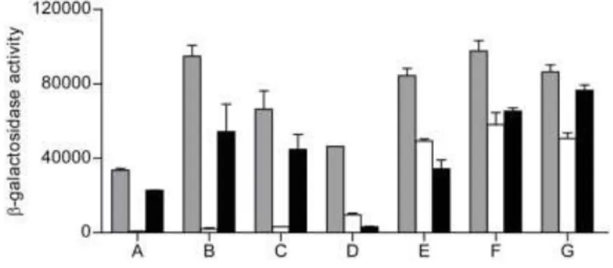
of the β-galactosidase specific activities (Figure 1). Seven pKT25-RIC transformants, 141
harbouring plasmids A to G, exhibited significant β-galactosidase activity indicative of a 142
specific interaction (Figure 1). Nucleotide sequencing followed by BLAST analysis was 143
used to identify the genes within the inserts of these plasmids. Sequencing data revealed 144
that plasmids A to C contain an ~2 kb E. coli DNA fragment upstream of the T18 Cya 145
domain, and that all cases included the complete efp and ecnA genes, and part of the ecnB 146
gene. The efp gene encodes the elongation factor EF-P, a translation factor that facilitates 147
the in vitro the formation of the first peptide bond during translation (19, 20). The gene 148
8
cluster ecnAB expresses two small cell-membrane associated entericidin lipoproteins, 149
forming EcnAB a toxin-antitoxin module that regulates a programmed bacterial cell death 150
under high osmolarity conditions, with EcnA acting as the antidote for the bacteriolytic 151
entericidin, EcnB (21). 152
The other four plasmids D to G also contained a ~2 kb insert located upstream of the T18 153
Cya domain, but in these cases the inserts carried the entire rhtA gene, encoding an inner-154
membrane transporter involved in resistance to homoserine/threonine (22), and the dps 155
gene, encoding the DNA-binding and iron-storage protein from starved cells (12). Like 156
RIC, E. coli Dps has been implicated in oxidative-stress protection, which raises the 157
possibility of a functional association between these two proteins that might be dependent 158
on their direct interaction. For this reason, the potential interaction between the two 159
proteins was investigated further in order to establish its validity and determine its 160
physiological purpose. 161
162 163
E. coli RIC protein interacts with Dps 164
165
To determine whether the interaction between the RIC protein and Dps, as identified 166
through the screening of the pUT18 library, is indeed genuine, further BACTH experiments 167
were performed. To enable such experiments, the gene encoding the RIC protein was 168
cloned into pUT18C and pUT18 vectors (to create T18-RIC and RIC-T18 fusions), and the 169
dps-coding region was introduced into the pKNT25 vector (to give Dps-T25 fusions), 170
following which the β-galactosidase activities of the corresponding co-transformants were 171
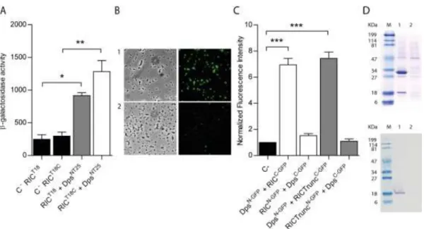
measured. High β-galactosidase activities were recorded for both sets of the RIC-Dps 172
9
BATCH combinations tested, with activities 4-6 times greater than those of the controls 173
(Figure 2A), indicative of interaction between the RIC protein and Dps within the cytosol 174
of E. coli. 175
A second approach was used to test the proposed RIC-Dps interaction, which involved a 176
Bimolecular Fluorescence Complementation (BiFC) assay. In this method, one of the two 177
proteins of interest is fused to the N-terminal half of the green fluorescent protein (GFP), 178
and the other protein of interest is fused to the C-terminal half; the assay depends upon an 179
interaction between the two proteins that promotes the reassembly of the two halves of GFP 180
such that emission of fluorescence is restored (23). Thus, GFP fusions (both the N- and C-181
terminal domains) were generated for both the RIC protein and Dps, and the fluorescence 182
intensity of the corresponding E. coli cells containing plasmids co-expressing the RIC and 183
Dps fusions was measured (Figure 2BC). The data showed that cells expressing RICC-GFP 184
and DpsN-GFP exhibit an approximately six-fold higher fluorescence relative to the control, 185
although transformants expressing RICN-GFP and DpsC-GFP presented fluorescence levels 186
similar to that of the control samples. 187
The RIC protein consists of two domains: a short N-terminal ‘ScdA_N’ domain of ~60 188
residues of unclear function with a highly-conserved pair of Cys residues (11); and a larger 189
C-terminal ‘hemerythrin’ domain of ~140 residues that forms a di-iron centre. We tested 190
the BiFC interaction between Dps and a truncated form of RIC that lacks the so-called first 191
Scd_N domain to determine which of the two RIC protein domains is responsible for the 192
observed interaction with Dps. The results showed that the degree of interaction between 193
the truncated RIC protein and Dps is similar to that observed when using the full-length 194
protein (Figure 2C). Thus, the interaction observed here between the RIC protein and Dps 195
appears to be mediated through the C-terminal hemerythrin domain of the RIC protein. 196
10
The interaction between RIC and Dps was also investigated by a pull-down assay. To this 197
end, cells containing plasmids that express non-labelled Dps and N-terminally His-tagged-198
RIC were treated with formaldehyde, as described in Methods, to promote in vivo cross-199
linking. The cell extract was loaded into a Ni-chelating column and the His-Tag RIC was 200
eluted at 100 mM of imidazole buffer. The fraction was analysed by denaturing SDS-201
PAGE, and Western blotting in which the E. coli Dps antibody was used. Also, cells 202
expressing only the non-labelled Dps were treated and analysed similarly to serve as 203
control. The results depicted in Figure 2D show that elution of His-Tag RIC occurred 204
together with a band that has a molecular mass correspondent to that of Dps. This band was 205
proved by Western-blotting to be the E. coli Dps (Figure 2D). Therefore, the pull-down 206
assays support the interaction between RIC and Dps. 207
208
Dps modulates the function of the RIC protein in maintaining Fe-S cluster status 209
The RIC protein has been linked to the resistance of E. coli to oxidative and nitrosative 210
stresses as its inactivation decreases the survival of E. coli upon exposure to hydrogen 211
peroxide or nitric oxide donors (4). Due to the interaction of the RIC and Dps proteins 212
shown above, we questioned whether Dps could contribute to the stress protection afforded 213
by the RIC protein. To test this possibility, a ΔdpsΔric double mutant was constructed and 214
the growth of E. coli wild type, Δric, Δdps, ΔdpsΔric mutants under oxidative and 215
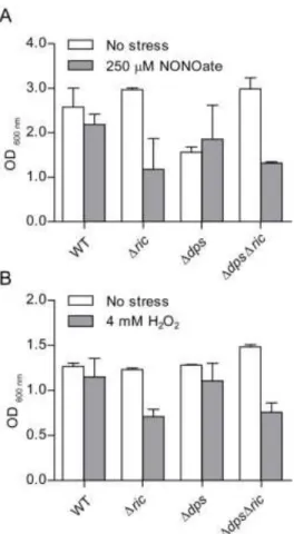
nitrosative stress conditions was tested (Figure 3). The growth experiments showed that 216
inactivation of ric resulted in impaired growth under stress conditions imposed by 4 mM 217
H2O2 or 250 µM spermine NONOate (Figure 3), which is consistent with previous reports 218
11
(4). However, the dps mutation had little impact on growth under these conditions. 219
Combining the Δdps mutation with the Δric mutation did not result in any further growth 220
reduction under the same stress conditions, i.e. the ΔdpsΔric strain grew similarly to the 221
Δric strain under the oxidative and nitrosative stress conditions employed (Figure 3). Thus, 222
Dps does not notably compensate for the lack of the RIC protein under peroxide or NO-223
induced stress. 224
Another characteristic of the E. coli ric mutant is the reduced endogenous activity of Fe-S 225
cluster-containing proteins, such as aconitase and fumarase, that contain solvent-exposed 226
Fe-S clusters with a marked sensitivity to redox and nitrosative stress (4). Therefore, the 227
possible contribution of Dps to this phenotype was explored by comparing the aconitase 228
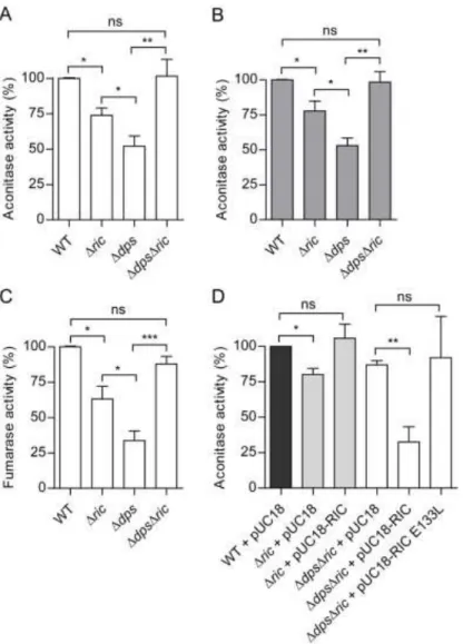
activity of the Δdps and ΔdpsΔric strains to that of the wild type and Δric mutant. The 229
results showed that the Δdps mutation caused a 50% reduction in aconitase activity in log 230
phase (Figure 4A), consistent with a role for Dps in maintaining Fe-S cluster status. As 231
expected, a similar effect was observed for the Δric mutant, although the reduction in 232
activity (30%) was only approximately half as great as that observed for the Δdps mutant 233
(Figure 4A). Surprisingly, the ΔdpsΔric mutant exhibited aconitase activity that was higher 234
than that of the corresponding single mutants and similar to that of the wild type (Figure 235
4A). These aconitase-activity effects were apparent in both the early-log and the post-236
exponential phase (OD600 0.6 and 2, respectively; Figure 4A and B), suggesting that the 237
phenotype is independent of growth stage (note that dps is stationary-phase induced). 238
Similar effects were observed when testing the activity of another Fe-S enzyme, namely 239
fumarase. The data showed a reduction of 70% in fumarase activity in the Δdps mutant 240
12
when compared to wild type during the early-log phase (OD600=0.6). Accordingly, in the 241
Δric mutant there was a reduction in fumarase activity of about 40% while the double 242
mutant ΔdpsΔric displayed a fumarase activity similar to that of the wild type (Figure 4C). 243
The restoration of aconitase and fumarase activity to wildtype levels in the double dps-ric 244
mutant (with respect to the corresponding single mutants) suggests that the negative impact 245
of the lack of the RIC protein on such activity is dependent on the presence of Dps (and 246
vice-versa), and this in turn indicates a hitherto unrecognised functional interdependence 247
for these two proteins. 248
The association of the above aconitase-activity effects with the RIC protein was confirmed 249
by complementation using a multicopy plasmid bearing the wild type ric gene under 250
control of its natural promoter. Complementation of the single Δric mutant led to the 251
recovery of aconitase activity to levels similar to those of the wild type (Figure 4D). More 252
importantly, provision of a wild type version of ric (in multicopy) caused a large (60%) and 253
significant reduction in the aconitase activity of the ΔdpsΔric double mutant (Figure 4D). 254
Thus, as anticipated, the ric-complemented double mutant exhibited the same phenotype as 255
the dps mutant. This confirms that the RIC protein is responsible for decreasing aconitase 256
activity in a dps- background. 257
To investigate whether the role of the RIC protein in lowering aconitase activity in the dps 258
mutant is dependent on a biochemically-functional version of the RIC protein, the ability of 259
a RIC protein variant (lacking a complete di-iron site due to an E133L substitution; (10)), 260
was used in the complementation experiments (Figure 4D). The resulting activity data 261
13
clearly show that he non-functional E133L-RIC variant does not enable a notable decrease 262
in aconitase activity when expressed in the ΔdpsΔric strain (Figure 4D). 263
In summary, the above data suggest that in the absence of Dps, the RIC protein has a 264
deleterious effect on aconitase and fumarase activities, but that such an effect is not 265
exhibited when Dps is present. This would imply that the interaction between Dps and the 266
RIC protein, as revealed here, acts to ensure that neither of these two proteins can 267
participate in processes that negatively impact the activity of these Fe-S enzymes. 268
269
RIC does not interact with other E. coli iron-storage proteins 270
Escherichia coli Dps is an iron-sequestering protein composed of 12 identical subunits 271
forming a shell surrounding a central cavity where up to ~500 ferric iron atoms can be 272
sequestered. As E. coli encodes two other iron-storage proteins, namely bacterioferritin 273
(Bfr) and ferritin (FtnA), the possibility that the RIC protein might interact with these other 274
iron-storage proteins was also investigated. Thus, corresponding BiFC experiments were 275
performed in cells carrying recombinant plasmids that express the RIC protein with either 276
Bfr or FtnA, as N- or C-terminal fusions to GFP domains. The resulting fluorescence 277
intensity data failed to support any protein-protein interaction between the RIC protein and 278
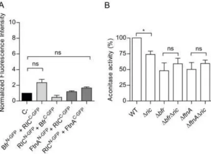
Bfr or FtnA (Figure 5A). 279
In a second set of experiments, the aconitase activity of wild type, Δric, Δbfr, ΔftnA, 280
ΔbfrΔric and ΔftnAΔric strains, grown to the exponential phase (OD600 of 0.6), was 281
determined. Similarly to the Δdps strain, the Δbfr and ΔftnA strains both displayed ~50% 282
14
lower aconitase activity levels (Figure 5B). But contrary to the effect of combining the 283
Δdps and Δric mutations, the combined absence of the RIC protein and the Bfr or FtnA 284
proteins resulted in aconitase activities similar to those present in the correspondent single 285
mutant strains (Figure 5B). Thus, the lower aconitase activity caused by the Δric mutation 286
is not additive with respect to the lower activity resulting from the Δbfr or ΔftnA mutations. 287
Further, it can be concluded that (unlike Dps) Bfr and FtnA do not interact with the RIC 288
protein, and that their absence does not result in a RIC-protein dependent decrease in 289
aconitase activity. 290
291
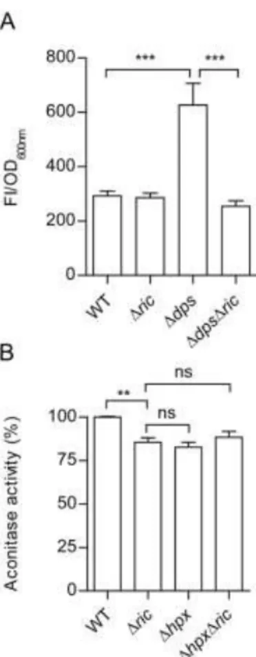
The RIC protein increases intracellular ROS levels when Dps is absent 292
Dps protects cells from oxidative stress due to its ability to couple the reduction of 293
hydrogen peroxide to water with the oxidation of free-ferrous iron to sequestered-ferric 294
iron. In addition, its association with DNA helps to prevent ROS-induced DNA damage 295
(24). This suggests that the role of Dps in preventing RIC-protein induced inhibition of 296
aconitase activity may arise from the ability of Dps to detoxify ROS that might be produced 297
by the di-iron centre of the RIC protein (e.g. through binding and reduction of oxygen). 298
Therefore, the ROS content of Δric, Δdps and ΔdpsΔric strains were compared with those 299
found in the wild type to determine whether the presence of the RIC protein, in the absence 300
of Dps, results in raised levels of ROS (Figure 6A). Data show that the wild type and Δric 301
mutant contain similar amounts of ROS while the Δdps strain had significantly higher (~2-302
fold) levels (Figure 6A). This is as expected given the known role of Dps in redox-stress 303
resistance (24). However, introduction of the ric mutation into the dps mutant eliminated 304
15
the increased intracellular ROS levels of the single Δdps mutant (Figure 6A). This suggests 305
that the raised ROS levels of the dps single mutant are a consequence of an increase in 306
RIC-protein-dependent ROS production which thus supports a role for Dps in interacting 307
with the RIC protein to restrict its release of ROS species. 308
To discover whether other elements of the redox-stress resistance response might also act to 309
lessen RIC-protein induced ROS production, the Δric mutation was introduced into a strain 310
(Δhpx) lacking capacity to degrade hydrogen peroxide due to inactivation of both catalase 311
genes as well as the alkyl-hydroperoxide reductase genes (Table 1; (25, 26)). Assay of the 312
resulting aconitase activity levels showed that the ΔhpxΔric quadruple mutant has activity 313
levels similar to those determined for the Δric and Δhpx mutants (Figure 6B). Therefore, 314
we concluded that the three major peroxidases (KatE, KatG, AhpCF) of E. coli are not 315
involved in countering any RIC-protein mediated ROS production, at least under conditions 316 where Dps is active. 317 318 Discussion 319
Aconitase and fumarase are enzymes of the TCA cycle that are prone to oxidative stress 320
damage. We previously showed that the di-iron RIC protein repairs these enzymes and is 321
able to transfer iron to Fe-S containing proteins (4–7). In the work described here, we 322
screened an E. coli BACTH library in order to identify proteins that interact with the RIC 323
protein and thus might be required to assist its function. As a consequence of our screening, 324
Dps emerged as a RIC protein interaction candidate. This suggested interaction was 325
supported by generation and analysis of additional Dps and RIC protein BACTH constructs 326
16
and by GFP complementation and pull-down assays. Dps belongs to the ferritin 327
superfamily which led us to investigate the possible interaction of RIC with the two other 328
ferritins present in E. coli, namely ferritin and bacterioferritin. However, none of these 329
proteins were found to interact with the RIC protein or to influence its activity in vivo. 330
We also observed that inactivation of the RIC protein resulted in lower aconitase and 331
fumarase activity, which is consistent with previous findings indicating that this protein 332
contributes to the protection of solvent accessible Fe-S clusters from ROS damage under 333
aerobic growth conditions (6). Similar results were herein obtained for the single mutant 334
strains of dps, ftnA and bfr, indicating that lack of any of these gene products results in 335
lower endogenous aconitase activity. The role of FtnA and Bfr in aconitase protection was 336
previously demonstrated as the two ferritins promote the reactivation of aconitase activity 337
following stress damage in Salmonella enterica serovar Typhimurium (27). In contrast with 338
our findings with E. coli, no loss of aconitase activity was observed for S. enterica ftnA or 339
bfr single mutants in the absence of stress; this discrepancy may be related to different 340
physiological roles and expression control of ferritins in Salmonella and E. coli species (27, 341
28). 342
A surprising result was the finding that the defective aconitase activity of the Δdps and Δric 343
single mutant strains was reversed when these two mutations were combined in the 344
ΔdpsΔric double mutant, such that activity was restored to that measured in the wild type. 345
This result, together with the lower amounts of ROS observed in the ΔdpsΔric mutant 346
compared to the Δdps mutant, suggests that the RIC protein is responsible for the 347
generation of ROS, but only in the absence of Dps and, thus, that the interaction of Dps and 348
the RIC protein serves to enable Dps to restrict ROS release (which is presumed to damage 349
17
the Fe-S cluster of aconitase and fumarase, and hence lower the observed activity of these 350
enzymes in a dps mutant) by the RIC protein. Interestingly, other redox-stress resistance 351
components (KatE, KatG and AhpCF) failed to impact the RIC-protein-mediated inhibition 352
of aconitase activity (at least in the presence of Dps). These results suggest that the effect of 353
Dps on the ROS-generation activity of the RIC protein is one that is highly specific and not 354
replicated by the other peroxide-consuming cytosolic factors examined. Indeed, the 355
findings relayed here indicate that a direct interaction is required to enable Dps to quench 356
the ROS-generating activity of the RIC protein. The exact mechanism involved in the 357
apparent quenching of RIC-protein-mediated ROS production by Dps is unclear; such 358
understanding will require in vitro reaction studies combining the Dps and RIC proteins. 359
However, two possible processes by which Dps could exert a ROS-quenching action upon 360
the RIC protein can be considered: Dps might sequester iron released from the di-iron site 361
of the RIC protein and thus restrict Fe-driven Fenton chemistry; or Dps could consume 362
hydrogen peroxide (or hydroxyl radicals; (15, 29)) generated by the RIC protein through 363
reaction at its di-iron site with molecular oxygen. 364
Although the absence of the RIC protein in the presence of Dps resulted in reduced 365
aconitase and fumarase activity, lack of RIC protein had no impact on ROS levels when 366
Dps was present. The reason for this effect is unclear but may indicate a role for the RIC 367
protein in supply of iron from Dps for Fe-S cluster repair and/or synthesis. 368
The proposed role of the RIC protein (4) is to repair damaged Fe-S clusters of [Fe-S]- 369
proteins, such as aconitase and fumarase, by donating iron from its di-iron centre leading to 370
the formation of an intermediate mononuclear iron centre that is prone to react with oxygen 371
to generate ROS such as hydrogen peroxide. In this process, the interaction with Dps would 372
18
fulfil two roles, namely by trapping ROS released by the RIC protein and providing a sink 373
for iron liberated from the di-iron centre of RIC. 374
In conclusion, we report an interaction between the Dps and RIC proteins of E. coli which 375
represents the first example of a protein that interacts with the ferritin-like Dps protein. In 376
addition, our results indicate that the Dps-RIC protein interaction contributes to the 377
function of RIC, which is one of the few known bacterial proteins involved in repair. 378
379
380
381
19 Materials and Methods
383
Bacterial strains and growth conditions 384
Escherichia coli strains used in this work are listed in Table 1, and were grown at 37 ᵒC. E. 385
coli XL2Blue and E. coli reporter strain DHM1 non-reverting adenylate cyclase deficient 386
(cya) were used as host strain and for detection of protein-protein interactions, respectively. 387
Construction of the E. coli double mutant strains was performed by bacteriophage P1-388
mediated transduction (30), and the corrected mutations were confirmed by PCR using 389
primers listed in Table 2. 390
E. coli cells were grown in LB medium under aerobic conditions in flasks containing a 1/5 391
volume of culture or under anaerobic conditions in rubber seal-capped flasks filled with 392
medium and extensively bubbled with nitrogen prior to growth. For the stress assays, cells 393
were grown, at 37 °C and 150 rpm, in M9B minimal medium (60 mM K2HPO4, 33 mM 394
KH2PO4, 7.6 mM (NH4)2SO4, 1.7 mM sodium citrate, 1 mM MgSO4 and 10 µM MnCl2, pH 395
7) supplemented with 10 µg/mL thiamine and 40 µg/mL L-arginine, L-leucine, L-proline, 396
L-threonine and 40 mM glucose. Cultures at an OD600 of 0.3 were either left untreated or 397
exposed to 4 mM H2O2 for 6 h or 250 µM spermine-NONOate for 9 h. 398
399
BACTH experiments 400
The Bacterial Adenylate Cyclase-based Two-Hybrid (BACTH) system assay (17) was used 401
to identify RIC-interacting proteins. E. coli RIC protein was fused to the C-terminal of 402
20
Bordetella pertussis Cya (adenylate cyclase) T25 domain (pKT25-RIC) and used to screen 403
an E. coli MC4100 gene library containing chromosomal fragments fused to the N-terminal 404
of B. pertussis Cya T18 domain. The DNA fragments were obtained by partial digestion 405
with Sau3AI and cloning into the BamHI site of pUT18 plasmids (18). About 1 µg of 406
pUT18BamHI DNA library was transformed together with pKT25-RIC into E. coli DHM1 407
cells by electroporation. Blue colonies present in AmpR CmR selective plates (L-agar with 408
5-bromo-4-chloro-3-indolyl β-D-galactopyranoside (X-Gal)) were identified after 409
incubation at 30 ᵒC for 36 h, and cells with the highest β-galactosidase were considered to 410
contain recombinant plasmids harbouring genes encoding polypeptides that interact with 411
the E. coli RIC protein. Twenty two colonies were obtained and the corresponding plasmids 412
were isolated, co-transformed with pKT25-RIC plasmid in E. coli DHM1, and the strength 413
of the protein-protein interactions observed was again estimated by quantification of the β-414
galactosidase activity. Seven isolates considered positive were named ‘A’ to ‘G’ (Figure 1), 415
and sequenced using primer T18Fw (Table 2). To identify the encoded genes, the sequences 416
were screened against the E. coli K-12MG1655 genome using BLAST. 417
Genes coding for the RIC protein and Dps were PCR amplified from E. coli K-12 genomic 418
DNA using the oligonucleotides described in Table 2, and cloned into pKT25 (fused to Cya 419
C-terminal T25 domain), pKNT25 (fused to Cya N-terminal T25 domain), pUT18 (fused to 420
Cya N-terminal T18 domain) and pUT18C (fused to Cya C-terminal T18 domain) 421
plasmids, and the enzyme Pfu DNA polymerase (Thermo Scientific). The resulting 422
recombinant plasmids encoded Dps or RIC with either a C- or N-terminally linked T25 or 423
T18 domain from the B. pertussis Cya protein. Two complementary plasmids, one carrying 424
a T25 fragment and the other a T18 fragment, were co-transformed into the E. coli DHM1 425
strain (cya-). E. coli DHM1 cells containing the ric-encoding pUT18 or pUT18C plasmids 426
21
were co-transformed with complementary pKTN25 empty plasmid that served as negative 427
controls. 428
In all cases, false positives were tested by co-transformation of E. coli DHM1 with 429
plasmids containing each gene and pKT25-TorD, which expresses E. coli TorD that binds 430
non-specifically to a wide variety of polypeptides (31). 431
For β-galactosidase activity determination (32), at least 3 representative colonies of each 432
transformation plate were inoculated, in duplicate, in LB medium, and following an 433
overnight growth at 37 ᵒC, transformant cultures were re-inoculated (at a 0.01 dilution) into 434
LB with ampicillin (100 μg/mL), kanamycin (50 μg/mL) and IPTG (0.5 mM). When 435
cultures reached an OD600=0.5 (approximately after 16 h of growth, at 30 ᵒC), 1 mL of each 436
culture was collected by centrifugation (5000 g, 5 min at 4 ᵒC). The pellets were lysed by 437
incubation with 100 µL BugBuster HT 1x (Novagen) at 37 ᵒC, for 30 min. Cellular debris 438
was then removed by centrifugation and the β-galactosidase activities were assayed in 20 439
µL suspensions in a microplate reader. The assays were initiated by addition of a reaction 440
mixture comprising: 0.27% β-mercaptoethanol (v/v) and 0.9 mg/mL ONPG (o-nitrophenyl-441
β-D-galactopyranoside) in buffer A (60 mM Na2HPO4.7H2O, 40 mM NaH2PO4.H2O, 1 mM 442
MgSO4.7H2O, 10 mM KCl). Reactions were incubated at 28 ᵒC, and the absorbance was 443
recorded at 420 nm at 2 min intervals, for 90 min. The β-galactosidase specific activity was 444
defined as ONP/min/milligram of protein. Interactions were considered positive for those 445
reactions where β-galactosidase activity was at least four times higher than the negative 446
control. 447
22
Bimolecular fluorescence complementation (BiFC) assays 449
BiFC assay was performed essentially as described previously (33). For this purpose, the 450
genes encoding RIC protein, a truncated version of the RIC protein (lacking the first 57 451
amino acid residues in N-terminal (10)), Dps, Bfr and FtnA were PCR amplified from 452
genomic DNA of E. coli K-12 using the oligonucleotides described in Table 2. Tthe DNA 453
fragments were cloned into vectors (pET11a-link-N-GFP and pMRBAD-link-C-GFP (33)) 454
that express the green fluorescence protein, GFP, to allow formation of corresponding N- or 455
C-terminal GFP fusions, respectively. Cloning was achieved using XhoI and BamHI sites 456
(for cloning into pET11a-link-N-GFP) or NcoI and AatII sites (for cloning into pMRBAD-457
link-C-GFP) sites, except for Dps for which SphI replaced NcoI. All recombinant plasmids 458
were sequenced confirming the integrity of the genes and the absence of undesired 459
mismatches. Cells harboring pET11a-link-N-GFP and pMRBAD-link-C-GFP served as 460
negative control. 461
E. coli BL21(DE3)Gold (Agilent) was co-transformed with the resulting recombinant 462
pET11a-link-N-GFP and pMRBAD-link-C-GFP vectors, in various combinations 463
(RIC/Dps, truncated-RIC/Dps, RIC/Bfr and RIC/FtnA), and plated on selective LB-agar. 464
Colonies were inoculated in LB medium, grown overnight, at 37 ᵒC and 150 rpm, and 465
plated onto inducing LB agar medium containing 20 µM IPTG and 0.2% of arabinose. 466
After an overnight incubation at 30 ˚C followed by two days incubation at room 467
temperature, colonies were suspended in PBS and spread onto 1.7% agarose slides. Cells 468
were examined for green fluorescence in a Leica DM6000 B upright microscope coupled to 469
an Andor iXon+ camera, using a 1000x amplification and a FITC filter. The images were 470
analysed using the MetaMorph Microscopy Automation and Image Analysis Software. 471
23 Pull-down and Western Blot assays
472
The genes encoding RIC and Dps were amplified from E. coli K-12 genomic DNA by 473
PCR, using the oligonucleotides listed in Table 2, cloned into pET28a and pACYCDuet-1 474
vectors, respectively, and sequenced which confirmed their integrity and the absence of 475
undesired mutations. E. coli BL21(DE3)Gold was transformed with the following pair of 476
plasmids : i) pET28a-RIC (expressing the RIC protein fused to a N-terminal His-Tag-RIC) 477
and pACYCDuet-1-Dps (expressing a non-labelled Dps); and ii) pET28a (empty vector) 478
together with pACYCDuet-1-Dps. Cells harbouring the later pair of recombinant plasmids 479
served as control samples. Cells were grown in LB medium, supplemented with 10 μM of 480
Fe and the appropriate antibiotics, at 30 ˚C to an OD600 of 0.3. At this time, 0.3 mM IPTG 481
was added to induce the expression of the His-tagged-RIC and Dps proteins, and after 4 h 482
the cross-linking agent formaldehyde (1% final concentration) was added to the cells. The 483
cross-linking reaction (34) was carried at 37 ˚C for 20 min, and the reaction was stopped by 484
incubation with glycine (final concentration of 0.5 M) at room temperature for 5 min. 485
Bacterial cells were harvested by centrifugation, washed twice with PBS and resuspended 486
in PBS. Cells were disrupted in a French Press (Thermo) and cell debris was removed by 487
centrifugation. The total protein concentration of the supernatants was determined by the 488
Pierce BCA Protein Assay Kit (Thermo Scientific). For the pull-down experiments, these 489
supernatants were loaded into Ni-Chelating Sepharose Fast Flow columns (GE Healthcare), 490
which were first washed with 10 mM Tris-HCl (pH 7.5), and the proteins were eluted with 491
imidazole containing buffers. The protein fractions were analysed by denaturing 12.5% 492
SDS-PAGE, in which the cross-linking promoted by formaldehyde was reversed by 493
heating, and analysed by Western blotting 494
24
For Western Blot analysis, samples that were first resolved by SDS-PAGE were transferred 495
to a nitrocellulose blotting membrane (GE Healthcare) in a Trans-blot semi-dry cell 496
apparatus (Bio-Rad). The membrane was blocked by addition of TBS (20 mM Tris-HCl pH 497
7.5, 500 mM NaCl) containing 5% of dried skimmed milk and incubation at room 498
temperature for 1 h. Then, the membrane was incubated with the primary antibody against 499
E. coli K-12 Dps (1:1000 dilution in TBS-T (TBS + 0.05% Tween-20) plus 5% of dried 500
skimmed milk). Following an overnight incubation at 4 ˚C, the membrane was washed with 501
TBS-T and incubated with the secondary antibody (anti-rabbit IgG-alkaline phosphatase 502
from Sigma) diluted 1:10000 in TBS-T + 5% of dried skimmed milk). The reaction 503
proceeded for 1 h at room temperature and the color was developed by addition of 10 μL of 504
NBT-BCIP (Sigma) in 10 mL buffer (100 mM Tris-HCl pH 9.5, 100 mM NaCl, 5 mM 505
MgCl2). 506
507
Enzyme activity assays and determination of endogenous ROS 508
E. coli wild type, Δric, Δdps and Δdps Δric strains that were transformed with either 509
pUC18, pUC18-RIC or pUC18-RIC-E133L (prepared described in (10)) were tested for 510
endogenous aconitase and fumarase activities. To this end, the E. coli cells strains were 511
grown in LB medium at 37 ᵒC, under aerobic conditions, to an OD600 of 0.6 and 2, as 512
indicated in each case. 513
For the aconitase assays, cells grown to the desired cell density were centrifuged, washed in 514
reaction buffer (50 mM Tris-HCl, 0.6 mM MnCl2, pH 8), and the pellets were frozen in 515
liquid nitrogen. The following experiments were performed under anaerobic conditions. 516
25
Prior to the activity assay, the cell pellets were resuspended in reaction buffer containing 517
0.5 mg/mL lysozyme and 0.2 mg/mL DNAse and incubated on ice for 10 min, and then 518
centrifuged at 9600 g for 10 min, at 4 ᵒC. The aconitase activity was determined in these 519
supernatants (50 μL) in reaction mixtures that also contained 200 µM NADP+, 1 U 520
isocitrate dehydrogenase and 30 mM sodium citrate (10), and by recording the formation of 521
NADPH at 340 nm. 522
For the fumarase activity assays (35), once the cells reached the desired cell density they 523
were centrifuged, washed with 50 mM sodium phosphate pH 7.3 buffer, and frozen in 524
liquid nitrogen. Cell pellets were resuspended in 2 mL of the same phosphate buffer, lysed 525
by five freeze-thaw cycles that used liquid nitrogen and a water bath at room temperature. 526
The resulting cell extracts were cleared by addition of sodium deoxycholate, to a final 527
concentration of 0.5%. Fumarase activity was determined under anaerobic conditions in 528
reaction mixtures that contained the cell lysates (10 μL) and 50 mM L-malate (which is 529
quickly converted to fumarate) and by following the consumption of fumarate at 240 nm. 530
531
Endogenous reactive oxygen species content was determined in E. coli wild type, Δric, 532
Δdps, ΔdpsΔric, Δbfr, ΔbfrΔric, ΔftnA, ΔftnAΔric strains (Table 1). Cells were grown 533
aerobically to an OD600 of 0.6, collected by centrifugation, resuspended in PBS, and 534
distributed in 96-well microtitre plates. Following the addition of dichloro-dihydro-535
fluorescein diacetate (10 µM DCFH-DA), the fluorescence was measured in a 536
spectrofluorimeter Varian Cary (Agilent) at λex = 485 nm and λem = 538 nm,, and for 2 h. 537
26
The Fluorescence Intensity (FI) was normalized in relation to the optical density of each 538 culture at 600 nm. 539 540 Acknowledgments 541 542
We thank Professor Hirotada Mori (Graduate School of Biological Sciences, Nara Institute 543
of Science and Technology, Ikoma, Nara, Japan) for provision of the mutants from the Keio 544
collection, and Professor James Imlay (Department of Microbiology, B103 CLSL, 601 S 545
Goodwin Ave, Urbana, IL 61801, USA) for the Hpx mutant. We also grateful to Professor 546
Tracy Palmer and Frank Sargent (Centre for Bacterial Cell Biology, Newcastle University) 547
for providing the BACTH E. coli library. 548
This work was financially supported by: Project LISBOA-01-0145-FEDER-007660 549
(Microbiologia Molecular, Estrutural e Celular) funded by FEDER funds through 550
COMPETE2020 - Programa Operacional Competitividade e Internacionalização (POCI) 551
and by national funds through FCT - Fundação para a Ciência e a Tecnologia" for grants 552
PTDC/BBB-BQB/5069/2014, and SFRH/BD/118545/2016 (LSOS). This project has also 553
received funding from the European Union’s Horizon 2020 research and innovation 554
programme under grant agreement number 810856. 555
556 557
27 References
558 559
1. Overton TW, Justino MC, Li Y, Baptista JM, Melo AMP, Cole J a, Saraiva LM. 560
2008. Widespread distribution in pathogenic bacteria of di-iron proteins that repair 561
oxidative and nitrosative damage to iron-sulfur centers. J Bacteriol 190:2004–2013. 562
2. Chow ED, Liu OW, O’Brien S, Madhani HD. 2007. Exploration of whole-genome 563
responses of the human AIDS-associated yeast pathogen Cryptococcus neoformans 564
var grubii: nitric oxide stress and body temperature. Curr Genet 52:137–148. 565
3. Harrington JC, Wong SMS, Rosadini C V., Garifulin O, Boyartchuk V, Akerley BJ. 566
2009. Resistance of Haemophilus influenzae to reactive nitrogen donors and gamma 567
interferon-stimulated macrophages requires the formate-dependent nitrite reductase 568
regulator-activated ytfE gene. Infect Immun 77:1945–1958. 569
4. Justino MC, Almeida CC, Teixeira M, Saraiva LM. 2007. Escherichia coli di-iron 570
YtfE protein is necessary for the repair of stress-damaged iron-sulfur clusters. J Biol 571
Chem 282:10352–10359. 572
5. Balasiny B, Rolfe MD, Vine C, Bradley C, Green J, Cole J. 2018. Release of nitric 573
oxide by the Escherichia coli YtfE (RIC) protein and its reduction by the hybrid 574
cluster protein in an integrated pathway to minimize cytoplasmic nitrosative stress. 575
Microbiology 164:563–575. 576
6. Justino MC, Almeida CC, Gonçalves VL, Teixeira M, Saraiva LM. 2006. 577
Escherichia coli YtfE is a di-iron protein with an important function in assembly of 578
iron-sulphur clusters. FEMS Microbiol Lett 257:278–284. 579
28
7. Nobre LS, Garcia-Serres R, Todorovic S, Hildebrandt P, Teixeira M, Latour J-M, 580
Saraiva LM. 2014. Escherichia coli RIC is able to donate iron to iron-sulfur clusters. 581
PLoS One 9:e95222. 582
8. Silva LO, Nobre LS, Mil-Homens D, Fialho A, Saraiva LM. 2018. Repair of Iron 583
Centers RIC protein contributes to the virulence of Staphylococcus aureus. 584
Virulence 9:312–317. 585
9. Todorovic S, Justino MC, Wellenreuther G, Hildebrandt P, Murgida DH, Meyer-586
Klaucke W, Saraiva LM. 2008. Iron-sulfur repair YtfE protein from Escherichia 587
coli: Structural characterization of the di-iron center. J Biol Inorg Chem 13:765–770. 588
10. Nobre LS, Lousa D, Pacheco I, Soares CM, Teixeira M, Saraiva LM. 2015. Insights 589
into the structure of the diiron site of RIC from Escherichia coli. FEBS Lett 590
589:426–431. 591
11. Lo F-C, Hsieh C-C, Maestre-Reyna M, Chen C-Y, Ko T-P, Horng Y-C, Lai Y-C, 592
Chiang Y-W, Chou C-M, Chiang C-H, Huang W-N, Lin Y-H, Bohle DS, Liaw W-F. 593
2016. Crystal Structure Analysis of the Repair of Iron Centers Protein YtfE and Its 594
Interaction with NO. Chem - A Eur J:1–10. 595
12. Almiron M, Link a J, Furlong D, Kolter R. 1992. A novel DNA-binding protein 596
with regulatory and protective roles in starved Escherichia coli. Genes Dev 6:2646– 597
2654. 598
13. Grant R a, Filman DJ, Finkel SE, Kolter R, Hogle JM. 1998. The crystal structure of 599
Dps, a ferritin homolog that binds and protects DNA. Nat Struct Biol 5:294–303. 600
14. Calhoun LN, Kwon YM. 2011. Structure, function and regulation of the DNA-601
29
binding protein Dps and its role in acid and oxidative stress resistance in Escherichia 602
coli: a review. J Appl Microbiol 110:375–386. 603
15. Zhao G, Ceci P, Ilari A, Giangiacomo L, Laue TM, Chiancone E, Chasteen ND. 604
2002. Iron and hydrogen peroxide detoxification properties of DNA-binding protein 605
from starved cells. A ferritin-like DNA-binding protein of Escherichia coli. J Biol 606
Chem 277:27689–27696. 607
16. Karas VO, Westerlaken I, Meyer AS. 2015. The DNA-Binding protein from starved 608
cells (Dps) utilizes dual functions to defend dells against multiple stresses. J 609
Bacteriol 197:3206–3215. 610
17. Karimova G, Pidoux J, Ullmann A, Ladant D. 1998. A bacterial two-hybrid system 611
based on a reconstituted signal transduction pathway. Proc Natl Acad Sci U S A 612
95:5752–5756. 613
18. Jack RL, Buchanan G, Dubini A, Hatzixanthis K, Palmer T, Sargent F. 2004. 614
Coordinating assembly and export of complex bacterial proteins. EMBO J 23:3962– 615
3972. 616
19. Blaha G, Stanley RE, Steitz TA. 2009. Formation of the first peptide bond: The 617
structure of EF-P bound to the 70S ribosome. Science 325:966–970. 618
20. Ganoza MC, Aoki H. 2000. Peptide bond synthesis: function of the efp gene product. 619
Biol Chem 381:553–559. 620
21. Bishop RE, Leskiw BK, Hodges RS, Kay CM, Weiner JH. 1998. The entericidin 621
locus of Escherichia coli and its implications for programmed bacterial cell death. J 622
Mol Biol 280:583–596. 623
30
22. Livshits VA, Zakataeva NP, Aleshin V V., Vitushkina M V. 2003. Identification and 624
characterization of the new gene rhtA involved in threonine and homoserine efflux in 625
Escherichia coli. Res Microbiol 154:123–135. 626
23. Magliery TJ, Wilson CGM, Pan W, Mishler D, Ghosh I, Hamilton AD, Regan L. 627
2005. Detecting protein-protein interactions with a green fluorescent protein 628
fragment reassembly trap: scope and mechanism. J Am Chem Soc 127:146–157. 629
24. Chiancone E, Ceci P. 2010. The multifaceted capacity of Dps proteins to combat 630
bacterial stress conditions: Detoxification of iron and hydrogen peroxide and DNA 631
binding. Biochim Biophys Acta 1800:798–805. 632
25. Park S, You X, Imlay J a. 2005. Substantial DNA damage from submicromolar 633
intracellular hydrogen peroxide detected in Hpx- mutants of Escherichia coli. Proc 634
Natl Acad Sci U S A 102:9317–9322. 635
26. Seaver LC, Imlay JA. 2004. Are respiratory enzymes the primary sources of 636
intracellular hydrogen peroxide? J Biol Chem 279:48742–48750. 637
27. Velayudhan J, Castor M, Richardson A, Main-Hester KL, Fang FC. 2007. The role 638
of ferritins in the physiology of Salmonella enterica sv. Typhimurium: A unique role 639
for ferritin B in iron-sulphur cluster repair and virulence. Mol Microbiol 63:1495– 640
1507. 641
28. Abdul-Tehrani H, Hudson AJ, Chang YS, Timms AR, Hawkins C, Williams JM, 642
Harrison PM, Guest JR, Andrews SC. 1999. Ferritin mutants of Escherichia coli are 643
iron deficient and growth impaired, and fur mutants are iron deficient. J Bacteriol 644
181:1415–1428. 645
31
29. Bellapadrona G, Ardini M, Ceci P, Stefanini S, Chiancone E. 2010. Dps proteins 646
prevent Fenton-mediated oxidative damage by trapping hydroxyl radicals within the 647
protein shell. Free Radic Biol Med 48:292–297. 648
30. Lennox ES. 1955. Transduction of linked genetic characters of the host by 649
bacteriophage P1. Virology 1:190–206. 650
31. Pommier J, Mejean V, Giordano G, Iobbi-Nivol C. 1998. TorD, a cytoplasmic 651
chaperone that interats with the unfolded trimethylamine N-oxide reductase enzyme 652
(TorA) in Escherichia coli. J Biol Chem 273:16615–16620. 653
32. Thibodeau SA, Fang R, Joung JK. 2004. High-throughput B-galactosidase assay for 654
bacterial cell-based reporter systems. Biotechniques 36:410–415. 655
33. Wilson CGM, Magliery TJ, Regan L. 2004. Detecting protein-protein interactions 656
with GFP-fragment reassembly. Nat Methods 1:255–262. 657
34. Ferreira E, Giménez R, Aguilera L, Guzmán K, Aguilar J, Badia J, Baldomà L. 2013. 658
Protein interaction studies point to new functions for Escherichia coli 659
glyceraldehyde-3-phosphate dehydrogenase. Res Microbiol 164:145–154. 660
35. Puchegger S, Redl B, Stoffler G. 1990. Purification and properties of a thermostable 661
fumarate hydratase from the archaeobacterium Sulfolobus solfataricus. J Gen 662
Microbiol 136:1537–1541. 663
36. Vine CE, Justino MC, Saraiva LM, Cole J. 2010. Detection by whole genome 664
microarrays of a spontaneous 126-gene deletion during construction of a ytfE 665
mutant: confirmation that a ytfE mutation results in loss of repair of iron-sulfur 666
centres in proteins damaged by oxidative or nitrosative stress. J Microbiol Methods 667
32 81:77–79.
668
37. Baba T, Ara T, Hasegawa M, Takai Y, Okumura Y, Baba M, Datsenko KA, Tomita 669
M, Wanner BL, Mori H. 2006. Construction of Escherichia coli K-12 in-frame, 670
single-gene knockout mutants: the Keio collection. Mol Syst Biol 2:1-11 671
38. Seaver LC, Imlay JA. 2001. Alkyl hydroperoxide reductase is the primary scavenger 672
of endogenous hydrogen peroxide in Escherichia coli. J Bacteriol 183:7173–7181. 673
39. Jang S, Imlay J a. 2010. Hydrogen peroxide inactivates the Escherichia coli Isc iron-674
sulphur assembly system, and OxyR induces the Suf system to compensate. Mol 675
Microbiol 78:1448–1467. 676
677 678
33 Table 1 –Strains and plasmids used in this study 679
E.coli Description Source
Strains
DHM1 F′, cya-854, recA1, endA1, gyrA96 (NalR), thi1, hsdR17, spoT1, rfbD1, glnV44(AS)
Euromedex
Wild type K-12 ATCC 23716 ATCC
Δric K-12 Δric::cat (36)
Δdps JS091 Δdps::kan (37)
Δbfr JW3298 Δbfr::kan (37)
ΔftnA MC4100 ΔftnA::spc (28)
ΔdpsΔric K-12 Δdps::kan, Δric::cat This study
ΔbfrΔric K-12 Δbfr::kan, Δric::cat This study
ΔftnAΔric K-12 ΔftnA::spc, Δric::cat This study
MG1655 F- WT (38)
SJ90 BW25113 Δric::cat (39)
LC106 (hpx) ΔahpCF′kan::′ahpḞ, Δ(katG17::Tn10)1, Δ(katE12::Tn10)1
(26)
ΔhpxΔric LC106 Δric1::cat This study
XL2 Blue recA1, endA1, gyrA96, thi-1, hsdR17, supE44, relA1, lac [F’ proAB+, lacIqZΔM15 Tn10 (Tetr)]
Agilent
BL21Gold(DE3) E. coli B, F-, ompT, hsdS (rB- mB-), dcm+, Tetr, gal, λ(DE3), endA, Hte
34 Plasmids
pUT18/pUT18C Vector that allows construction of in-frame fusions at the N-terminus/C-terminus of T18 fragment (amino acids 225-399 of CyaA)
(17)
pKT25/pKNT25 Vector that allows construction of in-frame fusions at the N-terminus/C-terminus of T25 fragment (amino acids 1-224 of CyaA)
(17)
pUT18/pUT18C-RIC RIC fused to T18 fragment in N/C-terminal This study pKT25/pKNT25-RIC RIC fused to T25 fragment in N/C-terminal This study pUT18/pUT18C-Dps Dps fused to T18 fragment in N/C-terminal This study pKT25/pKNT25-Dps Dps fused to T25 fragment in N/C-terminal This study pUT18-Zip Leucine zipper fused to T18 fragment in the
N-terminal
(17)
pKT25-Zip Leucine zipper fused to T25 fragment in the C-terminal
(17)
pUT18-TorD TorD fused to T18 fragment in N-terminal (18) pKT25-TorD TorD fused to T25 fragment in C-terminal (18)
BamHI pUT18 plasmid that contains chromosomal
fragments obtained by partial digest of the MC4100 chromosomal DNA with Sau3A1 and cloned into de BamHI site
(18)
35
pUC18-RIC Vector for expression of RIC (4)
pUC18-RIC-Glu133Leu Vector for expression of RIC-Glu133Leu (10) pET11a-link-GFP Vector for expression of fusions with N-
terminal fragment of GFP
(33)
pMRBAD-link-GFP Vector for expression of fusions with C- terminal fragment of GFP
(33)
pET11a-RIC-GFP RIC fused to N-terminal GFP fragment This study pMRBAD-RIC-GFP RIC fused to C-terminal GFP fragment This study pET11a-Dps-GFP Dps fused to N-terminal GFP fragment This study pMRBAD-Dps-GFP Dps fused to C-terminal GFP fragment This study pET11a-Bfr-GFP Bfr fused to N-terminal GFP fragment This study pMRBAD-Bfr-GFP Bfr fused to C-terminal GFP fragment This study pET11a-FtnA-GFP FtnA fused to N-terminal GFP fragment This study pMRBAD-FtnA-GFP FtnA fused to C-terminal GFP fragment This study pET11a-RICTrunc-GFP Truncated RIC fused to N-terminal GFP
fragment
This study
pMRBAD-RICTrunc-GFP Truncated RIC fused to C-terminal GFP fragment
This study
pET-28a Expression vector Novagen
pET-28a-RIC(HisTag) Vector for expression of N-terminal Poly-HisTag-RIC
This study
pACYCDuet-1-Dps Vector for expression of Dps This study
36 Table 2 – Oligonucleotides used in this study 681
Primer Name Sequence
Construction of plasmids used in BACTH
ric_Fw GAGGTGTCGACTATGGCTTATC ric_Rv CTTTTAGGATCCTCACCCGCC dps_Fw GTTAATTACTGGGATCCAACATCAAGAGG dps_Rv TCCTGTCAGGTACCCGCTTTTATC T18_Fw CATTAGGCACCCCAGGCTTTAC T18_Rv GAGCGATTTTCCACAACAAGTC T18C_Fw CATACGGCGTGGCGGGGAAAAG T18C_Rv AGCGGGTGTTGGCGGGTGTCG T25_Fw ATGCCGCCGGTATTCCACTG T25_Rv CGGGCCTCTTCGCTATTACG NT25_Fw CACCCCAGGCTTTACACTTTATGC NT25_Rv CAATGTGGCGTTTTTTTCCTTCG
Construction of plasmids used in BiFC
ric_xhoFw GAATGAGGTCTCGAGTATGGCTTATC ric_bamRv GCGCAATGGGATCCAGCTTTTAGA ric_ncoFw GAGGTATCAGCCATGGCTTATCG ric_aatRv CCAGCTTTTAGACGTCTCACCC dps_xhoFw CGTTAATTACTCGAGCATAACATCAAG dps_bamRv GTACTAAGGATCCGCACCATCAGC
37 dps_sphFw CAAGAGGATATGCATGCATGAGTACCGCTA dps_aatRv CATCAGCGATGGGACGTCTCGATGTTAG bfr_xhoFw GAGTGGAAGCGCTCGAGTCAAAAAATG bfr_bamRv GGAGGGTTCTGGATCCCGACACG bfr_ncoFw GAAGGAGTCAAACCATGGAAGGTGATAC bfr_aatRv CGGACGTCCCTTCTTCGCGGATC truncric_xhoFw CTTTAAGAAGGCTCGAGACATATGGCTG truncric_ncoFw GGAGATATACCCATGGCTGAACAAC ftna_xhoFw CAAATATAACCTTTCTCGAGCACTATC ftna_bamRv TGAAACGGATCCAGTAAACCTGC ftna_ncoFw GAGCACTACCATGGTGAAACCAGAAAT ftna_aatRv CGGAGAGGACGTCTTTTGTGTGTC
Construction of plasmids used for protein expression
pric_ndeFw AAGAATGAGGTATCACATATGGCTTATCGC
pric_ecoriRv GGCTGTTTATTGGTAAGAATTCGGCTGCTG
pdps_ndeFw GAGGATATGAACATATGAGTACCGC
pdps_kpnRV GTACTAAAGTTCGGTACCATCAGCG
Double mutant construction confirmation
Conf_dps_Fw CAGAATAGCGGAACACATAGC
Conf_dps_Rv GATGCACTAAATAAGTGCGTTG
Conf_bfr_Fw CTCTTCAAAGAGTGGAAGCG
Conf_bfr_Rv GATCTCTTATTAACCGGGAGG
38 Conf_ftnA_Rv ACCGATCAGAGTAAGATTTGC Conf_ric_Fw AAGAATGAGGTATCACATATGGCTTATCGC Conf_ric_Rv GGCTGTTTATTGGTAAGAATTCGGCTGCTG 682 683
39
Figure Legend
684
Figure 1: E. coli RIC interactions investigated by BACTH. β-Galactosidase activities of 685
E. coli DHM1 cell lysates expressing, separately, plasmids A to G, which were extracted 686
from the BamHI library and co-transformed with the pKTN25-RIC (grey bars), pKTN25 687
empty plasmid (white bars), and pKTN25 fused to TorD (black bars). Each bar represents 688
the mean value ± standard error from results of at least three independent cultures. 689
690
Figure 2: E. coli RIC interacts with Dps. 691
A) Bacterial two-hybrid assay: the interaction of the RIC protein, linked to the C- (white 692
bar) or N-terminus (grey bar) of the T18-Cya domain and expressed from pUT18 or 693
pUT18C, respectively, was evaluated in E. coli DHM1 co-transformed with pKTN25 694
containing a Dps linked to the N-terminus of the T25-Cya domain. E. coli cells harbouring 695
simultaneously empty pKTN25 and pUT18/pUT18C vectors expressing ric fusions served 696
as negative controls (black bars). 697
B-C) BiFC assays. Cells were co-transformed with vectors expressing either RICC-GFP 698
(pMRBAD-link-C-GFP-RIC) or RIC-TruncatedC-GFP (pMRBAD-link-C-GFP-RICTrunc) 699
with DpsN-GFP (pET11a-link-N-GFP-Dps). The inverse configurations were also included. 700
B) Cells expressing RICC-GFP and DpsN-GFP were analysed by light microscopy (bright field, 701
left upper panel) and fluorescence microscopy (right upper panel). Lower panels depict 702
images of cells co-transformed cells with empty vectors. Images were acquired using a 703
100x objective and a FITC filter was used for the acquisition of the fluorescence images. C) 704
Fluorescence quantification was performed using MetaMorph Microscopy Automation and 705
40
Image Analysis Software. Fluorescence values for negative control (empty plasmid 706
vectors) were normalized to 1. 707
D) Pull-down assays. Lane 1: Cells expressing pET28a-RIC (RIC protein linked to a N-708
terminal His-Tag-RIC (~30 KDa)) and pACYCDuet-1-Dps (non-labelled Dps (~18 KDa)). 709
Lane 2: Cells expressing pET28a (empty vector) and pACYCDuet-1-Dps (non-labelled Dps 710
(~18 KDa)). Protein fractions were eluted from the Ni-chelating column at 100 mM 711
imidazole and analysed by SDS-PAGE (upper panel) and Western blotting using the anti-E. 712
coli Dps antibody (bottom panel). 713
Values are means ± standard error of at least three independent cultures analysed in 714
duplicate ***p<0.0005 (One-way ANOVA multiple comparison test). 715
716 717
Figure 3: Growth of E. coli Δric, Δdps and ΔdpsΔric strains under nitrosative and 718
oxidative stress. E. coli wild type, Δric, Δdps and ΔdpsΔric strain were grown under 719
anaerobic (A) and aerobic (B) conditions. At OD600=0.3, cells were left untreated (white 720
bars), treated (grey bars) with 250 µM spermine NONOate for 7 h (A), or treated with 4 721
mM H2O2 for 5 h (B). Error bars are ± SD for experiments carried out at least three times. 722
723
Figure 4: Aconitase and fumarase activity of E. coli Δric and Δdps mutant strains. 724
Aconitase activity of E. coli wild type, Δric, Δdps and ΔdpsΔric strains grown aerobically 725
to OD600 of 0.6 (A, D) or 2.0 (B). Fumarase activity of E. coli wild type, Δric, Δdps and 726
ΔdpsΔric strains grown aerobically to OD600 of 0.6 (C). Complementation experiments of 727
aconitase activity (D) were performed using Δric and ΔdpsΔric strains transformed with 728