R E S E A R C H
Open Access
Comparison between two methods of scorpion
venom milking in Morocco
Naoual Oukkache
1, Fatima Chgoury
1, Mekki Lalaoui
1, Alejandro Alagón Cano
2and Noreddine Ghalim
1*Abstract
Background:The present study compared two methods used successfully in a large-scale program for the collection of scorpion venoms, namely the milking of adult scorpions via manual and electrical stimulation. Results:Our immunobiochemical characterizations clearly demonstrate that regularly applied electrical stimulation obtains scorpion venom more easily and, most importantly, in greater quantity. Qualitatively, the electrically
collected venom showed lack of hemolymph contaminants such as hemocyanin. In contrast, manual obtainment of venom subjects scorpions to maximal trauma, leading to hemocyanin secretion. Our study highlighted the
importance of reducing scorpion trauma during venom milking.
Conclusions:In conclusion, to produce high quality antivenom with specific antibodies, it is necessary to collect venom by the gentler electrical stimulation method.
Keywords:Scorpion venoms, Manual stimulation, Electrical stimulation, Antivenom, Lethality
Background
Envenomation by scorpion stings is widespread in several countries of the world. In Morocco, the two most
danger-ous types, the black scorpion(Androctonus mauretanicus
mauretanicus: Amm) and the yellow scorpion (Buthus occitanus tenutanus: Bot)are responsible for the majority of stings [1-4].
Scorpion venom contains many proteins, peptides and other compounds, several of which are biologically active and found to be particularly useful in physiological and pharmacological research as investigatory tools [5]. Hence, there is a strong demand to obtain venom from various species of scorpions for research purposes. A variety of techniques have been described to carry out the extraction of venom from scorpions [6,7].
Numerous methods of collecting venom from scor-pions have been described. The venom can be obtained by different methods such as manual extraction, electric stimulation and maceration [8].
The choice of a venom-extraction technique is very important. Moreover, to obtain high-quality venom that does not contain hemolymph contaminants and is usable
for producing antivenom with a specific neutralizing antibody, the milking method is one of the parameters that should be taken into consideration.
In the current work, scorpion venom samples obtained by two different methods (manual and electrical stimula-tion) were compared as to protein content, biochemical characteristics and lethality in order to establish the best methodology for milking and use as animal antigen to produce antivenom with specific neutralizing antibodies.
Methods Venoms
Venom obtained manually
Androctonus mauretanicus(Amm) andButhus occitanus tunetanus (Bot) scorpion venoms were manually milked as described by Louis [9]. This method employs manual stimulations of the abdomen to release venom. The venoms of many scorpion specimens were pooled, lyophilized and stocked at−20°C until use.
Venom obtained electrically
Amm and Bot venoms were collected by the electrical
stimulation method described by Ozkan and Filazi [10]. A series of regular currents were applied to shock the scorpion until the venom was ejected. For that purpose, we immersed the body of the scorpion in a saline
* Correspondence:[email protected]
1Laboratory of Venoms and Toxins, Pasteur Institute of Morocco, 1 Place
Louis Pasteur, Casablanca 20360, Morocco
Full list of author information is available at the end of the article
solution for better electrical conduction and gave a shock with electrode. We used a simple 12-volt battery. The venom droplet was recovered in a Petri dish after which the extract was kept frozen until use. Venom was recovered using distilled water and centrifuged (10,000 g). The supernatant was lyophilized (freeze dried), and then kept at−20°C until use.
Measurement of protein concentration
Protein concentrations were determined by absorbance measurements at 280 nm, assuming an extinction coeffi-cient (E1%280) of 10. For each venom type, we prepared a solution of venom with a final concentration of 5 mg/mL (as determined by A280 nm).
Gel electrophoresis of venoms
Electrophoretic analysis of venoms was performed on 15% polyacrylamide gel in the presence of SDS under reducing conditions. All samples were dissolved in a
sample buffer (50 mM Tris–HCl, pH 6.8, 0.1 M DTT,
10% glycerol, 2% SDS, and 0.1% bromophenol blue). A constant electric current of 70 mA was applied for two hours. After migration, the gel was stained with Coomassie Brilliant Blue R250.
Spectral analysis
The absorbance spectra of venoms were obtained by reading the optical density range between 220 and 600 nm, using a Beckman spectrophotometer.
Determination of median lethal dose (LD50)
Lethal potency of venoms (in micrograms of dry weight per mouse) was determined as recommended by the World Health Organization [11]. Groups of five mice were used per venom dose; venom concentration was di-luted in 150 mM NaCl and injected in final volume of 500μL by intravenous route.
Percent mortality was recorded 24 hours after injec-tion. The median lethal dose was determined using the Software package Prism 5 GraphPad, Inc., according to the provided algorithm. Briefly a non-linear curve fitting (variable slope) was generated using the four-parameter logistical equation; constraints were imposed on minimum (0% mortality) and maximum (100% mortality) values, and no weighing was used. The same package was used to cal-culate median doses (Anova). Plots were generated using KaleidaGraph 4.03 (Synergy Software).
Immunization of horses and antivenom production The antivenom was produced through horse immunization
by manually collectedAmm venom. Following the
immu-nization program, horses were bled. Plasma proteins were fractionated by precipitation with ammonium sulfate and immunoglobulins enzymatically digested to produce F(ab’)2
fragments. The fractions containing the F(ab’)2 fragments
were extensively dialyzed against saline solution.
Ethics committee approval
All the procedures involving animals were in accordance with the ethical principles in animal research adopted by the World Health Organization [11].
Western blot analysis
To investigate the antivenom cross-reactivity to venom components, polyacrylamide gel electrophoresis (12.5%), supplemented with sodium dodecyl sulfate (SDS), was
used to separate venom components (1μg of venom per
well), using venoms collected by manual stimulation
fromAmmand by electric stimulation fromAmmandBot.
Then, separated venom proteins were electrophoretically blotted onto a nitrocellulose membrane. Membrane was blocked with 5% skim milk-phosphate-buffered solution
and incubated in the presence of polyspecific F(ab')2
Amm scorpion antivenom (Pasteur Institute of Morocco;
1:200 dilution), for one hour at room temperature. After the washing step, the membrane was incubated with a horseradish peroxidase-labeled goat anti-horse IgG (Sigma-Aldrich, USA) at 1:1,000 dilution for one hour at room temperature. Immunoblotted proteins were revealed using a substrate of 0.05% 4-chloro-1-naphthol in 15% methanol, in the presence of 0.03% hydrogen peroxide.
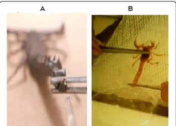
Results Venom colors
The primary characteristic associated with venom
qual-ity is the color. We compared the color of theAmmand
Bot venoms obtained in both manners. Venom obtained
by electrical stimulation is white and does not turn blue after milking (Figure 1). To the contrary, the manually collected venom rapidly becomes blue after milking.
Venom biochemical characteristics
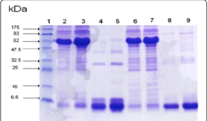
The SDS-PAGE separation and Coommassie staining of the venom proteins were investigated. Electrophoresis analysis indicates that the venom sample obtained by electrical stimulation showed one major band of protein with molecular weights averaging 6.5 kDa
(Figure 2–lanes 2, 3, 6 and 7). Venom obtained
manu-ally showed two protein bands: the first very dense with an average molecular mass of 75 kDa corresponding to the molecular weight of hemocyanin and the second in-distinct corresponding to the molecular weight of scor-pion toxins (Figure 2–lanes 4, 5, 8 and 9). Our results demonstrated that electrically collected venoms contain only proteins normally corresponding to venom toxins. However, venoms collected by manual stimulation con-tain a high percent of hemolymph and small amounts of toxins (Figure 2).
Spectral analysis
The absorption spectra of proteins are currently charac-teristic of all scorpion venoms. We used this method to compare the two differentially milked venoms. Results show that venoms collected by the manual method have three absorption peaks: one at 280 nm and two bands
were recorded near ultraviolet (220–380 nm) with a
maximum at 340 nm and the other in the visible region
(520–600 nm). However, venoms collected by electrical
stimulation present a single absorption peak at 280 nm corresponding to the absorption wavelength of proteins (data not shown).
Indeed, protein content of venom obtained electri-cally was very high (absorption peak is larger compared to venom collected by manual milking). The high hemo-cyanin content within manually obtained venom was
demonstrated by its characteristic spectral profile and absorption in ultraviolet and visible wavelengths.
Determination of median lethal Dose (LD50)
Toxicity of both manually and electrically milked venoms was tested in one group of five mice and median lethal
dose was determined. Results revealed respective LD50
values of 12.5 μg/mouse and 4.9 μg/mouse for manual
and electrical milkedAmm venom. Likewise, LD50s were
32.6 μg/mouse and 11μg/mouse for manually and
elec-trically milked Bot venoms (Table 1). Mice showed the
same envenomation symptoms following intravenous injec-tion of both venom samples (manual and electric milking).
The results were reported in Table 1. Median lethal dose values highlighted that toxicity was lower in the manually collected venoms than those obtained by elec-trical stimulation (very toxic signifies high activity). The activity of venoms collected by the electrical method was estimated to reach three fold greater values than for venoms collected manually.
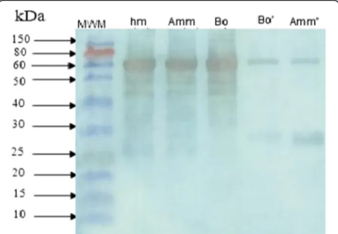
Western blot analysis
Androctonus mauretanicus mauretanicus antivenom was
produced through horse immunization with the Amm
venom obtained by manual stimulation and subsequently used to study the immunochemical characteristics of the two differentially milked venoms. Our results showed that this antivenom strongly recognized the venom obtained by manual stimulation. Western blotting data (Figure 3) clearly revealed proteins with molecular weight of 74 kDa which are highly immunogenic.
Discussion
Scorpion venom contains a short sequence of neuro-toxin polypeptides consisting of simple, low-molecular-weight proteins that have lethal and paralytic effects. Venom toxicity varies according to several factors such as genus, species, age, physiology, feeding state and re-gion of the scorpion. Then, major difficulties are related to standardizing venom quality [8]. To develop an anti-venom that neutralizes the toxic effects of anti-venoms as much as possible, we must have a high quality of venom with a high toxic activity (LD50) (containing a large amount of
toxins). This activity changes according to the method-ology of collecting the venom [6].
Figure 2Electrophoretic profile of venoms on polyacrylamide gel in the presence of SDS in reducing conditions.Lane 1: molecular mass markers, lane 2:Ammmanual method (MM) (10μg),
lane 3:AmmMM (20μg), lane 4:Ammelectric method (EM) (10μg),
lane 5:AmmEM (20μg), lane 6:BotMM (10μg), lane 7:Bot MM
(20μg), lane 8: Bot EM (10μg), lane 9:BotEM (20μg).
Table 1 Comparison of median lethal dose (LD50) of venom obtained manually and those obtained electrically
Injection way LD50(μg venom/mouse)
Ammvenom Botvenom
Manual method
Electric method
Manual method
Electric method
Intravenous (μg venom/mouse)
The antivenom plays an important role in the treatment of envenomation by scorpion stings. The present work found that the best antigen preparation for immunization is non-hemolymph-contaminated venom designed to pro-duce antivenom with a high neutralizing capacity. Thus, it is important to have an effective venom-milking method-ology. It is well established that the LD50 of scorpion
venom can vary even if the venom was extracted by using a single method. For instance, Ismailet al. [12], found that
the LD50 of A. crassicauda venom obtained by electric
stimulation was 0.64 mg/kg, whereas Latoxan Laboratory
reported an LD50 of 0.87 mg/kg for the same venom
obtained by the same method [13]. However, Altinkurt and Altan [14] reported that the LD50 of A. crassicauda
venom was 11.5 mg/kg by the maceration method. Ozkan and Filazi [10] found the toxicity of milked venom to be eight times higher than that of venom obtained by macer-ation of telsons.
Latifi and Tabatabai [15] reported that the mean amount of venom obtained by electric stimulation was 0.3 mg per scorpion (A. crassicauda), whereas 0.5 mg of venom was obtained by telson maceration.
In the present study, we found that the venom extracted by the manual method contains undesirable substances of hemolymph origin. Moreover, the venom obtained by electric stimulation is highly toxic with an LD50three-fold
greater than its manually collected counterpart.
Our biochemical investigations showed that venoms collected by manual stimulation have an additional band of 75 kDa that is absent in the electrophoretical profile of electrically obtained venom. Besides the spectrophotometric
absorption at 280 nm (protein absorption), the absorption profiles show that the venoms obtained manually have two absorption peak regions (at 220–380 nm and 520–600 nm), which are absent in the venoms extracted by electric stimu-lation. According to the literature, hemocyanin is a respira-tory pigment that gives scorpion hemolymph its blue color. As a percentage, it is almost the exclusive component of the hemolymph. Hemocyanin possesses a high molecular mass, and extracellular copper containing glyco-protein that displays the important function of oxygen-carrying proteins freely dissolved in the hemolymph of many mol-lusks and arthropods [16]. In Arthropods, hemocyanins occur mainly in the subphyla Crustacea and Chelicerata. They consist of hexameric or multihexameric complexes containing up to eight 70–75 kDa polypeptide chains with different functional and structural properties [17]. Two absorption spectra have been described, the first recorded in the near ultraviolet [265–365 nm] and the second in the visible range [400–700 nm].
The present results indicated that low-molecular-weight proteins played an important immunogenic role in the production of high-quality antivenom. Lethality and protein patterns showed variability according to the method used. Toxicity of venom obtained by the manual method was lower than when obtained by electric stimula-tion. Therefore, toxicity variation depends on the employed milking method, which strongly reaffirms its importance to the lethality of the venom. Venom obtained manually showed lower toxicity. Indeed, the venom obtained by manual stimulation produced similar hemolymph and Western blot profiles (similar electrophoretical feature). The most abundant protein band migrated at 75 kDa mo-lecular weight and corresponds to hemocyanin. The large amount of hemolymph and small amounts of toxins pro-vide the most likely explanation for its low toxicity. More-over, the corresponding antivenom contains small amounts of specific antibodies that are capable of binding toxins and subsequently of neutralizing the lethality of the venom. This result confirms the data presented above and high-lights the correlation among protein content, absorbance, toxicity and electrophoresis profile. Our result revealed that hemocyanin retrieved from the manually collected venom is a contaminant protein of hemolymph origin.
One of the interesting points is the low toxicity of the manually collected venom, a result that confirms the ob-servations published by Inceoglu et al. [18] that scor-pions, when initially stimulated, secrete a small quantity of transparent venom denominated prevenom. If secre-tion continues, cloudy dense venom, white in color, is subsequently released. The prevenom contains a com-bination of salt and several peptides that modulate ionic channels and elicit significant pain and toxicity because of a massive local depolarization. Indeed, the authors revealed that venom collected manually corresponds to Figure 3Western blot analysis of scorpion venoms by 12.5%
the first venom type (prevenom), whereas the electrically milked venom corresponds to the second type, with high concentration of toxins.
Conclusion
In this study, we clearly demonstrated that there is a high hemocyanin contamination in scorpion venom obtained by the manual stimulation method. Indeed, the corresponding antivenom produced from the venom obtained manually presents a high percentage of specific antibodies that neutralize the hymolymph molecules and few specific anti-bodies that neutralize scorpion toxins.
The venoms obtained by electrical stimulation may con-tribute to the production of a better antivenom with a higher neutralization potency. As to its advantages in rela-tion to the manual method, electrical stimularela-tion not only enables the collection of nearly 100% of the venom, but also yields more venom than manual stimulation with a higher content of toxins.
Competing interests
The authors declare that they have no competing interest.
Authors’contributions
ON performed the chemical tests. CF was responsible for the venom manually obtained and its preparation. LM, director of the Pasteur Institute of Morocco, established the conditions for this study and took part by reading and correcting the manuscript. AA and ON set up the sampling technique for electrically obtained venom and contributed with technical analysis. GN provided the scorpions, supervised the venom collection and preparation, and was responsible for drafting the present manuscript and for the editorial corrections. All authors read and approved the final manuscript.
Acknowledgements
We are grateful to Dr Jean-Philippe Chippaux for valuable suggestions and helpfulness during the preparation of the manuscript. We are indebted to Dr Boussadda (responsible for the Animal Unit of the Pasteur Institute of Morocco). This research was supported by the Pasteur Institute of Morocco, the Bioclon Institute (Mexico) and the Institute of Biotechnology, National Autonomous University of Mexico.
Author details
1Laboratory of Venoms and Toxins, Pasteur Institute of Morocco, 1 Place
Louis Pasteur, Casablanca 20360, Morocco.2Institute of Biotechnology, National Autonomous University of Mexico, Cuernavaca, Mexico.
Received: 2 October 2012 Accepted: 13 November 2012 Published: 28 March 2013
References
1. Ghalim N, El-Hafny B, Sebti F, Heikel J, Lazar N, Moustanir R,et al:Scorpion envenomation and serotherapy in Morocco.Am J Trop Med Hyg 2000,62(2):277–83.
2. El Hafny B, Ghalim N:Évolution clinique et taux circulants du venin dans les envenimations scorpioniques au Maroc.Bull Soc Pathol Exot 2002,95(3):200–4.
3. Charrab N, Soulaymani R, Mokhtari A, Semlali I, El Oufir R, Soulaymani A:The epidemiological situation of scorpion stings in the Beni Mellal province.
Santé Publique2009,21(4):393–401.
4. Bencheikh RB, Soulaymani A, Semlali I, Tamim OK, Zemrour E, Eloufir R,et al:
Les piqûres et les envenimations scorpioniques au niveau de la population de Khouribga (Maroc).Bull Soc Pathol Exot2005,98(1):36–40.
5. Oukkache N, Rosso JP, Alami M, Ghalim N, Saïle R, Hassar M,et al:New analysis of the toxic compounds from theAndroctonus mauretanicus mauretanicusscorpion venom.Toxicon2008,51(5):835–52.
6. Wee MCE, Wong PTH, Cheah LS, Gopalakrishnakone P, Low KSY:The black scorpionHeterometrus longimanus: pharmacological and biochemical investigation of the venom.Toxicon1993,31(10):1305–14.
7. du Plessis JL:Collection of venom from southern African scorpions.
Toxicon2005,45(5):681–82.
8. Gopalakrishnakone P, Cheah J, Gwee MCE:Black scorpion (Heterometrus longimanus) as a laboratory animal: maintenance of a colony of scorpion for milking of venom for research, using a restraining device.Lab Anim 1995,29(4):456–8.
9. Louis J:Venin et antivenin du scorpion Marocain :Androctonus mauretanicus.Med Armées1976,4:429–34.
10. Ozkan O, Filazi A:The determination of acute lethal dose-50 (LD50) levels of venom in mice, obtained by different methods from scorpions,
Androctonus crassicauda(Olivier 1807).Acta Parasitol Turcica2004,28(1):50–3.
11. World Health Organization:Progress in the characterization of venoms and standardization of antivenoms.Geneva: WHO offset Publication; 1981:58. 12. Ismail M, Abd-Elsalam MA, al-Ahaidib MS:Androctonus crassicauda(Olivier),
a dangerous and unduly neglected scorpion--I. Pharmacological and clinical studies.Toxicon1994,32(12):1599–618.
13. Ozkan O, Kar S, Güven E, Ergun G:Comparison of proteins, lethality and immunogenic compounds ofAndroctonus crassicauda(Olivier, 1807) (Scorpiones: Buthidae) venom obtained by different methods.J Venom Anim Toxins incl Trop Dis2007,13(4):844–56.
14. Altinkurt O, Altan M:Pharmacological effects of the scorpion
(Androctonus crassicauda) venom from Urfa environment on laboratory animals and the antagonistic effects of streptomycin to most of these effects.J Fac Pharmacol Ankara1980,10(1):41–61.
15. Latifi M, Tabatabai M:Immunological studies on Iranian scorpions venom and antiserum.Toxicon1979,17(6):617–20.
16. Burmester T:Origin and evolution of arthropod hemocyanins and related proteins.J Comp Physiol B2002,172(2):95–107.
17. Jaenicke E, Decker H, Gebauer W, Markl J, Burmester T:Identification, structure, and properties of hemocyanins from diplopod Myriapoda.
J Biol Chem1999,274(41):29071–4.
18. Inceoglu B, Lango J, Jing J, Chen L, Doymaz F, Pessah IN,et al:One scorpion, two venoms: prevenom ofParabuthus transvaalicusacts as an alternative type of venom with distinct mechanism of action.Proc Natl Acad Sci USA2003,100(3):922–7.
doi:10.1186/1678-9199-19-5
Cite this article as:Oukkacheet al.:Comparison between two methods
of scorpion venom milking in Morocco.Journal of Venomous Animals and
Toxins including Tropical Diseases201319:5.
Submit your next manuscript to BioMed Central and take full advantage of:
• Convenient online submission
• Thorough peer review
• No space constraints or color figure charges
• Immediate publication on acceptance
• Inclusion in PubMed, CAS, Scopus and Google Scholar
• Research which is freely available for redistribution