i
LAIN URIEL OHLWEILER
PARTICULARIDADES DO ESPERMATOZÓIDE E DA CÉLULA SOMÁTICA NA INTERAÇÃO COM O OOPLASMA: O BOVINO COMO MODELO NA FIV E O
SUÍNO COMO MODELO NA CLONAGEM
LAGES – SC
ii
UNIVERSIDADE DO ESTADO DE SANTA CATARINA – UDESC
CENTRO DE CIÊNCIAS AGROVETERINÁRIAS – CAV
PROGRAMA DE PÓS-GRADUAÇÃO EM CIÊNCIA ANIMAL
LAIN URIEL OHLWEILER
Particularidades do espermatozóide e da célula somática na interação com o ooplasma: o bovino como modelo na FIV e o suíno como modelo na clonagem
Dissertação apresentada ao Curso de Mestrado em Ciência Animal, Área de Concentração em Reprodução Animal, do Centro de Ciências Agroveterinárias da Universidade do Estado de Santa Catarina (CAV-UDESC), como requisito para obtenção de grau de Mestre em Ciência Animal.
Orientador: Alceu Mezzalira
LAGES – SC
iv AGRADECIMENTOS
A Deus, por ter me concedido a vida, a saúde e a oportunidade de ter encontrado colegas, professores e oportunidades que proporcionassem minhas conquistas.
Aos colegas de laboratório Norton, Gustavo, Alysson, Renata, Nerissa, Mariana, Fernanda e tantos outros que colaboraram. Aos professores que proporcionaram condições para que este trabalho fosse executado e foram mais do que incentivadores Daniela dos Santos Brum, Fabio Gallas Leivas, Vilceu Bordignon, e ao doutor e orientador Alceu Mezzalira.
Aos meus familiares: mãe, Hilga Ignes Ohlweiler, pai, Flávio Ohlweiler e ao meu irmão Fabiano Ohlweiler.
A minha colega de laboratório, parceira incansável, co-orientadora e namorada, Joana Mezzalira.
Ao Dr. Mathew Wheeler, por ter proporcionado acesso a base de dados bibliográficos da Universidade de Illinois, em Urbana-Champaign.
Aos frigoríficos Fox, Verdi e Pamplona, nas pessoas de dona Irani, Sr. Ariel Verdi, Sr. Valdecir Ferreira, Sr. Junior Pamplona e Sr. Valssionir, entre tantos outros, que infelizmente não estão aqui listados, mas que nos proporcionam o mais importante de nosso trabalho, a matéria prima.
v
vi
Ficha catalográfica elaborada pela Bibliotecária Renata Weingärtner Rosa – CRB 228/14ª Região
(Biblioteca Setorial do CAV/UDESC)
Ohlweiler, Lain Uriel
Particularidades do espermatozóide e da célula somática na interação com o ooplasma: bovino como modelo na fiv e o suíno como modelo na clonagem / Lain Uriel Ohlweiler; orientador: Alceu Mezzalira . – Lages, 2012.
72f.
Inclui referências.
Dissertação (mestrado) – Centro de Ciências Agroveterinárias / UDESC.
1. Hand-made cloning . 2. Clonagem inter-espécie. 3. Ativação embrionária. 4. Qualidade dos gametas. I. Título.
vii RESUMO
PARTICULARIDADES DO ESPERMATOZÓIDE E DA CÉLULA SOMÁTICA NA INTERAÇÃO COM O OOPLASMA: BOVINO COMO MODELO NA FIV E O
SUÍNO COMO MODELO NA CLONAGEM
O desenvolvimento embrionário depende da adequada interação nucleo-citoplasmática, o que é influenciado pelo tipo de célula doadora e pela qualidade do oócito receptor na clonagem, assim como por características dos espermatozóides e oócitos na fecundação in vitro (FIV). O
primeiro estudo foi constituído de dois experimentos. O primeiro experimento avaliou o tipo de célula doadora de núcleo (células fibroblásticas - FIB vs. células mesenquimais derivadas de adipócitos - ADMSC), com diferentes citoplastos receptores (suíno – reconstruído com dois hemi-citoplasto suínos; mosaico – reconstruído com um citoplasto suíno e um citoplasto bovino; bovino – reconstruído com dois hemi-citoplastos bovinos), no desenvolvimento de embriões suínos, clonados por transferência nuclear de células somáticas (TNCS). Os cultivos celulares foram estabelecidos a partir de dois suínos de raças ameaçadas de extinção (casco de mula e moura), sendo os embriões reconstruídos por clonagem manual e cultivados in vitro
por 7 dias, em meio PZM-3. Os grupos mosaico e bovino apresentaram produção embrionária menor que o grupo suíno (5,5; 1,9 e 18,0%, respectivamente). O grupo ADMSC-mosaico do animal moura apresentou produção embrionária intermediaria em relação ao controle e ao bovino, e superior ao grupo FIB-mosaico do mesmo animal. A porcentagem de blastômeros fragmentados em embriões clivados e mórulas foi superior nos grupos mosaico e bovino, em relação ao grupo suíno. A dinâmica de fusão, observada conforme a migração mitocondrial entre os citoplastos, foi diferente em função do citoplasto empregado. No segundo experimento foi investigado o efeito do inibidor de desacetilases “Scriptaid” no desenvolvimento embrionário in vitro dos grupos suíno, mosaico e bovino, utilizando-se
células fibroblastos do animal moura. Os embriões reconstruídos foram expostos a 500 nM de Scriptaid por 12 h, iniciando a partir da ativação, sendo então cultivados em PZM-3 por 7 dias. A taxa de produção de blastocistos do grupo controle (9,2 vs. 17,3%) e mosaico (1,0 vs. 9,2%) aumentou com o uso de Scriptaid (p < 0,05), enquanto a proporção de fragmentos em mórulas reduziu no grupo mosaico (9,8 vs. 2,8%) (p < 0,05). No entanto, o uso de Scriptaid não aumentou a produção embrionária no grupo bovino. No segundo estudo, constituído de três experimentos, avaliou-se a influência de distintas qualidades de gametas na produção embrionária por FIV, em bovinos. Nos experimentos 1 e 3, foram avaliadas as taxas de produção embrionária no sétimo dia de cultivo. No experimento 2, dois touros de comprovada eficiência na produção embrionária in vivtro, foram utilizados na FIV de oócitos de qualidade
viii
ambas qualidades. Não foi observado nenhum efeito de touro ou oócito na produção embrionária. Os resultados permitem concluir que o uso de moduladores da reprogramação como Scriptaid e tecnologias como a ICSI são alternativas adequadas para incrementar, ao menos em condições particulares, o desenvolvimento embrionário.
ix ABSTRACT
PARTICULARITIES OF SPERM AND SOMATIC CELLS IN THEIR INTERACTIONS
WITH THE OOPLASM: THE IVF AS A BOVINE MODEL AND THE INTER-SPECIES
CLONING AS A PORCINE MODEL
x
xi
LISTA DE TABELAS
Page Article 1
Table 1. In vitro development of porcine SCNT embryos reconstructed from distinct
donor cells and recipient cytoplasts………...…..……. 27 Table 2. In vitro development of control, mosaic and inter-species porcine SCNT
embryos treated with Scriptaid …….……… 33 Table 3. Average nuclei number in morulas and blastocysts, and proportion of cells in the inner cell mass of clone blastocysts produced in control, mosaic and inter-generic porcine SCNT, treated or not with Scriptaid………..…… 34
Article 2
Table 1. Cleavage and blastocyst rates obtained after IVF and IVC using sperm from two bulls and oocytes of good* or poor** quality …..……….….. 53 Table 2. Maturation rates of good* or poor** quality oocytes fertilized with semen from either bull A or bull B. Penetration rates and developmental status of zygotes immediately after in-vitro fertilization……….…… 53
Table 3. Sperm penetration rates after SUZI procedure within 1 h or 2-3 h after sperm processing………... 54 Table 4. Cleavage and blastocyst rates obtained after IVF and ICSI using sperm
xii
LIST OF FIGURES
Page Article 1
Figure 1. Cleavage, fragmented blastomeres, morulae and blastocyst rate in fibroblast cells, ADMSC and in the average of both cells reconstructed with porcine, mosaic and bovine cytoplasts, independent of the animal donor of the cells ...…………... 28 Figure 2. Representative pictures showing the progress of fusion and mitochondrial
migration evaluated in porcine, mosaic and bovine cytoplasts. Hoechst staining represents the donor cell location, which stays close to the staining mitochondria original position. Mito-tracker staining represents the migration of mitochondrial from one of the recipients cytoplasts, always reconstructed close to the donor cell. Reconstructed embryo immediately before fusion (A), with starting the mitochondria migration until 10 minutes after fusion (B), with mitochondria located in the opposite cytoplast (C), mitochondria starting to return to the original cytoplast and re-homogenate in the cytoplasm (D), mitochondrial homogeneous distribution (E). Original Magnification x400…………... 30 Figure 3. Mitochondrial distribution after fusion of the reconstructed embryo.
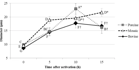
Presence of stained mitochondria in both cytoplasts in porcine (a), mosaic (b) and bovine (c) reconstructed embryos. Presence of stained mitochondria in just one cytoplast in porcine (a`), mosaic (b`) and bovine (c`) reconstructed embryos.…... 32 Figure 4. Pronuclear diameter (µm) 0, 5, 10 and 15 hours after activation of porcine
xiii
LIST OF ABREVIATURES
AC Corrente Alternada (Alternate Current)
ANOVA Análise de Variância (Analysis of Variance)
ART Assisted Reproductive Technologies Bl Blastocisto (Blastocyst)
BSA Albumina sérica bovina (Bovine Serum Albumin)
CIV (IVC) Cultivo in vitro (in vitro culture)
COC Complexos cumulus-oócito (cumulus-oocyte complexes)
DC Corrente Contínua (Direct Current)
FCS Soro fetal bovino (fetal calf serum)
FIV (IVF) Fecundação in vitro (in vitro fertilization)
HMC Clonagem manual (Handmade Cloning)
HM Meio de manipulação (Hepes-buffered medium - M-199)
ICSI Injeção intra-citoplasmática de espermatozóides (Intracytoplasmic sperm injection)
ICM Massa celular interna (inner cell mass)
MII Metáfase II (Metaphase II)
MIV (IVM) Maturação in vitro (in vitro maturation)
mRNA RNA mensageiro (Messenger RNA)
mSOFaa modified Synthetic Oviductal Fluid medium supplemented with amino acids
PIV (IVP) Produção in vitro (in vitro production)
6-DMAP 6-Dimetil aminopurina (6-DymethylAminoPurine)
SUZI Injeção sub-zona de espermatozoide (sub-zona sperm injection)
TCN Total cell number
xiv
TNCS (SCNT) Transferência Nuclear de Células Somáticas (somatic cell nuclear transfer)
TNCSi/iSCNT Transferência nuclear de célula somática inter-espécie (inter-specie somatic cell nuclear transfer)
OT Transferência de ooplasma (ooplasm transfer)
xv SUMMARY
Page
1 INTRODUCTION... 1
2 LITERATURE REVIEW... 6
2.1 Gametes manipulation: evolution of the techniques of embryo production and the embryo development biology understandment ... 6
2.1.1 IVF dissemination and its obstacles ... 8
2.1.2 Cloning: current status and impairing factors ………..…... 9
2.2 Strategies to increase efficiency of Assisted Reproductive Technologies... 11
2.2.1 Intracytoplasmic sperm injection (ICSI) ... 11
2.2.2 Sub-zona sperm injection (SUZI) …….…... 11
2.2.3 Ooplasm transfer (OT)……….…... 12
2.2.4 Inter-species somatic cell nuclear transfer (iSCNT)... 13
2.2.4.1 The reprogramming capacity from distinct cell types ...…... 15
3 ARTICLE 1……….………... 16
4 ARTICLE 2………..………..…………... 43
5 GENERAL CONCLUSIONS .………... 62
1 1. INTRODUCTION
Epigenetic alterations involving changes in global DNA methylation and histone modifications are necessary to give to the embryo the ability to initiate the transcription of the genome followed by the development of two different cell lineages present in the blastocyst (FULKA et al., 2008). These events occur in all embryos derived from any methodology. The main difference is that the fertilized embryo starts with two haploid nuclei while the nuclear transfer embryo start with a diploid nucleus.
The oocyte is the unique common factor that is involved in any embryo production modality. The previous statement is true for in vivo embryo production, in vitro fertilization
2
Known molecular factors hidden from the classical gamete classification as well as the inadequate factors during IVF as particularities observed in morphology and behaviour of sperm (HANSEN et al., 2009) and oocytes (KHURANA and NIEMANN, 2000; HANSEN et al., 2009) generally are closely related with failures in embryo production. Some of the events that occur during the normal development of IVF embryos are the adequate interaction and synchrony between male and female pronucleus (LAURINČÍK et al., 1998) and the adequate embryo genome activation. The sperm ability to activate correctly the oocyte, inducing the oocyte cleavage, not necessarily is related with the capacity of sperm to de-methylate adequately and develop a good quality male pronucleus. The opposite are also true, the plenum oocyte activation is not guarantee of development of a good quality female pronucleus. Experimentally, to overpasses deficiencies of embryo production, researchers use pre-tested semen to assure the sires are proven for in vitro fertility, and select oocytes based
on morphological good quality, for the IVF procedures (SIRARD et al., 2006). However, mainly in commercial IVF programs that are established and profitable for the bovine, the specific breeding wanted causes as these variables cannot be controlled by the simple selection of only the good gametes.
3
efficiency, one of the best established aspects is that after SCNT, the genome of the donor cell does not respond to the active de-methylation activity of the oocyte cytoplasm, and in most cases, the DNA methylation level remains higher than in normal embryos (HAN et al., 2003; BEAUJEAN et al., 2004). Jin et al., (2007) showed that the use of nucleus derived from mesenchymal stem cells can increase the development of porcine embryos. However, the strategy of using cells less differentiated as donor of nucleus is not capable alone of increase the efficiency of cloning (MARTINEZ-DIAZ et al., 2010). By the other hand, the use of nucleus with different competence of reprogramming is a good strategy to study the nucleus-cytoplasmic interaction and activity.
A proposed strategy to overpass this situation is the treatment with histone deacetylase inhibitors (HDACi) such as Trichostatin-A (TSA) and Scriptaid, which stimulate nascent mRNA synthesis, increases histone acetylation, and enhance the transcriptional activity and histone acetylation.
The TSA treatment has no increment on bovine fertilized embryos production (IKEDA et al., 2009; OLIVEIRA et al., 2010) and rat fertilized or ICSI embryo production (YOSHIZAWA et al., 2010). However, it increases the development until blastocyst stage in cloning in pigs (LI et al., 2008; YAMANAKA et al., 2009; BEEBE et al., 2009; MARTINEZ-DIAZ et al., 2010), cattle (ENRIGHT et al., 2003; DING et al., 2008; AKAGI et al., 2011; SRIRATTANA et al., 2012), rabbits (SHI et al., 2008), mouse (BUI et al., 2010), and increase production of offspring in mice (KISHIGAMI et al., 2006, 2007), the weight at birth pig cloning tended to be higher (MARTINEZ-DIAZ et al., 2010) and the pregnancy rate in bovines (SRIRATTANA et al., 2012). The in vitro and in vivo beneficial effects of TSA
4
However, TSA treatment has the adverse effect to be teratogen (SVENSSON et al., 1998) and more severe, might causes placenta megaly in mouse (KISHIGAMI et al., 2006).
Scriptaid, which is less toxic than TSA has also shown a significantly improvement in the production of cloned embryos in pigs (ZHAO et al., 2009, 2010), mice (VAN THUAN et al., 2009) and bovines (AKAGI et al., 2011, WANG et al., 2011). However, Scriptaid treatment also did not show beneficial effect in parthenogenetic and IVF embryo production of porcine (ZHAO et al., 2009) and bovine (BARRETA, 2012).
One of the species that show the better constancy about in vitro and in vivo embryo
development in IVF and SCNT is the bovine. This is one of the reasons why the bovine cytoplasts are also one of the most used to study the inter-specie somatic cell nuclear transfer (iSCNT). One of the early attempts of iSCNT using the enucleated bovine oocyte as recipient cytoplast was reported by Dominko et al. (1999). The interest for study iSCNT increased in the last years because of the appeal of preserving endangered animals (that are threatened with extinction), and also by the excellent potential to study nucleus-cytoplasmic interactions, as well as the embryo development.
As the nucleus-cytoplasmic compatibility are problematic in iSCNT embryos, a high
in vitro developmental block from 8- to 16-cell stage has already been observed in the pig
(YOON, 2001), horse (LI et al., 2002; SANSINENA, 2002), llama (SANSINENA et al., 2003), Siberian tiger (HASHEM et al., 2007), monkey (LORTHONGPANICH et al., 2008; SONG et al., 2009), marbled cat (Pardofelismarmorata) and flat-headed cat
(Prionailurusplaniceps) (THONGPHAKDEE et al., 2010), and Tibetan antelope (ZHAO et
5
embryos, where they found the stage of 8- to 16 cells to show a significantly low level of ATP production. The strategy of combining cytoplasts of one species with a high availability of oocytes with cytoplasts of a species with low availability (i.e in risk of extinction) appears as a good technique to try to supply nucleus-cytoplasmic deficiencies caused by the incompatibility. Moreover, it also offers more cytoplasm content to support the embryo development. This idea is more viable in the hand-made cloning (HMC) technique, proposed by PEURA et al. (1998) and modified by VAJTA et al. (2001), where the association of two cytoplasts is easily performed.
6 2. LITERATURE REVIEW
2.1 GAMETES MANIPULATION: EVOLUTION OF EMBRYO PRODUCTION TECNIQUES AND THE UNDERSTANDING OF EMBRYO DEVELOPMENT BIOLOGY
On early 1900s, Hans Spemman performed experiments considered elegant and genial, using amphibians as a model (TAGARELLI et al., 2004). His pioneering studies involved embryo splitting for studying events of early embryo development. Those have been indeed the first reports of clone production, where genetically identical individuals have been first artificially through embryo splitting. The same researcher started nuclear transfer studies about the year of 1920, and had been awarded with a Nobel Prize of Physiology/Medicine later, on 1935.
Hans Spemman tried to answer, for the first time, the following question: “Do cell nuclei change during development?” Using increasing development stages nuclear transfer with amphibian eggs (TAGARELLI et al., 2004) he was aiming to determine when exactly the developmental potential of nuclei would become restrict. At that point, many years ago, he was disseminating the principles of animal cloning. Those principles happen to be the base for studies on cell differentiation and totipotency.
After Spemman death, in 1941, studies on animal cloning had only been continued as late as in the 1950s, by Robert Briggs and Thomas King (BRIGGS and KING, 1952), using frogs as a model.
With a few understanding of in vivo embryo development added by a slight success in
7
al. (1982) reported the birth of the first calf derived from in vitro fertilization (IVF). Few
years later, in 1986, the first offspring derived from ovine blastomers was reported (WILLADSEN, 1986), again using in vivo maturated oocytes. The successful results with the
ovine have been soon reported for the bovine, when Prather et al. (1987) and Robl et al. (1987) reported the birth of the first calves derived from blastomeres nuclear transfer (NT). This reinforced the process was possible to repeat, at that time, as long as totipotent or multi-potent cells were used.
Even with reports of positive results on in vitro fertilization and cloning by nuclear
transfer, the efficiency was still considerably low to start considering to offer the technique in large scale. By the way, Lu et al. (1987) reported the birth of twin calves after in vitro
maturation, fertilization and culture. This has been certainly a huge step towards the ability to control, in vitro, almost all embryo developing stages.
At this point, the use of blastomeres was still an impairing factor for cloning, due to the reduced number of identical offspring that could be eventually produced, derived from a single embryo. The search for an executable technique, to be used with undifferentiated cells has led to the use of cells derived from day-9 embryonic disc, from passages 6 to 13. Again, the results reported the birth of viable offspring (CAMPBELL et al., 1996). This improvement raised the possibility of using a higher number of identical cells, and maintained for a longer period of time.
8
1997). This has opposed the biological dogma that persisted from as early as the XX century, and said a differentiated cell, already specialized, could not go back to its totipotent status.
The achievements reported above were not devoid of criticism and skepticism, though reduced by the reporting of the first mouse offspring derived from differentiated (cumulus) cells. The female mouse was born on 1997, (WAKAYAMA et al., 1998). Few time later, the first calf derived from fetal somatic cells was born (CIBELLI et al., 1998), being this report increased in importance as the calves were transgenic. During the same year, the first calf derived from adult differentiated cells nuclear transfer was born (KATO et al., 1998), providing a more solid pathway for researchers to continue.
2.1.1 IVF dissemination and its obstacles
9
2.1.2 Cloning: current status and impairing factors
Even with a considerable low efficiency of cloning (WESTHUSIN et al., 2001; RENARD et al., 2002), few years after the first somatic cell cloning offspring were born, a number of companies started to offer cloning services. A considerable number of companies, not long time later, failed.
Currently, the main role played by cloning is to be used as a tool for producing transgenic animals, and also to supply the demand for cloning superior animals. Recently, the biotechnology promise is the regular delivery of transgenic animals for use in biomedical drugs production, and not only as models for research. The first encouraging results on transgenic animal production were reported by Hammer et al. (1985), with micro-injection of rabbits, sheep and pigs pronuclear stage embryos. The efficiency of this protocol ranges around 10 to 15% among the animals produced. In 1997, Schnieke et al. (1997) reported the birth of the first transgenic sheep produces by NT, with the great efficiency of 100% of transgenic among animals born. In this context, cloning of genetically modified animals has become extremely applicable, although not yet efficient. Animals with the ability to secrete specific molecules by their saliva, blood, urine and mainly, in milk, are highly needed by biomedicine and agriculture.
10
NT is not only an important step to reduce economical and time-wasting losses, but will represent another important breakthrough toward elimination of such problems.
Two of the determining factors for cloned embryo development are the nuclear donor and also the recipient cytoplast (usually an oocyte or an early zygote). Their interactions are, in fact, the most important factor or success.
It is known that different cell lines derived from the same donor are responsible for producing distinct embryo developing rates. Moreover, the same cell type from distinct donors may generate distinct embryo production efficiencies. This information illustrates how important it is to understand how nucleus-cytoplasm interactions occur and what exactly is the role played by them during nuclear reprogramming and embryo genome activation.
The recipient cytoplasm plays a key role in reprogramming the genome donor, however the ooplasmic components that are responsible for post-fertilization reprogramming events is not likely to be enough to alter the key-points for nuclear differentiation after SCNT (BIRD, 2002). This process is also known as “erasure” of somatic differentiation status (CEZAR, 2003).
11
2.2 Strategies to increase efficiency of assisted reproductive technologies
With the aim of increasing embryo production efficiency among the different species, different assisted reproductive technologies (ARTs) have emerged. Besides they can overpass specific particularities intrinsic to each species, they have also been responsible for raising new knowledge regarding the embryo development events.
2.2.1 Intra-cytoplasmic sperm injection (ICSI)
About a century ago, G. L. Kite was one of the pioneers in the field of microinjected spermatozoa and the complete result of his experiment was never published (HIRAMOTO, 1962). These studies re-started a half of a century later with Y. Hiram to using sea urchin eggs (HIRAMOTO, 1962). Some year after the ICSI technique has been proposed with the main objective in attempt to overpass the negative aspects of non-fertilization or polisher (HAFEZ, 2003). However, this technique also presents some disadvantages, such as the need for an additional chemical activation for species such as equine, porcine and ruminants. Still, the need for additional chemical activation may eventually reinforce the opportunity for an adequate oocyte activation.
2.2.2 Sub-zonal sperm injection (SUZI)
12
the injection of 6 to 12 spermatic cells per oocyte, resulting in a high polispermy rate (LIPPI et al., 1993).
The main importance of SUZI has been the possibility of evaluating more narrowly the particularities of sperm-oocyte interactions and its further effects on embryo development (LI et al., 2003).
2.2.3 Ooplasm transfer (OT)
Other than ICSI and SUZI techniques that advocated mainly for reverting problems related to male gametes, the OT has been proposed in 1980 decade (MUGGLETON-HARRIS and WHITTINGHAM, 1982) aiming to supply oocyte deficiencies. The purpose of this technique is to transfer a small ooplasm amount (ranging from 10 to 30% of total oocyte volume), from an excellent/good quality, to a low quality oocyte, in an attempt to aid the embryo development with this good cytoplasm boost (HAWES et al., 2001). Although controversial, the OT technique (JOHN et al., 2002; DONG et al., 2006) has already been used for human ARTs where it solved early- and mid- pregnancy losses. It has also been used in inter-species cloning (SANSINENA et al., 2011).
13
2.2.4 Inter-species somatic cell nuclear transfer (iSCNT)
What impairs most the maintenance of a wide range of genetic variability is the momentary commercial interest for determined strains and/or species of animals. The maintenance of animals that carry a high genetic value, and that are for many times threatened with extinction is very expensive and currently almost non-viable, due to their current low applicability. Thus, genetic banks generated from their cells are of a huge importance for preservation of genetic variability, and for reducing costs that would be extremely high for keeping these herds away from inbreeding.
The maintenance and also recovery of animal species can also be questioned due to the fact that it goes against the natural selection evolutionary process. However, it is still anyway more interesting to keep a genetic bank with gametes and cells stored for long term, even if the utility of such genomes is not promptly visualized. This enhances the opportunity to insert important characteristics that would be otherwise simply lost to industrial breeding.
When a certain species in already threatened with extinction, the availability of cytoplasts (i.e. oocytes) is severely reduced. To this, the solution appears as the application of inter-species cloning by nuclear transfer, for recovering and preserving not only the genotypes, but also the epigenetic variability of these animals.
14
oocyte activation, poor respiratory efficiency of embryos and also lack of the minimum ATP level availability, mainly from the stage when cells start to become compact. This corroborate to failure on genome activation that per se would already be a serious cause of embryo losses. Therefore, according to the state above, the SCNT between different species with the aim of understanding the distinct events that play a role on the technique efficiency is per se a tool to understand the events affected by its application. Due to establishing a relationship with early embryo developing events, there has been an increasing interest from the scientific community for the iSCNT.
Although wild animals cloning has already reported encouraging results using recipient cytoplasts from distinct species, not only for embryo production up to the blastocyst stage, but also with alive, although not viable offspring (VOGEL, 2001; FOLCH et al., 2009). Little is known regarding what total or partial mitochondrial heteroplasmy involves in these embryos, fetuses of born animals. Also little is known regarding their nuclear reprogramming process. Due to the fact that so many uncontrolled factors affect iSCNT success, we can infer that the results are positively surprising and that cloning provides great expectations on preservation of threatened, or even extinct, species.
15
2.2.4.1 The reprogramming capacity from distinct cell types
Fibroblasts are the most common cell type used for cloning. They are relatively simple to obtain and their population is wide. As fibroblasts are differentiated cells they are very stable in culture, conversely they are known to be difficult to be reprogrammed exactly to its stability, what makes the role played by the oocyte, during reprogramming, way more difficult.
Recently, mesenchymal stem cells have showed to be an important alternative to replace fibroblasts on SCNT. Such cell type presents a high tissue repairing and healing capacity, as well as self-renewal ability (HUANG et al., 2011). As they provide high differentiation plasticity in different cell lines the interest on them not only for cloning, but also for stem cells therapy, has been significantly increased. The mesenchymal cells can be obtained from different tissues, being the most used sources the bone marrow and the fat tissue, where it is found a higher population of them.
16 3. ARTICLE 1:
THE ROLE OF THE OOPLASM TYPE AND THE DEACETYLASE INHIBITOR SCRIPTAID IN PORCINE INTER-SPECIES SOMATIC CELL NUCLEAR TRANSFER
ABSTRACT
The adequate embryo development in cloning depends on proper nucleus-cytoplasmic interactions, what is influenced by the donor cell type and recipient oocyte quality. In this study the first experiment investigated the effect of the donor cell type (fibroblastic-like cells - FIB vs. adipocyte-derived mesenchymal stem cells - ADMSC), and host cytoplast (porcine – reconstructed with 2 hemi-porcine cytoplasts; mosaic – reconstructed with hemi-porcine and hemi-bovine cytoplast; bovine – reconstructed using 2 hemi-bovine cytoplasts), on development of porcine somatic cell nuclear transfer (SCNT) embryos. Somatic cell cultures were established from two different pigs, both of endangered breeds (Mule-foot and Moura). Embryos were reconstructed by hand-made cloning and cultured for 7 days in PZM-3. Mosaic and inter-species groups produced lower blastocyst rates than controls. The group ADMSC-mosaic from Moura animal showed embryo development rate intermediate between the control and inter-species groups, and higher than the FIB-mosaic group of the same animal. The percentage of fragmented blastomeres in cleaved embryos and morulas from the mosaic and bovine groups were higher than in the control. The dynamic of fusion was different according the cytoplasm reconstruction, observed by mitochondria staining. In the second experiment we investigated the effect of the histones de-acethylase inhibitors “Scriptaid” on the in vitro development of the porcine, mosaic and bovine SCNT reconstruction models. Activated embryos were exposed to 500 nM Scriptaid during 12 h, and then washed and cultured in PZM-3 for 7 days. The blastocyst rate in the control and mosaic groups was increased by Scriptaid treatment, and the proportion of fragmented morulae reduced in mosaic. However, Scriptaid treatment did not increase embryo development for the bovine group. The results evidenced that the use of the homologous cytoplasm complementation for a certain type of cells help the embryo development of the iSCNT embryos. In a general point of view, for the mosaic reconstruction the ADMSC cells have a better embryo development than the fibroblast cells. The use of reprogramming modulator as Scriptaid is an adequate alternative to provide at least a partial increase in embryo development.
17 Introduction
The ooplasm constitutes an unique mixture of factors that is critical for a successful reprogramming of the haploid maternal and paternal genomes at fertilization, as well as of the diploid somatic cell genome after somatic cell nuclear transfer (SCNT). As oocytes are more readily available in some species, such as cattle and pigs, than in others, the possibility of producing inter-species or even inter-generic SCNT embryos is promising for basic and applied research. Few live inter-species clones have been produced (LANZA et al., 2000; LOI et al., 2001; GÓMEZ et al., 2004, 2008, 2009; KIM et al., 2007; FOLCH et al., 2009; YANG et al., 2010) and all of these had been produced from cells reprogrammed after transfer into enucleated oocytes, that is, ooplasm, of the same genus. Inter-generic SCNT has so far only resulted in pregnancies that did not go to term (DOMINKO et al., 1999; CHEN et al., 2002; YIN et al., 2006). Recent research on inter-species (between different species of the same genus) and inter-generic SCNT (between different genii) has focused on improving the developmental rates of the embryos by modifying single steps of the SCNT protocol and the subsequent culture conditions (HASHEM et al., 2007; ZHAO et al., 2007). Only few reports studied initial interaction between the recipient ooplasm and the donor genome required for adequate reprogramming and initiation of embryonic development (WANG et al., 2009).
18
cloning, due to a number of factors affecting embryo reprogramming, and a deficient operation of histones soon after embryo genome activation (MARTINEZ-DIAZ et al., 2010) arises as one of the main reasons for failure. Not only the inter-species and inter-generics cloning is an important tool to provide alternatives for endangered species, it is also one of the most exciting tools for studding early development events and the roles played by the ooplasm during this complex circuitry that rules mechanical and molecular pathways of genome activation and embryo reprogramming. This study aimed to evaluate inter-species ooplasm interactions on early porcine clone embryos as well as to elucidate peri- fusion and activation events, in an attempt to increase the global efficiency of cloning.
2 Materials and Methods
2.1 Chemicals
Unless otherwise indicated, chemicals were purchased from Sigma Chemical Co. (St. Louis, MO, USA).
2.2 Experimental design
19
after activation), the proportion of fragmented blastomeres present in cleaved embryos and morulas, as well as the total cell number in the blastocysts. Mitochondrial migration through the fused cytoplasts as well as pronuclei swelling in the three groups of cytoplasts, were additionally evaluated.
In an attempt to increase the inter-species cloning development, the experiment 2 used the deacethylases inhibitor, Scriptaid treatment for the three groups of cytoplasts (porcine, mosaic and bovine) using fibroblast cells of the Moura animal. The same evaluations of the experiment 1 were performed, as well as the estimation of the blastocysts inner cell mass proportion.
2.3 Animal welfare
All animal procedures were approved by the Animal Care and Use Committee of Santa Catarina State University, and were in compliance with the guidelines from the Brazilian Council of Animal Care.
2.4 Nuclear donor cells collection and handling
20
10,000 IU/mL penicillin G, 10mg/mL streptomycin sulfate, and 10% fetal calf serum (FCS) (Nutricell, SP, Brazil). Cells were cultured up to 90% confluence, being either passage or cryopreserved for further utilization. Cryopreservation was performed in 0.25 mL plastic straws in culture medium with 10% of dimethyl sulphoxyde.
For cloning, cells were thawed at least 72 hours before the cloning, and only cells at a high confluence (> 85%) and up to the fourth passage were used.
2.5 Oocyte preparation
2.5.1 Porcine oocytes
21
maturation medium without LH, FSH, and dbcAMP for an additional 17 h under the same conditions.
2.5.2 Bovine oocytes
Bovine COCs were aspirated from 2–8 mm of diameter follicles from ovaries collected from a local abattoir and transported up to 6 hours to the laboratory. Only COCs completely surrounded by cumulus cells, with homogeneous or with an evenly granulated cytoplasm were selected. Groups of 50 COCs were cultured in four-well dishes containing 0.4 mL of maturation medium using four-well dishes in a humidified atmosphere of 5% CO2 and 95% air at 38.5 °C for 18 hours. Maturation medium consisted of TCM199 (TCM-199 with Earle’s salt, 25mM HEPES), 10% of estrous mare serum (EMS), supplemented with 26.2 mM of sodium bicarbonate, 0.2 mM of sodium pyruvate, 0.01 UI/mL of FSH, 0.5 mg/mL of LH, 10 ng/mL of EGF, 10,000 IU/mL of penicillin G and 10mg/mL of streptomycin sulfate.
2.6 Nuclear transfer, oocyte activation, and embryo culture
Matured porcine oocytes were submitted to successive pipetting in HEPES-buffered M-199 with 20% FCS (HM20) for cumulus cells removal, and the oocytes were cultured in HM20
supplemented with 0.4 µg/mL demecolcine and 0.05M sucrose for 40 to 60 min. After rinsed
in HM20, the partial zonae digestion was performed by exposure to 0.19% pronase in HEPES-buffered M-199 with 0.01% PVA and 25% FCS during 30 sec. After a recovery interval of at least 30 minutes in HM20, the oocytes with a loose and thin zona pellucida were
incubated in 2.5 µg/mL cytochalasin B in HM20, in groups of up to 3 oocytes in 5 µL drops
22
bisbenzmide 33342 (Hoechst) in HM20, being each hemi-oocyte placed into 2 µL drops in
the same medium into a 60 mm Falcon Petri dish under mineral oil. The selection of cytoplasts was performed under ultra violet (UV) light in an epifluorescence microscope. Selected porcine cytoplasts were removed from Hoechst screening dish and maintained in HM20 until the embryo reconstruction.
The bovine COCs were submitted to successive pipetting in HM with 10% FCS (HM10), for cumulus cells removal, followed by the selection of maturated oocytes by the presence of the first polar body. Zona pellucida removal was performed in 0.5% pronase in HEPES-buffered M-199 with 0.01% PVA during 30 sec. Zona-free oocytes were rinsed several times in HM10,
incubated in 5 µg/mL cytochalasin B in HM10 in groups of up to 3 oocytes in 5 µL drops
under oil and hand-bisected (Ultrasharp Splitting Blade, Bioniche, Athens, GA). The cytoplast selection was performed upon the same procedures described previously. After selected, the cytoplasts were removed from the Hoechst screening dish and maintained in HM20 until the embryo reconstruction.
Embryos reconstruction was performed by the brief exposure of two cytoplasts (porcine + porcine - porcine group; porcine + bovine – mosaic group; or bovine + bovine – bovine group) and a somatic cell to 500 µg/mL phytohaemoagglutinin (PHA) solution in HEPES -buffered M-199 with 0.01% PVA. The embryo reconstruction technique was based in de Ribeiro et al. (2009). Briefly, a somatic cell was attached to one cytoplast, followed by the adhesion of a second cytoplast to the first, creating a 180° arrangement and recovering the original cytoplasm volume. In the mosaic reconstruction, the cell was attached to the porcine cytoplast.
23
pre-fusion AC pulse, followed by two 1.6 kV/cm DC pulses for 24 µsec, in a BTX 453
chamber (BTX Instruments, Genetronics, San Diego, CA) coupled to an electro-fusion apparatus (BTX Electro Cell Manipulator 2001, Biotechnologies & Experimental Research Inc., San Diego, CA). After electro-fusion, the oocytes were cultured in porcine zygote medium (PZM-3) for 1 h. Fusion was assessed and only completely fused embryos were activated.
The SCNT embryos were activated through a brief exposure to ionomycin (10 µM/5 min) in HM20 followed by exposure to 6-dimethyl aminopurine (2mM/3 h) in PZM-3. Zygotes were cultured using the well-of-the-well (WOW) system, described by Vajta et al., (2001), and modified by Feltrin et al., (2006). In vitro culture was under atmosphere of 5% CO2, 5% O2 and 90% N2 at 38.5°C. The cleavage and blastocyst rates of SCNT embryos were determined at 48 h and 7 days after activation, respectively.
2.7 Mitochondrial migration analysis during the fusion/activation process and pronuclear
development evaluation
One cytoplast (porcine in porcine and mosaic reconstruction, bovine in bovine reconstruction) was stained with 200 nM of MitoTracker®Green FM (M-7514, Molecular Probes, Inc.) in HM20 for 45min at 38.5° C. Then cytoplasts were washed trice in HM20 and then maintained in HM20 until used on embryo reconstruction.
In all reconstructed groups, the cytoplast stained was the one attached to the somatic cell (Figure 2A).
24
The mitochondrial dynamics analysis was performed 0-60 min after oocytes fusion, with intervals of 5 min with concomitant analysis of the nuclear status and position by labeling
with 10 µg/ml of Hoechst. After 60 min of fusion, the mitochondrial distribution was analyzed until 180 min, with intervals of 30 min. The structures used on this analysis were not activated.
After activation, the pronuclear growing was analyzed with 0, 5, 10 and 15h after fusion. Mitochondrial and nuclear fluorescence was assessed using an epifluorescent inverted microscope under UV light, using a x40 objective and filters at ex = 490 nm and em> 516 nm for Mito tracker, and ex = 355 nm and em = 465 nm for Hoechst.
2.8 Scriptaid treatment
After activation porcine, mosaic and bovine SCNT zygotes reconstructed with Moura breed fibroblasts were separated into two groups and cultured in PZM-3 supplemented or not with 500nM Scriptaid for a period of 12 h starting after ionomycin treatment. Oocytes were then washed three to five times in PZM-3 and cultured as described previously.
2.9 Embryo fragmentation analysis, cell density and inner cell mass proportion
In the experiment 1 and 2, cleaved structures from day 2 of culture that did not progress on
development were stained with 15 µg/mL Hoecsht in HM10 at day seven of culture, being the
25
carried out, by gently pipetting. At least 30 cleaved embryos and 3 morulas were analyzed in each group.
The estimation of the total cell number (TCN) as well as the proportion of cells in the inner cell mass (ICM) per blastocyst were performed by differential staining, based on Mezzalira et al. (2011). Briefly, blastocysts from each group were incubated in Dulbecco’s
phosphate-buffered saline (DPBS) solution containing 10 µg/mL propidium iodide and 1 mg/mL Triton
X-100 for 40 sec at room temperature. Then, embryos were fixed in absolute ethanol
containing 15 µg/mL Hochest for additional 7 min. Fixed embryos were placed on in a 2-µL
glycerol droplet between slide and coverslip, for immediate evaluation under the epifluorescent inverted UV-microscope.
Statistical analysis
All data were analyzed using the JMP software (SAS Institute Inc. Cary, NC, USA). Cleavage, blastocyst and ICM:TCN ratios were analyzed by the Chi-square test. All other data including morula and blastocyst TCN, fragmentation in cleaved embryos and morulas were analyzed by one-way analysis of variance (ANOVA). Fragmentation in cleaved embryos and morulas and the proportion of cells in ICM were normalized by the Arco sine of the square root. The means were compared by Tukey test for TCN in blastocysts. Percentages of development to the morula and blastocysts stages were based on total of cultured zygotes. A value of p ≤ 0.05 was considered statistically significant.
3. Results
3.1 Embryo development features from the two pig breeds and two cell types, post-fusion
26
3.1.1 Embryo development
The detailed results of embryo development are illustrated in table 1. The cleavage rates for all groups were higher than 80%. Some particularities were found in the fibroblast groups, as well as between the different cell types within the same animal (table 1). For the Mule-foot, the porcine and bovine reconstructions showed lower cleavage than the mosaic. Still for the Mule-foot, the fibroblasts were responsible for higher cleavage rates than the ADMSC cells, the opposite was observed for the bovine reconstruction.
For the Moura breed, the cleavage of porcine and mosaic reconstructions were higher than the bovine, being the fibroblasts responsible for higher cleavage in the porcine reconstruction, whereas the ADMSC cells were for thebovine reconstruction.
For both animals, no differences were observed on cleavage among the three types of embryo reconstruction.
The percentage of fragmented blastomeres in the Mule-foot did not differ among the cytoplast reconstruction types, within each cell type. However, the ADMSC cells showed less fragmentation than the fibroblasts, for porcine and bovine reconstructions.
Moura fragmentation rate was increased according to the proportion of inter-species cytoplasts used for reconstruction, for both cell types. This animal has also showed a lower fragmentation when ADMSC cells were used for porcine and bovine reconstructions.
The reconstruction affected the percentage of morula production only for the Moura breed. When using fibroblasts as cell donors, the bovine reconstruction produced less morulas than the other two groups.
Also, for both Moura and Mule-foot, the use of ADMSC provided higher morula rates than the fibroblasts, when reconstruction was the bovine cytoplast. For Moura, this behaviour has also observed when the reconstruction was with the mosaic cytoplast. The proportion of enucleated blastomeres in morulas did not differ between the porcine reconstructions, despite of the cell line used (mean = 2.1%). Moreover, the porcine were lower than the mosaic (mean = 10.8%) and bovine (mean = 11.0%) reconstructions. The mosaic and bovine proportion of enucleated blastomeres in morulas also did not differ between the reconstruction and the cell types.
27
The Moura fibroblasts have also produced more blastocysts when porcine cytoplasts were used. It was only the association of ADMSC, Moura and mosaic reconstruction that showed an intermediated blastocyst rate, between the bovine and porcine cytoplasts reconstructions. This association has also provided a higher blastocyst rate than the association fibrolasts, Moura and mosaic cytoplast reconstruction.
In summary it was observed that only when fibroblasts were used, the cleavage of bovine reconstruction reduced, in comparison to the porcine and mosaic. Then it was analyzed based in the average of both cells, this difference disappeared (Figure 1).
The percentage of fragmented blastomeres present in embryos reconstructed with fibroblasts increased significantly, according to the higher proportion of inter-species cytoplast. For the embryos derived from ADMSC and also for the average of both cells, fragmentation rate was the lowest for the porcine reconstruction.
Table 1. In vitro development of porcine SCNT embryos reconstructed from distinct donor
cells and recipient cytoplasts
Cleavage Blastocysts
Cell donor
animal Cell Type Cytoplast
Fused
n (replicates) n (%) Fragmented blastomeres (%)
Morulas
n (%) n (%) Total of nuclei (n) Porcine 132 (5) 121 (91.7)b 16.5 ± 3.7* 23 (17.4) 24 (18.2)a 36.8 ± 3.9 (11)
Mosaic 128 (5) 125 (97.7)a* 22.8 ± 5.3 29 (22.7) 8 (6.3)b 28.3 ± 7.5 (3)
Fibroblast
Bovine 122 (5) 107 (87.7)b* 28.7 ± 3.9* 20 (16.4)* 4 (3.3)b 24.0 ± 7.5 (3)
Porcine 121 (5) 113 (93.4) 8.3 ± 4.5* 28 (23.1) 25 (20.7)A 43.8 ± 3.3 (15)
Mosaic 126 (5) 116 (92.1)* 18.9 ± 5.0 22 (17.5) 4 (3.2)B
Mulefoot
ADMSC
Bovine 123 (5) 117 (95.1)* 19.8 ± 4.4* 34 (27.6)* 3 (2.4)B 30.0 ± 7.5 (3)
Porcine 149 (6) 142 (95.3)x* 17.1 ± 4.7x* 26 (17.4)x 20 (13.4)x 34.8 ± 3.7 (12)
Mosaic 151 (6) 137 (90.7)x 20.9 ± 4.9x 26 (17.2)x* 3 (2.0)y*
Fibroblast
Bovine 139 (5) 115 (82.7)y* 37.7 ±4.5y* 13 (9.4)y* 0 (0.0)y
Porcine 119 (4) 105 (88.2)* 9.4 ± 5.4Y* 24 (20.2) 25 (21.0)X 39.5 ± 2.7 (23)XY
Mosaic 143 (4) 127 (88.8) 19.1 ± 4.7XY 41 (28.7)* 15 (10.5)Y* 31.8 ± 3.5 (14)Y
Moura
ADMSC
Bovine 138 (4) 128 (92.8)* 25.6 ± 5.0X* 32 (23.2)* 3 (2.2)Z 62.0 ± 7.5 (3)X
a,b; A,B; x,y,z; X,Y,Z distinct letters indicate a difference between cytoplasts within the same animal and cell type.
* indicates a difference between cell type within the same cell donor animal and cytoplast.
28
the porcine. When only ADMSC or the average of both cells was considered, no difference has been observed among the morula rates of the three cytoplasts reconstruction types.
For the fibroblasts, the blastocyst rate was lower for the mosaic and the bovine, in comparison to the porcine. However, the ADMSC as well as the average of both cell types showed a significant decrease in the blastocyst rate, as the proportion of inter-species cytoplasm increased (Figure 1).
29
3.1.2 Mitochondrial dynamics after fusion
After fusion, all the cytoplasts reconstructions displayed the same phases of mitochondrial migration, however with different time-points, according to the cytoplasts combination. The only stage that was common for all the groups was observed at ten minutes after fusion, and ranged from the onset of mitochondrial migration until their placement in the opposite cytoplast (Figure 2, B). The following stage was the presence of stained mitochondria in both cytoplasts (from 15 – 30 minutes in porcine, up to 15 minutes in mosaic, and from 15 – 30 minutes in bovine; figures 3 A, B and C, respectively).
After the onset of migration to the opposite cytoplast, mitochondria polarized, totally in the opposite cytoplast (figure 2, C), for different time-points in the different groups (15 – 45 minutes in the porcine, 15 - 120 minutes in the mosaic, and 15 – 60 minutes in the bovine; figures 3A`, 3B` and 3C` respectively).
Mitochondria started to return to the original cytoplasm (Figure 2, D) approximately 45 minutes after fusion for the porcine and the bovine, and 120 minutes after fusion for the mosaic.
The onset of a homogeneous distribution of stained mitochondria in both cytoplasts (Figure 2, E) was observed approximately 60 minutes after fusion for the porcine and bovine, and 120 minutes after fusion for the mosaic cytoplasts reconstruction.
The mitochondrial migration to the opposite cytoplast, polarization on the opposite cytoplasm and return to the original cytoplasm occurred in the same time-points described above when the stained cytoplast was the one attached in the opposite site of the cell.
3.1.3 Pronuclear swelling patterns
The porcine cytoplast reconstruction allowed a pronuclear swelling with the highest
diameter average after 10h of activation (24.1 µm) and nuclear re-condensation at 15h after
activation (16.2 µm).
The mosaic and bovine embryos showed a significant pronuclear swelling from zero to 5h after activation, and no more significant increase in diameter average was observed until the 15h (Figure 4). However, at 15h post activation the average diameter of the mosaic (21.7
µm) was higher than the porcine and bovine (respectively 16.2 and 16.7 µm), and also close
30
31
Figure 2. Representative pictures showing the progress of fusion and mitochondrial migration evaluated in porcine, mosaic and bovine cytoplasts. Hoechst staining represents the donor cell location, which stays close to the staining mitochondria original position. Mito-tracker staining represents the migration of mitochondria from one of the recipients cytoplasts, always reconstructed close to the donor cell. Reconstructed embryo immediately before fusion (A), with starting the mitochondria migration until 10 minutes after fusion (B), with mitochondria located in the opposite cytoplast (C), mitochondria starting to return to the original cytoplast and re-homogenate in the cytoplasm (D), mitochondrial homogeneous distribution (E). Original Magnification x400.
3.2 Embryo development of porcine, mosaic and bovine cytoplasts reconstruction, either
treated or not with Scriptaid
The Scriptaid treatment did not play a role in cleavage and morula rates in comparison with the non-treated embryos (table 2). Also, the proportion of fragments in cleaved structures did not differ to the Scriptaid treatment. Howsoever, the morulas produced in mosaic reconstruction and treated with Scriptaid showed a lower number of anucleated blastomeres, being this proportion similar to the fragments present in the porcine (table 3). The treatment with Scriptaid increased the blastocyst production from the porcine and mosaic cytoplasts reconstruction (table 2). The Scriptaid treatment provided also a similar blastocyst rate between the Scriptaid-mosaic and the non-Scriptaid porcine group (table 2).
32
33
Figure 4. Pronuclear diameter (µm) 0, 5, 10 and 15 hours after activation of porcine clone embryos produced with porcine (2 x 50% of porcine), mosaic (50% of porcine + 50% of bovine) and bovine (2x50% of bovine) recipient cytoplasts. Different letters (A, B, C) denote a difference (P < 0.05) in porcine cytoplasts, (D, E) denote a difference (P < 0.05) in mosaic cytoplasts, (F, G) denote a difference (P < 0.05) in bovine cytoplasts. Different symbols (*, †) denote a difference (P < 0.05) between cytoplasts in the same time-point after activation.
Table 2. In vitro development of control, mosaic and inter-specie porcine SCNT embryos
treated with Scriptaid Specie of
cytoplast Scriptaid treatment Fused n (replicates) Cleavage n (%) Morula n (%) Blastocyst n (%) - 130 (5) 122 (93.8)ab 35 (26.9)ab 12 (9.2)b Porcine
+ 156 (5) 149 (95.5)a 46 (29.5)a 27 (17.3)a - 97 (4) 92 (94.8)ab 27 (27.8)ab 1 (1.0)c Mosaic
+ 98 (4) 88 (89.8)abc 25 (25.5)ab 9 (9.2)b - 86 (3) 75 (87.2)bc 17 (19.8)ab 1 (1.2)c Bovine
34
Table 3. Average nuclei number in morulas and blastocysts, and proportion of cells in the inner cell mass of clone blastocysts produced in control, mosaic and inter-generic porcine SCNT, treated with or not with Scriptaid
Morulas Blastocysts
Specie of cytoplast
Scriptaid
treatment Average number
of nuclei anucleatedblastomeres % of
Average number of nuclei
Proportion (%) of cells in ICM:TCN - 17.6 ± 0.6 (10)ab 1.7 ± 1.4 b 61.6 ± 9.2 (7)ab 20.3 ± 4.1 (7)b Porcine
+ 18.2 ± 0.6 (11)a 0.5 ± 1.4 b 68.0 ± 9.2 (7)a 32.4 ± 4.1(7)a - 15.5 ± 1.2 (3)bc 9.8 ± 2.6 a
Mosaic
+ 17.1 ± 0.7 (8)ab 2.8 ± 1.6 b 33.7 ± 12.2 (4)b - 14.8 ± 0.8 (6)c 11.0 ± 1.8 a
Bovine
+ 17.0 (2) 0.0
a,b,c distinct letters in the same column differ (p ≤ 0.05).
4. DISCUSSION
Dominko et al. (1999) showed that the bovine cytoplast supports development of embryos from various mammalian species. Sansinena et al. (2002) suggested that the bovine cytoplast seems to be capable of supporting a few mitotic divisions, but heteroplasmy or mitochondrial incompatibilities may affect nuclear-ooplasm events occurring at the time of genomic activation. As the nucleus-cytoplasmic compatibility are problematic in iSCNT embryos, a high in vitro developmental block from 8- to 16-cell stage has already been
observed in the pig (YOON, 2001), horse (LI et al., 2002; SANSINENA, 2002), llama (SANSINENA et al., 2003), Siberian tiger (HASHEM et al., 2007), monkey (LORTHONGPANICH et al., 2008; SONG et al., 2009), marbled cat (Pardofelismarmorata),
flat-headed cat (Prionailurusplaniceps) (THONGPHAKDEE et al., 2010), and Tibetan
35
and most enzymes related to energy production come from the ooplasm in SCNT embryos (STOJKOVIC et al., 2001). According Kenyo and Moraes (1997), mitochondrial are incapable of maintaining proper respiration levels when the species providing the nuclei and mitochondria were evolutionarily more than 16 million years apart from each other, and Wang et al. (2009) confirmed these iSCNT embryos in the stage of 8- to 16 cells. In our study we confirm the fact of the developing block at morula stage (Table 1). Also, the lack of distinct nucleolar proteins related to transcription localization in intergeneric embryos indicates failures in sequence-specific interactions between the ooplasm and chromatin of another genus, what suggest a possible reason why the intergeneric SCNT embryos never reached the full term (ØSTRUP et al., 2011). Our elevated rate of morula production in the inter-species groups, independent of the cell used that did not developed to blastocyst (figure 1) confirm the molecular deficiency in such embryos, and also a high development blocking in the morula stage.
36
the initial steps of nucleologenesis was demonstrate by Maddox-Hyttel et al. (2007). What we observed related with this fact is that the growing of the pronucleus from the distinct groups of cytoplasts followed distinct pattern, probably caused by the incorrect crosstalk between nucleus and cytoplast. At least in part the Scriptaid treatment changed this scenario to an increment in the blastocyst production of mosaic embryos, showing the necessity of at least a little amount of specie-specific connection between nucleo-cytoplast for show an efficient result of and epigenetic mediator as Scriptaid.
It is possible that the mRNAs and organelles stored in the porcine cytoplast of the intra-specie group used for reconstruct the mosaic embryos only was effectively activated with Scriptaid treatment. In the porcine group, even the proportion of cells in ICM increased with the use of Scriptaid (table 3) with a close to the observed in in vivo blastocysts founded by Rath et al.
(1995) (32%), and higher than the observed by Tao et al. (1995) and Fuente and Allan King, (1997) (22- to 24%).
37
porcine homologous cytoplast factors. What evidence the major ability of reprogramming of the ADMSC is that when the fibroblasts where treated with Scriptaid, giving more competence to this nucleus, they also showed a higher embryo production then the bovine group (Table 2).
Not homogeneous at the time of activation – 1h after fusion (Figure 3), and sometimes far of the nucleus at this time (Figure 2C, mosaic), is probably that the non effect of the porcine mitochondria in the moment of the oocyte activation was responsible of the low embryo development in mosaic group in the first experiment.
In conclusion, the oscillations in embryo production observed in intra and inter-specie SCNT corresponds with to variations in mitochondrial migration during fusion and mainly to the pro-nucleus growing. The iSCNT embryo production has differences according the cell donor type and the level of homologous species cytoplast used as recipient. The efficiency of intra and inter-specie SCNT according embryo production and embryo quality can be increased with the use of Scriptaid.
5. REFERENCES
CHEN, D.; WEN, D.; ZHANG, Y.; SUN, Q.; HAN, Z.; LIU, Z.; SHI, P.; LI, J.; XIANGYU, J.; LIAN, L.; KOU, Z.; WU, Y.; CHEN, Y.; WANG, P.; ZHANG, H. Interspecies Implantation and Mitochondria Fate of Panda-Rabbit Cloned Embryos. Biology of reproduction, v. 67, p. 637–642, 2002.
38
M. A.; MEZZALIRA, A.; BERTOLINI, M. Developmental potential of bovine hand-made clone embryos reconstructed by aggregation or fusion with distinct cytoplasmic volumes. Cloning and Stem Cells, v. 11, p. 377-386, 2009.
DOMINKO, T.; MITALIPOVA, M.; HALEY, B.; BEYHAN, Z.; MEMILI, E.; MCKUSICK, B.; FIRST, N.L. Bovine oocyte cytoplasm supports development of embryos produced by nuclear transfer of somatic cell nuclei from various mammalian species. Biology of Reproduction, v. 60, p. 1496–1502, 1999.
FELTRIN, C.; FORELL, F.; SANTOS, L. C.; RODRIGUES, J. L.In vitro bovine embryo development after nuclear transfer by HMC using a modified WOW culture
system.Reproduction Fertility and Development, v. 18, p. 126, 2006, [abstract].
FOLCH, J.; COCERO, M. J.;CHESNÉ, P.; ALABART, J. L.; DOMÍNGUEZ, V.; COGNIÉ, Y.; ROCHE, A.; FERNÁNDEZ-ÁRIAS, A.; MARTÍ, J. I.; SÁNCHEZ, P.; ECHEGOYEN, E.; BECKERS, J. F.; SÁNCHEZ BONASTRE, A.; VIGNON, X. First birth of an animal from an extinct subspecies (Capra pyrenaicapyrenaica) by cloning.Theriogenology, v. 71, p. 1026–1034, 2009.
FUENTE, R. D. L.; ALLAN KING, W. Use of chemically defined system for the direct comparison of inner cell mass and trophectoderm distribution in murine, porcine and bovine embryos. Zygote, v. 5, p. 309-320, 1997.
GÓMEZ, M. C.; POPE, C. E.; GIRALDO, A.; LYONS, L. A.; HARRIS, R. F.; KING, A. L.; COLE, A.; GODKE, R. A.; BETSY L. Birth of african wildcat cloned kittens born from domestic cats dresser. Cloning and Stem Cells, v. 6, n. 3, p. 247-258, 2004.
GÓMEZ, M. C.; POPE, C. E.; KUTNER, R. H.; RICKS, D. M.; LYONS, L. A.; RUHE, M.; DUMAS, C.; LYONS, J.; LÓPEZ, M.; DRESSER, B. L.; REISER, J. Nuclear transfer of sand cat cells into enucleated domestic cat oocytes is affected by cryopreservation of donor cells full access.. Cloning and Stem Cells, v. 10, n. 4, p. 469-484, 2008.
39
HASHEM, M. A; BHANDARI, D. P.; KEUN KANG, P., CHUN LEE, B. Cell Cycle Analysis and Interspecies Nuclear Transfer of In Vitro Cultured Skin Fibroblasts of the Siberian Tiger (Panthera Tigris Altaica). Molecular reproduction and development, v. 74, p. 403–411, 2007.
JIN, H. F.; KUMAR, B. M.; KIM, J.G.; SONG, H. J.; JEONG, Y. J.; CHO, S. K.; BALASUBRAMANIAN, S.; CHOE, S. Y.; RHO, G. J. Enhanced development of porcine embryos cloned from bone marrow mesenchymal stem cells. Int J DevBiol, v. 51, p. 85-90, 2007.
KENYON, L.; MORAES, C. T. Expanding the functional human mitochondrial DNA database by the establishment of primate xenomitochondrialcybrids.Proc. Natl. Acad. Sci., v. 94, p. 9131–9135, 2007.
KIM, M. K.; JANG, G.; OH, H. J.; YUDA, F.; KIM, H. J.; HWANG, W. S.; HOSSEIN, M. S.; KIM, J. J.; SHIN, N. S.; KANG, S. K.; LEE, B. C. Endangered Wolves Cloned from Adult Somatic Cells. Cloning and Stem Cells, v. 9, n. 1, p. 130 -137, 2007.
LANZA, R. P.; CIBELLI, J. B.; DIAZ, F.; MORAES, C. T.; FARIN, P. W.; FARIN, C. E.; HAMMER, C. J.; WEST, M. D.; DAMIANI, P. Cloning of an endangered species (Bosgaurus) using interespecies nuclear transfer. Cloning, v. 2, n. 2, p. 79-90, 2000.
LI, X.; MORRIS, L. H. A.; ALLEN, W. R.In Vitro Development of Horse Oocytes Reconstructed with the Nuclei of Fetal and Adult Cells.Biology of Reproduction, v. 66, p. 1288-1292, 2002.
LOI, P.; PTAK, G.; FULKA, J. JR.; CAPPAI, P.; CLINTON, M. Genetic rescue of an endangered mammal by cross-species nuclear transfer using post-mortem somatic cells. Nature Biotechnology, v.19, p. 962–964, 2001.
LOI, P.; GALLI, C.; PTAK, G. Cloning of endangered mammalian species: any progress? Trends in biotechnology, v. 25, n. 5, p. 195-200, 2007.
40
MADDOX-HYTTEL, P.; SVARCOVA, O.; LAURINCIK, J. Ribosomal RNA and nucleolar proteins from the oocyte are to some degree used for embryonic nucleolar formation in cattle and pig. Theriogenology, v. 68, Supplement 1, S63-S70, 2007.
MARTINEZ-DIAZ, M. A.; CHE, L.; ALBORNOZ, M.; SENEDA, M. M.; COLLIS, D.; COUTINHO, A. R. S.; EL-BEIROUTHI, N.; LAURIN, D.; ZHAO, X.; BORDIGNON, V.. Pre- and postimplantation development of swine-cloned embryos derived from fibroblasts and bone marrow cells after inhibition of histone deacetylases. Cellular Reprogramming, v. 12, p. 85-94, 2010.
MEZZALIRA, J. C.; OHLWEILER, L. U.; DA COSTA GERGER, R. P.; CASALI, R.; VIEIRA, F. K.; AMBRÓSIO, C. E.; MIGLINO, M. A.; RODRIGUES, J. L.; MEZZALIRA, A.; BERTOLINI, M. Production of bovine hand-made cloned embryos by zygote–oocyte cytoplasmic hemi-complementation. Cellular Reprogramming, v. 13, p. 65-76, 2011.
ØSTRUP, O.; STREJCEK, F.; PETROVICOVA, I.; LUCAS-HAHN, A.; MOROVIC, M.; LEMME, E.; PETERSEN, B.; LAURINCIKOVA, N.; NIEMANN, H.; LAURINCIK, J.; HYTTEL, P. Role of Ooplasm in Nuclear and Nucleolar Remodeling of Intergeneric Somatic Cell Nuclear Transfer Embryos during the First Cell Cycle. Cellular Reprogramming, v 13, n 2, 2011.
PFEIFFER, T.; SCHUSTER, S.; BONHOEFFER, S. Cooperation and competition in the evolution of ATP-producing pathways. Science, v. 292, p. 504 –507, 2001.
RATH, D.; NIEMANN, H.; TAO, T.; BOERJAN, M. Ratio and number of inner cell mass and trophoblast cells of in vitro and in vivo produced porcine embryos. Theriogenology, v. 43, p. 304, 1995.
SANSINENA, M. J. R.; REGGIO, B. C.; DENNISTON, R. S.; GODKE, R. A. Nuclear transfer embryos from different equine cell lines as donor karyoplasts using the bovine oocyte as recipient cytoplast. Theriogenology, v. 58, p. 775-777, 2002.