André Filipe Afonso de Sousa Fonseca
Epigenetic of colorectal cancer:
a focus on microRNAs
UNIVERSIDADE DO ALGARVE
Departamento de Ciências Biomédicas e Medicina
2018
i
André Filipe Afonso de Sousa Fonseca
Epigenetic of colorectal cancer:
a focus on microRNAs
Master in Oncobiology - Molecular Mechanisms of Cancer
This work was done under the supervision of:
Pedro Castelo-Branco, Ph.D
Vânia Palma Roberto, Ph.D
UNIVERSIDADE DO ALGARVE
Departamento de Ciências Biomédicas e Medicina
2018
iii
Epigenetic of colorectal cancer:
a focus on microRNAs
Declaração de autoria do trabalho
Declaro ser a autora deste trabalho, que é original e inédito. Autores e trabalhos consultados estão devidamente citados no texto e constam da listagem de referências incluída.
“I declare that I am the author of this work, that is original and unpublished. Authors and works consulted are properly cited in the text and included in the list of references.”
______________________________________
iv
Copyright © 2018 André Filipe Afonso de Sousa Fonseca
A Universidade do Algarve reserva para si o direito, em conformidade com o disposto no Código do Direito de Autor e dos Direitos Conexos, de arquivar, reproduzir e publicar a obra, independentemente do meio utilizado, bem como de a divulgar através de repositórios científicos e de admitir a sua cópia e distribuição para fins meramente educacionais ou de investigação e não comerciais, conquanto seja dado o devido crédito ao autor e editor respetivos.
v
“If you want to succeed as bad as you want to breathe then you will be successful!”
vii Agradecimentos
Em primeiro lugar gostaria de agradecer ao meu orientador, o professor doutor Pedro Castelo-Branco por me ter acolhido no seu laboratório, por depositar em mim a sua confiança e acreditar na minha capacidade para realizar o seguinte trabalho. Gostaria também de lhe agradecer por partilhar comigo toda a sua sabedoria e todas as suas ideias que me permitiram adquirir um conhecimento inimaginável ao longo da realização desta tese.
Agradeço à minha co-orientadora, à doutora Vânia Palma Roberto por me ter acompanhado ao longo deste percurso, por toda a incansável ajuda que me forneceu e pela disponibilidade e paciência que teve para comigo. Estou extremamente agradecido por todo os conceitos que partilhou comigo, imprescindíveis para a concretização deste trabalho.
Gostaria também de agradecer à professora doutora Ana Marreiros por todo o conhecimento que me dispôs, pela disponibilidade que apesar de escassa era sempre valiosa. Acima de tudo, gostaria de agradecer pela boa disposição que apresentou diariamente, o que me proporcionou sempre imensas alegrias e sorrisos, melhorando substancialmente a experiência de concretização desta tese.
Um imenso obrigado à minha colega Sara Ramalhete e ao meu colega André Mestre, sem os quais esta tese não teria o mesmo sabor. Estou profundamente agradecido por toda a ajuda que me forneceram e por todo o tempo que passámos juntos.
Um obrigado muito especial à minha colega Joana Dias Apolónio que é umas das pessoas mais simpática, divertida e humilde que tive o prazer de conhecer, que se encontrou sempre disposta a ajudar-me ao longo jornada.
Por último gostaria de agradecer aos meus pais por me incentivarem a persistir e a dar o meu melhor ao longo desta etapa e pela paciência que só uns pais têm para aturar a impaciência de um filho.
ix Abstract
Colorectal cancer (CRC) is one of the most commonly diagnosed cancers and a frequent cause of cancer related deaths worldwide. Despite recent advances, CRC characterization still exhibits a lot to unveil, especially regarding the epigenetic contribution of miRNAs on disease initiation and progression.
MicroRNAs (miRNAs) are a class of small (21-23 nucleotides) endogenous non-coding RNAs that regulate gene expression at a post-transcriptional level. MiRNAs are known to regulate about 2/3 of our genes and since their discovery they have been implicated in almost every physiological process. Thus, it is not a surprise to find alterations of miRNA expression linked to several pathological conditions, including CRC. However, the role of miRNAs in CRC carcinogenesis is far from being completely understood. A clearer comprehension of these players in CRC would contribute to better understand this pathology and unveil new biomarkers for CRC detection and/or clinical management.
Here we propose to study the patterns of miRNAs behaviour throughout disease progression. Our main goal was to identify stage specific miRNAs alterations that could act as novel biomarkers. For that, we aimed to investigate (i) miRNAs expression and methylation patterns across CRC development; (ii) the targets of the differentially expressed miRNAs in order to better understand their role in CRC progression; (iii) stage specific alterations that could characterize patients within each stage of disease and provide a more accurate patient subclassification, and (iv) miRNA expression and methylation as prognostic value for CRC patients.
We found that the major miRNA deregulation events occur during Normal to Stage I transition, and are generally maintained throughout disease progression. Importantly, we show that alterations in both miRNAs expression and methylation were able to distinguish normal from malignant tissue and to predict patient’s outcome, which evidences their potential as CRC diagnostic and prognostic biomarkers.
xi Resumo
O cancro pode ser definido como um conjunto de doenças extremamente complexas cuja heterogeneidade dificulta o seu combate. Caracterizado por uma proliferação celular descontrolada, subjacente a uma invasão de tecidos adjacentes ou órgãos distantes (metastização), o cancro é hoje em dia um problema mundial de saúde.
Dos diversos tipos de cancros identificados atualmente, o cancro colorretal (CCR) é um dos cancros mais proeminentes a nível mundial, afectando cerca de 1.23 milhões de indivíduos e contabilizando a morte de 600 mil pessoas anualmente em todo o mundo. Apesar da extensa investigação realizada no âmbito do combate a esta patologia, o CCR é o terceiro cancro mais frequente e a quarta principal causa de morte por cancro a nível mundial. Uma das principais dificuldades registadas no combate a esta doença deve-se ao facto dos métodos de rastreio atualmente empregues na prática clínica revelarem-se pouco sensíveis/específicos ou altamente invasivos. Assim sendo, torna-se evidente a necessidade de uma ferramenta de rastreio que não seja invasiva mas que permita simultaneamente identificar a doença num estadio inicial com elevada especificidade/sensibilidade. Adicionalmente, após o diagnóstico é fundamental determinar como o paciente irá progredir e ultimamente definir o seu prognóstico. Com o intuito de contornar estes obstáculos, têm aumentado os estudos que focam a descoberta de biomarcadores que permitam identificar precocemente esta doença e/ou “prever” o prognóstico dos pacientes.
pacientes.
Biomarcadores são componentes celulares ou alterações moleculares que refletem modificações a nível celular/tecidual sugestivas de um estado patológico. Ao longo dos últimos anos, diversos biomarcadores de CCR têm sido descortinados com o intuito de identificar e tratar esta doença. Os biomarcadores atualmente em uso têm a vantagem de poderem ser identificados na corrente sanguínea ou nas fezes, consistindo por isso num processo não invasivo (ou menos invasivo) e ultrapassando assim um dos principais obstáculos das técnicas standard utilizadas hoje em dia. Alguns biomarcadores atualmente utilizados na prática clínica são o
Carcinoembryonic antigen (CEA), Carbohydrate antigen 19-9 (CA19-9), Tissue polypeptide specific antigen (TPS), Tumor-associated glycoprotein 72 (TAG72) e o Tissue inhibitor of metalloproteinases-1 (TIMp-1)). Contudo, a maioria dos biomarcadores utilizados atualmente
xii
especificidade. Assim, é urgente novos estudos que visem a descoberta e identificação de biomarcadores mais precisos e capazes de dar resposta às necessidades clínicas atuais.
Nesse sentido, mais recentemente verificou-se um aumento do número de estudos que pretendem identificar marcadores moleculares alternativos, como biomarcadores epigenéticos. Alterações epigenéticas são modificações que regulam a expressão genética sem alterar a sequência do DNA. Três grandes eventos epigenéticos são conhecidos atualmente: modificação de histonas, metilação do DNA e regulação por RNAs não codificantes, entre os quais se encontram os microRNAs (miRNAs), piwi-interacting RNAs (piRNAs), entre outros. Contudo, no CCR, as alterações epigenéticas mais extensivamente descritas englobam a metilação do DNA e os RNAs não codificantes, mais propriamente os miRNAs. A metilação do DNA consiste na adição de um grupo metilo (CH3) no carbono 5 de um nucleótido de citosina, que consequentemente tem impacto na regulação génica. Relativamente aos miRNAs, estes são pequenos RNAs não codificantes que regulam a expressão genética ao nível pós-transcricional por ligação ao 3’UTR dos seus genes alvo.
Ao longo dos últimos anos o número de miRNAs identificados que se encontram alterados no CCR tem crescido de forma exponencial, sugerindo um forte envolvimento dos mesmos nesta patologia. Para além disso, os miRNAs são extremamente estáveis quando excretados pelas células que os expressam, permitindo a sua deteção de forma menos invasiva, nomeadamente, em fluídos biológicos como sangue ou soro, ou até mesmo em fezes. Esta característica torna assim os miRNAs biomarcadores ideais para a identificação desta doença. Complementarmente, alterações na expressão dos miRNAs têm sido demonstradas estar não só envolvidas na génese mas também na progressão do CCR. MiRNAs encontrados desregulados em CCR compreendem os miRNAs: 21and 29b, 20a, 92a, 203, miR-145, miR-17-3p, entre outros.
Desta forma, a caracterização e a compreensão dos padrões de expressão dos miRNAs e mecanismos subjacentes ao desenvolvimento do CCR parece essencial para entender o papel dos mesmo no contexto patológico. Seguindo esta linha de pensamento, neste trabalho propomo-nos então a caracterizar os padrões de expressão e de metilação dos miRNAs na iniciação e progressão do CCR. O nosso principal objetivo é desvendar se estas alterações poderiam servir como uma ferramenta não só de diagnóstico mas também de prognóstico de pacientes com CCR. Para além disso, procurámos caracterizar os genes/vias de sinalização regulados pelos miRNAs
xiii
encontrados diferencialmente expressos a fim de perceber o seu modo de ação no contexto do CCR.
Os nossos resultados demonstram que tanto alterações na expressão como na metilação dos miRNAs ocorrem numa fase muito precoce do desenvolvimento do CCR, nomeadamente durante a transição de tecido normal para o estadio I. Para além disso, ambos os valores de expressão e de metilação são mantidos constantes ao longo da progressão da doença. Adicionalmente, os nossos resultados evidenciam explicitamente que miRNAs encontrados diferencialmente expressos ao longo da progressão de CRC se encontram maioritariamente sub-expressos.
No entanto, as nossas análises sugerem que tanto os miRNAs sub-expressos como os sobre expressos interagem com genes frequentemente alterados durante a progressão de CRC tais como p53, APC, WNT3A e KRAS. Estas interações podem então sugerir um possível envolvimento destes miRNAs no controlo da expressão destes genes durante o processo de carcinogénese. Adicionalmente, verificámos que a vasta maioria das vias de sinalização reguladas tanto pelos miRNAs sobre expressos como pelos miRNAs sub-expressos são equivalentes.
Relativamente ao potencial clinico, os nossos resultados demonstram que tanto alterações na expressão como na metilação dos miRNAs têm bons valores de diagnóstico num estadio precoce da doença (estadio I). De facto, certas CpGs e diversos miRNAs conseguiram distinguir pacientes normais de tumorais em estadio I com sensibilidades e especificidades de 100 %.
Por fim as nossas análises demonstram que quer as alterações de expressão quer as alterações de metilação de miRNAs são possíveis biomarcadores de prognóstico. De facto alguns dos painéis desenvolvidos neste trabalho conseguiram distinguir de forma bastante inequívoca pacientes com melhor prognostico daqueles com pior pronósticos tanto a nível de sobrevivência como de recorrência de doença.
Embora sejam necessários mais estudos, este trabalho evidencia claramente que os padrões de expressão e metilação dos miRNAs no CCR podem constituir importantes ferramentas no âmbito da clinica num futuro próximo.
Palavras-chave: Cancro colorectal, alterções epigenéticas, metilação do DNA, microRNAs,
xv
Index of Contents
Agradecimentos ... vii
Abstract ... ix
Resumo ... xi
Index of figures ... xix
Index of tables ... xxi
Index of Annexes ... xxiii
Abbreviations ... xxv
CHAPTER 1 - INTRODUCTION: ... 1
1.1 Cancer ... 1
1.2 Colorectal cancer (CRC) ... 3
1.2.1 Molecular Pathogenesis of Colorectal Cancer ... 4
1.2.2 Histopathological classification of colorectal cancer ... 6
1.2.3 Screening, diagnosis and prognosis ... 7
1.2.4 Biomarkers in CRC ... 9 1.3 Epigenetics ... 9 1.3.1 Histone Modifications ... 10 1.3.2 DNA Methylation ... 11 1.3.3 Non-coding RNAs ... 13 1.3.3.1 MicroRNAs ... 13 1.4 Epigenetic biomarkers ... 16
1.4.1 DNA methylation as potential biomarkers in CRC ... 16
1.4.2 MiRNAs as potential biomarkers in CRC ... 18
1.5 Investigating miRNA expression and DNA methylation of miRNA genes ... 19
1.5.1 RNA-sequencing ... 20
1.5.2 Illumina Infinium HumanMethylation450K array ... 22
CHAPTER 2 - AIMS: ... 25
CHAPTER 3 – Methodology: ... 27
3.1 Data collection ... 27
3.1.1 The Cancer Genome Atlas (TCGA) ... 27
xvi
3.1.3 DNA Methylation and patient data collection ... 28
3.1.4 TCGAbiolinks package ... 28
3.2 TCGA data processing - Preparing data for analysis ... 28
3.2.1 - Missing data treatment ... 28
3.2.2 Sample selection and stratification ... 29
3.2.3 Outlier detection and removal ... 30
3.3 MiRNA Expression and DNA methylation Analysis... 30
3.3.1 Normal distribution assessment - Shapiro-Wilk test ... 31
3.3.2 Two sample t-test ... 32
3.3.3 Levene’s test ... 32
3.3.4 Wilcoxon-Mann-Whitney test ... 33
3.3.5 Multiple testing correction ... 33
3.4 Biomarker Analysis ... 34
3.4.1 Receiver Operating Characteristic (ROC) curves ... 34
3.4.2 Kaplan-Meier ... 35
3.4.2.1 Logrank test ... 35
3.4.2.2 Cox proportional hazards model ... 36
3.5 MiRNA functional analysis ... 36
3.5.1 MiRNAs target genes analysis... 36
3.5.2 Function and pathway enrichment analysis ... 36
3.6 Bibliographic research analysis ... 37
3.7 Study pipeline ... 37
CHAPTER 4 – Results: ... 41
4.1 - Differentially expressed miRNAs as potential CRC biomarkers ... 41
4.1.1 MiRNA deregulation is an early event in tumorigenesis ... 41
4.1.2 Deregulated miRNAs target genes that are often altered in CRC ... 45
4.1.3 Upregulated and Downregulated miRNAs target similar pathways ... 48
4.1.4 Identification of novel miRNAs as diagnostic biomarkers for early stage CRC ... 50
4.1.5 Identification of miRNAs with potential prognostic value in CRC ... 55
4. 2 - DNA methylation of miRNAs as potential CRC biomarkers. ... 61
4.2.1 MiRNA methylation is an early event in tumorigenesis ... 61
xvii
4.2.3 MiRNA methylation alterations as early diagnostic tools in CRC ... 66
4.2.4 MiRNA methylation alterations as prognostic tools in CRC ... 67
CHAPTER 5 – Discussion: ... 77
5. 1 Expression of miRNAs is a valuable tool for diagnosis and prognosis of CRC ... 77
5.2 Methylation of miRNA genes are potential epigenetic biomarkers for CRC management ... 82
5.3 Limitations of our study ... 86
CHAPTER 6 – Conclusion: ... 87
Bibliography ... 89
xix Index of figures
Figure 1. 1 Hallmarks of cancer ... 1
Figure 1. 2 Colorectal cancer estimated incidence and mortality rates for the year 2012 in Portugal. ... 3
Figure 1. 3 Histological scheme of polyp formation within the large intestine walls. ... 4
Figure 1. 4 Morphological and molecular changes implicated in colorectal cancer development 5 Figure 1. 5 Post-translational histone modifications regulate chromatin compaction. ... 11
Figure 1. 6 Schematic representation of DNA methylation. ... 13
Figure 1. 7 MiRNA biogenesis. ... 15
Figure 1. 8 Outline of the Illumina workflow. ... 21
Figure 1. 9 The Infinium Assay for Methylation ... 23
Figure 3. 1 Boxplot with outliers ... 30
Figure 3. 2 Study Pipeline. ... 39
Figure 4.1 MiRNAs differentially expressed in each stage of disease. ... 42
Figure 4. 2 Non-hierarchical heatmap of 11 Normal Tissue samples and 321 Primary Tumor samples across the four stages of CRC based on the total 230 miRNAs found differentially expressed between the four stages of disease. ... 43
Figure 4. 3 Pie chart of Tumor (T) vs. Normal (N) miRNAs expression status. ... 43
Figure 4. 4 Log2 (fold-change) values for the miRNAs found differentially expressed in each stage ... 44
Figure 4. 5 Venn diagram of differentially expressed miRNAs correlated to the stages of disease throughout colorectal cancer progression ... 45
Figure 4. 6 Venn diagram of the target genes of down- and upregulated miRNAs... 46
Figure 4. 7 Pie charts depicting pathway subgrouping for both upregulated (A) and downregulated (B) miRNAs. ... 48
Figure 4. 8 Pie chart of the Stage I differentially expressed miRNAs distributed in accordance to the stratification suggested by Khouli in 2009 ... 50
Figure 4. 9 MiRNA expression profiling and diagnostic accuracy for stage I differentially expressed miRNAs... 51
xx Figure 4. 10 Bibliographic search for the 213 miRNAs as potential diagnostic biomarkers. ... 52 Figure 4. 11 Best miRNA panel for prognosis of Stage II patients. ... 57 Figure 4. 12 Best miRNA panel for prognosis of Stage III patients. ... 59 Figure 4. 13 CpGs differentially methylated in each stage of disease. ... 62 Figure 4. 14 Venn diagram of differentially methylated CpGs correlated to the stages of disease
throughout colorectal cancer progression. ... 63
Figure 4. 15 Non-hierarchical heatmap of 45 Normal Tissue samples and 373 Primary Tumor
samples into the four stages of CRC based on the total 439 CpGs found differentially methylated between the four stages of disease. ... 64
Figure 4. 16 Pie chart of Tumor (T) vs. Normal (N) CpGs methylation status. ... 65 Figure 4. 17 CpGs sites location for the 439 differentially methylated CpGs. ... 65 Figure 4. 18 Stage I differentially methylated CpGs distributed in accordance to the stratification
suggested by Khouli in 2009... 66
Figure 4. 19 Best CpGs panel for prognosis of Stage II patients. ... 70 Figure 4. 20 Best CpG panel for prognosis of Stage III patients. ... 72 Figure 4. 21 Best CpG panel for prognosis of Stage IV patients ... 75
xxi Index of tables
Table I Colorectal cancer classification according to local invasion depth (T), lymph node
involvement (N), and presence of distant metastases (M). ... 6
Table II Union International Against Cancer stage classification of colorectal cancer. ... 7 Table III Distribution of Colorectal Cancer patients samples by groups ... 29 Table IV Genes involved in the CRC carcinogenic process targeted by both upregulated and
downregulated miRNAs... 47
Table V List of pathways found in the subgroups “Cancer related alterations”, “Cellular
pathways” and “Cellular functions”... 49
Table VI List of the 70 miRNAs not previously associated with colorectal/colon/rectal cancers.
... 53
Table VIII Stage II differentially expressed miRNAs with good prognostic value for both OS
and RFS. ... 56
Table VIII Stage III differentially expressed miRNAs with good prognostic value for both OS
and RFS. ... 58
Table IX Stage IV differentially expressed miRNAs with good prognostic value for OS. ... 60 Table X Stage II differentially methylated CpGs with good prognostic value for both OS and
RFS. ... 67
Table XI Stage III differentially methylated CpGs with good prognostic value for both OS and
RFS. ... 71
Table XII Stage IV differentially methylated CpGs with good prognostic value for both OS and
xxiii Index of Annexes
Annex 1 Detailed patient information for Colon and Rectal cancer patients used in miRNA
expression analysis... 101
Annex 2 Detailed patient information for colon and Rectal Cancer patients used in the DNA
xxv Abbreviations
A - Adenine
AD - Anderson-Darling Ago2 -Argonaute 2
AJCC - American Joint Committee on Cancer APC - Aadenomatous polyposis coli
AUC – Area under the curve BP- Base pairs
C - Cytosine
CA19-9 - Carbohydrate antigen 19-9 CEA - Carcinoembryonic antigen CRC – Colorectal cancer
DAVID - Database for Annotation, Visualization and Integrated Discovery DNA - Deoxyribonucleic Acid
DNMTs - DNA methyltransferases
EMT - Epithelial-to-mesenchymal transition FAP - Familial adenomatous polyposis FOBT – Fecal Occult Blood Test FPR – False positive rate
G - Guanine
GA - Genome analyzer GI – Gastrointestinal H – Histone
HATs - Histone acetyltransferases HDACs - Histone deacetylases
xxvi
HDMs -Histone demethylases
HMTases- Histone methyltransferases
HNPCC - Hereditary non-polyposis colon cancer HR –Hazard ratio
IARC - International Agency for Research on Cancer KEGG - Kyoto Encyclopedia of Genes and Genomes KM – Kaplan-Meier
KRAS - Kirsten rat sarcoma viral oncogene homolog KS - Kolmogorov-Smirnov
LF - Lilliefors
LncRNA - long non-coding RNAS MiRNAs - MicroRNAs
MTIs - microRNA-target interactions NCI - National Cancer Institute NcRNAs - Non-coding RNAs NGS - Next-generation sequencing
NHGRI - National Human Genome Research Institute OS - Overall survival
PiRNAs - Piwi-interacting RNAs Pre-miRNAs - precursor miRNAs Pri-miRNAs - Primary miRNAs
PTEN - Phosphatase and tensin homolog PTM - Post-translational Modifications Q - Quartile
xxvii
RLC - RISC loading complex RFS - Recurrence free survival RNA - Ribonucleic acid
RNAseq – RNA sequencing
ROC - Receiver Operating Characteristic RUNX3 - Runt-related transcription factor 3 SEPT9 – Septin 9
SBS – Sequencing by synthesis SnoRNAs - Small ncRNAs
SnoRNAs - Small non-coding RNAs SW - Shapiro-Wilk
T - Timin
TAG72 - Tumor-associated glycoprotein 72 TCGA - The Cancer Genome Atlas
TGFβ - Transforming growth factor beta
TGFBR - Transforming growth factor beta receptor TIMp-1 - Tissue inhibitor of metalloproteinases-1 TNM - Tumour-node-metastasis
TPR – True Positive rate
TPS - Tissue polypeptide specific antigen TRBP - RNA (tar)-binding protein TSG - Tumor suppressor gene TSP1 - Thrombospondin 1 TSS – Transcription start site U - Uracil
xxviii
UCSC - University of California, Santa Cruz cancer UTR - Untranslated region
VIM –Vimentin
WGA - whole-genome amplification Wnt - Wingless/Integrated
1
CHAPTER 1 - INTRODUCTION:
1.1 Cancer
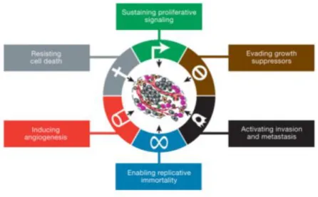
Cancer is a class of complex and heterogeneous diseases that share common features and are characterized by an abnormal and uncontrolled proliferation of cells that ultimately invade surrounding tissues or spread to distant organs (metastasize)1,2. In 2000, Douglas Hanahan and Robert Weinberg proposed the existence of six fundamental properties transversal to all cancers essential for the carcinogenic process2. These properties designated “hallmarks of cancer” included: sustaining proliferative signaling, evading growth suppressors, resisting cell death, enabling replicative immortality, inducing angiogenesis and activating invasion and metastasis (Figure 1.1)2. Eleven years later Hanahan and Weinberg revisited the hallmarks of cancer in an updated publication named “Hallmarks of cancer: the next generation” and four new hallmarks were proposed: deregulating cellular energetic, avoiding immune destruction, tumour-promoting inflammation and genome instability and mutations3. These publications revolutionized the comprehension of cancer and potentiated new perspectives in the approach to this condition.
Figure 1. 1 Hallmarks of cancer. (A) Illustration of the six hallmarks of cancer first established by Douglas Hanahan and Robert Weinberg in 2000 in the publication “Hallmarks of Cancer”. From
2
Cancer develops when normal cells lose their ability to control proliferation due to the accumulation of genetic and epigenetic changes4. These alterations disrupt the normal function of cells leading to abnormal or damaged cells which start to grow out of control resulting in the formation of masses of tissue called tumor1. As tumors develop they can eventually progress into a cancerous state, however not all tumors do so. The last types of tumors are known as benign tumors, as they do not spread or invade nearby tissues and thus are considered non-cancerous1,5. On the other hand malignant tumors or cancerous tumors spread into nearby tissues, and, as they grow, can travel to distant places in the body through the blood or the lymphatic system and form new tumors far from the original tumor site5.
There are numerous types of cancers reported in humans, however all begin with defective cells that are the result of either somatically acquired alterations due to environmental exposures and errors during DNA, or inherited (hereditary) alterations6,7. Among the different types of cancers, one of the most prominent is colorectal cancer (CRC). CRC is the third most commonly diagnosed cancer worldwide and the fourth most common cause of cancer related death globally8. Every year, over 1.23 million individuals are diagnosed with CRC and about 600 000 succumb to this disease. Incidence is approximately 30% higher in men than women, with approximately 814,000 cases reported in men and 664,000 cases in women every year. This makes CRC the third most commonly diagnosed cancer in man and the second in women8. Regardless, in both sexes CRC incidence is generally low for patients younger than 50 years old, however it strongly increases with age9.
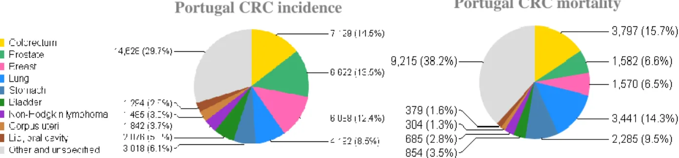
In Portugal CRC rises as the most frequent diagnosed cancer and the main cause of cancer related deaths. According to the last data estimated by the International Agency for Research on Cancer (IARC) in 2012, CRC was predicted to account for 14.5 % of all incident cancers in Portugal and responsible for 15.7% of all deaths by cancer (Figure 1.2).
3
1.2 Colorectal cancer (CRC)
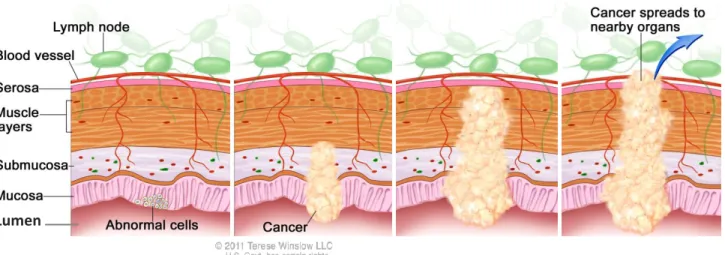
Colorectal cancer (CRC) is a malignant tumor that usually begins as an abnormal tissue growth in the cells of the colon or rectum which combined constitute a segment of the large intestine, the terminal portion of the gastrointestinal (GI) system9. These small growths designated as polyps, are projections of tissue that emerge from the innermost layer of the colon (the mucosa) into the lumen (hollow center) of the colon (Figure 1.3)9,10 . Polyps are as non-cancerous (benign) growths, however as they slowly grow over time, normally over a period of 10 to 20 years, they can eventually become cancerous (malignant)11,12. Whether a polyp changes into a cancerous state or not depends on the type of polyp it is11. Hyperplastic and inflammatory polyps are the most common growths yet in general they are not pre-cancerous. On the other hand adenomatous polyps also known as adenomas, although less common, have the ability to transit into a malignant state, and thus are often called as pre-cancerous conditions11. Adenomas arise from glandular cells which produce mucus that lubricate the inside of the colon and rectum9. Although all adenomas have the potential to become cancerous, fewer than 10% are estimated to progress into a cancerous state13. As an adenoma becomes larger, the likelihood of becoming cancerous increases, and when that transition happens it is referred to as adenocarcinoma. Among the five subtypes of CRC (adenocarcinomas, carcinoid tumors, gastrointestinal stromal tumors, lymphomas and sarcomas), adenocarcinomas are the most predominant making up about 95% of all CRCs14.
Portugal CRC incidence Portugal CRC mortality
Figure 1. 2 Colorectal cancer estimated incidence and mortality rates for the year 2012 in Portugal. Pie charts illustrating the different cancer estimated incidence (left) and mortality (right) rates in the Portuguese population for the year 2012. From GLOBOCAN project data, available at International Agency for Research on Cancer (IARC) (http://globocan.iarc.fr/Pages/fact_sheets_population.aspx).
4
CRC is considered to be an environmental disease, were cultural, social, and lifestyle factors heavily influence the risk of disease15,16. In fact sporadically occurring CRC cases which results from complex interactions between gene susceptibility and environmental factors account for the vast majority (75% - 80%) of all CRCs in the population17. Nevertheless, like many other cancers CRC also exhibits a heritable component, as familial adenomatous polyposis (FAP) and hereditary non-polyposis colon cancer (HNPCC or Lynch syndrome) account for close to 20% of all colorectal cancer incidence16,17.
1.2.1 Molecular Pathogenesis of Colorectal Cancer
CRC is a multistage process that results from the progression of a sequential accumulation of genetic mutations18. According to Fearon and Vogelstein, genetic events during the adenoma to carcinoma sequence lead to the development of CRC through specific genetic changes in oncogenes and tumor suppressor genes19. The accumulation of mutations in key tumor suppressor genes or oncogenes deregulates the cellular homeostatic functions affecting a wide range of cellular functions from proliferation, migration, differentiation, adhesion, cell death, to DNA stability and repair, causing the transformation of normal cells into cancer cells
Lumen
Figure 1. 3 Histological scheme of polyp formation within the large intestine walls. The four main layers that compose the large intestine from the outer portion to the inner portion (lumen): Serosa, Muscle layer, Submucosa and Mucosa. Polyps emerge in the mucosa layer and protrude to the lumen. However, as polyps grow they can invade the surrounding layers, spread into the blood vessels and metastasize. Adapted from Terese Winslow LCC, Medical And Scientific Illustration.
5
(Figure 1.4)18,20. Mutations typically alter the gene product by changing the amino acid sequence of proteins which leads to truncated or dysfunctional proteins or by altering the quantity of protein produced. Common mutation in the context of CRC include inactivation of the tumor suppressor gene Adenomatous polyposis coli (APC) which leads to activation of the Wingless/Integrated (Wnt) pathway, a common mechanism (which occurs in approximately 70% of adenomas) for initiating the adenoma to carcinoma sequence20,21. Subsequently mutations in genes such as Kirsten rat sarcoma viral oncogene homolog (KRAS) or TP53 (p53) also contribute to the progression of adenomas into carcinomas. CRC development can also involve mutations in the Transforming growth factor beta (TGFβ) signaling pathway as mutations in type II TGFβ receptor (TGFBR2) gene occur in approximately 30% of CRCs. Additionally, mutations affecting other TGF signaling pathway members, including SMAD2, SMAD4, Runt-related transcription factor 3 (RUNX3) and Thrombospondin 1 (TSP1) have been reported in colorectal cancers. Furthermore Serine/threonine-protein kinase B-raf (BRAF), phosphatidylinositol-4,5-bisphosphate 3-kinase catalytic subunit alpha (PIK3CA), Phosphatase and tensin homolog (PTEN) and β-catenin alterations have been associated with CRC carcinogenesis20.
Figure 1. 4 Morphological and molecular changes implicated in colorectal cancer development. Accumulation of specific genetic mutations in oncogenes and tumor suppressor genes contribute to adenoma-carcinoma progression. Mutations in APC, β-catenin and other players of this pathway are postulated to occur early in the carcinogenic process mediating the transition of single neoplasm cells to early adenoma. Meanwhile mutations involving KRAS, PTEN and p53 tend to occur later on leading to the emergence of carcinoma. Adapted from Terzić, et al., 2010.
6
Since Fearon and Vogelstein formulated the multi-step events of the molecular pathway of CRC formation involving oncogenes and tumour suppressor genes, there have been considerable advances in the understanding of colorectal carcinogenesis. Recent studies have shown that epigenetic alterations are an alternative mechanism in carcinogenesis22. Most of CRCs have epigenetic abnormalities such as over-expression of miR-21 , and hypermethylation of hsa-miR-129 and hsa-miR-137 that coexist with classical genetic changes such as APC, p53,
KRAS and β-catenin mutations22
1.2.2 Histopathological classification of colorectal cancer
Colorectal cancers are classified according to a tumour-node-metastasis (TNM) staging system established by the American Joint Committee on Cancer (AJCC)23,24. This system describes the anatomical extent of the disease based on the assessment of three features: local invasion depth (T), lymph node involvement (N), and presence of distant metastases (M) (Table I)25.
Table I Colorectal cancer classification according to local invasion depth (T), lymph node involvement (N), and presence of distant metastases (M). From Brenner, 2014.
7
Combined, these features produce an overall stage classification, the Union Internationale Contre le Cancer stage classification (UICC) that ranges from stage 0 to stage IV (Table II) and provides the basis for therapeutic decisions25.
Although stage classification according to TNM and UICC provides valuable prognostic information and guides clinicians therapeutic decisions, the response and outcome of individual patients to therapy or treatment is not predicted25.
1.2.3 Screening, diagnosis and prognosis
Patients with CRC are often asymptomatic in the early stages of the disease13. When symptoms appear, CRC has already grown or spread. Due to this particularity, early stage CRC detection is not always feasible. CRC patient survival is significantly affected by the stage of disease at diagnosis, as patients diagnosed in early stage have 5-year survival rates of 90%, which decrease to less than 10% when diagnosed at later stages (when distant metastasis develop)15. In fact if polyps are detected in an early stage they can be removed and cancer may be prevented26. This implies that an early detection of this disease is imperial for the success of the treatment, thus reducing both morbidity and mortality17.
8
Early detection of CRC is manageable through screening techniques which involves the detection and removal of pre-cancerous growths or early stage cancer, before the manifestation of any symptoms (in patients who have no symptoms)27.The idea is to detect the disease in a curable state, before it has a chance to grow or spread which makes treatment easier to manage, less expensive, and more likely to be successful9,27. Currently, the most predominantly used screening modalities for CRC, are Fecal Occult Blood tests (FOBTs) and colonoscopy28.
FOBTs are non-invasive screenings methods that can detect microscopic amounts of blood in feces indicating bleeding from the gastrointestinal tract29. FOBTs are the commonly used methods for CRC screening worldwide and the primary choice in most screening programs in Europe17. Given the non-invasive nature and low cost, it is one of the most accepted techniques by the population17. However, despite being widely used due to their simplicity, low cost and non-invasive nature, FOBTs have a suboptimal diagnostic accuracy suffering from low sensitivities and low specificities30.
Colonoscopy on the other hand is the most reliable method for CRC screening. It offers the opportunity to visualize the entire colonic mucosa and provides the ability to remove colon polyps and potentially prevent CRC31. For this reason it is considered to be the gold-standard screening test for CRC32. Nevertheless this technique is highly invasive, expensive and presents a high risk of complications such as perforation, bowel tears and bleeding when compared to other screening tests26. Moreover, the quality of the colonoscopy depends on the bowel preparation, which many patients find unpleasant33. All of this factors lead to a poor patient compliance34.
There is undeniable evidence that individuals who do not comply with the current screening programs have higher risk of developing cancer34. Therefore simpler, more efficient and less invasive screening methods that would improve compliance and ultimately decrease the incidence and mortality of this disease are needed. Thus, a drive to identify new screening methods that can overcome the limitations of the current techniques has stimulated a considerable interest in researching for potential molecular markers (biomarkers)35.
Moreover, specific histological characteristics and pathological staging of the tumors are necessary to accurately assess CRC patient prognosis36. However, individual to therapy and survival times of patients within the same stage of CRC are very heterogeneous, highlighting the necessity for a more precise system to assess patient prognosis36.
9
1.2.4 Biomarkers in CRC
Currently, biomarkers play an important role in the detection and treatment of patients with CRC37. A biomarker is a cellular biochemical or molecular alteration found in body fluids or in tissues that serves as an indicator of a biological process, pathogenic process, or pharmacological responses to a therapeutic intervention38. In cancer, a biomarker can be either a molecule secreted by the tumor or a specific response of the body to the presence of the tumor such as an antibody38. Biomarkers can act as a precious tool for cancer detection, diagnosis, and patient prognosis and can influence treatment choice39,40.
Although several molecular biomarkers (Carcinoembryonic antigen (CEA), Carbohydrate antigen 19-9 (CA19-9), Tissue polypeptide specific antigen (TPS), Tumor-associated glycoprotein 72 (TAG72), Tissue inhibitor of metalloproteinases-1 (TIMp-1)) are able to detect CRC and determinate the progression of the disease there is still a great way to go to implement these biomarkers as first line of screening, prevention and treatment of CRC patients as they usually lack sensitivity/specificity or are unable to detect cancer at early stages of disease41. Recently it has been demonstrated that epigenetic changes contribute to tumor progression, by influencing key transformation steps in CRC development (disrupting pivotal signaling pathways or affecting genes that regulate DNA repair and cellular proliferation)42. Epigenetic alterations appear to occur very early in the adenoma to carcinoma sequence, making them ideal screening biomarkers43. Recently, several studies have provided the identification of a variety of specific epigenetic alterations as potential clinical biomarkers for CRC patients30,44.
1.3 Epigenetics
Epigenetics are environmentally-mediated, frequent, powerful and widespread changes that affect gene expression without modifying the DNA sequence45. The epigenetic regulation of gene expression occurs in normal tissues and plays an important role in embryonic development, imprinting, and tissue differentiation. However, when disturbed, epigenetic mechanisms may lead to the development of several pathologies such as cancer46,47. Contrarily to gene mutations, epigenetic alterations can be reverted. This feature provides an opportunity to correct these epigenetic changes, and possibly to help combat disease progression or development48. Epigenetic modifications include histone post-translational modifications (PTMs), DNA
10
methylation, and gene regulation through non-coding RNAs, especially post-transcriptional regulation by microRNAs (miRNAs)45.
1.3.1 Histone Modifications
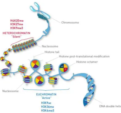
Post-translational histone modifications constitute an epigenetic mechanism that affects the compaction state of chromatin which influences the structure and folding (packaging) of the DNA, thereby affecting gene expression42,49. These modifications orchestrate the unraveling of the chromatin into a “open” or active chromatin state accessible for transcription (euchromatin), or into, a “closed” or inactive chromatin inaccessible for transcription (heterochromatin) modifying DNA accessibility leading to transcription regulation30. Histones are structures that package and order the DNA into structural units called nucleosomes. The nucleosomes are the fundamental units of the DNA and are composed of octamers of four core histones (H3, H4, H2A, H2B) around which 147 base pairs of DNA are wrapped50(Figure 1.5). The core histones are characterized by the presence of N-terminal tails that are subjected to extensive post-translational modifications such as acetylation, methylation, phosphorylation, ubiquitination, sumoylation, citrullination and ADP-ribosylation30. Among the various histone modifications the most extensively characterized in CRC are histone acetylation and methylation51.
Histone modifications, recently recognized as “histone code”, have been proposed to play an important role in the establishment of gene silencing during tumorigenesis52. However much is still to uncover regarding the contribution of post-translational histone modifications to the CRC carcinogenic process45.
11
1.3.2 DNA Methylation
DNA methylation occurs when a methyl group is covalently added to the 5-position of the pyrimidine ring of Cytosines (C) bases that are preceded by Guanine (G) nucleotides, typically designated CpG dinucleotides20. Approximately 80 % of all CpG sites are found dispersed genome-wide, while a small portion of CpGs can be found concentrated in specific regions called CpG islands50. CpG islands are genomic region of approximately 1–2 kilobases (kb) located in approximately 70% of all promoter regions of human genes, playing an important role in transcriptional regulation51. In healthy cells, the genome-wide scattered distributed CpG sites are heavily methylated, whereas the CpGs located in the promoter regions are unmethylated. However, CpG islands often become aberrantly methylated in cancer cells affecting not only the expression of protein coding genes but also the expression of various non-coding RNAs, some of which play a role in malignant transformation20,53.
Figure 1. 5 Post-translational histone modifications regulate chromatin compaction. Histone`s N-terminal tails are susceptible to PTMs that can lead either to an active (euchromatin) or silent (heterochromatin) conformation. These modifications can directly mediate the DNA binding of proteins necessary for transcription and thus impact gene expression. Well characterized histone modifications and the respective effect on chromatin conformation are shown. Adapted from
12
Generally, methylation of CpG islands in promoter regions is associated with transcriptional repression. In these regions, DNA methylation contributes to chromatin conformation changes influencing gene expression by affecting DNA exposure to transcription factors binding (Figure 1.6)20. In fact, epigenetic silencing of various tumor suppressor genes by hypermethylation of their promoters has been observed in a diversity of cancers, including CRC22. One of the best-characterized epigenetic events in tumor progression is the sporadic hypermethylation of the promoter of the mismatch repair gene MLH1, associated with approximately 12% of all CRCs cases30. Yet, recent evidences have demonstrated some exceptions to this classical view of DNA methylation repressor effect, as promoter DNA hypermethylation has been associated with increased gene expression54. Interestingly, global DNA hypomethylation may also play an important role in CRC development, possibly through genomic instability, however this process is far from being well understood35.
The addition of a methyl group to C5 position of cytosine is catalyzed by a family of enzymes designated DNA methyltransferases (DNMTs)50. Eukaryotes have three different DNMTs, DNMT1 is considered the maintenance DNA methyltransferase being responsible for mimicking the methylation pattern of the unreplicated strand of DNA onto the newly generated DNA strand55. Conversely DNMT3A and DNMT3B are responsible for de novo methylation which refers to the methylation of DNA without the use of a DNA template that carries an existing methylation pattern51.
DNA methylation plays a significant role in normal cells as it is involved in securing DNA stability through transcriptional silencing of genetic elements such as repetitive nucleotide sequences and endogenous transposons. Furthermore, DNA methylation contributes to gene imprinting, X-chromosome inactivation, homeostasis maintenance and genomic adaption in response to environmental stimuli besides other biological activities46,55. However, changes in DNA methylation were shown to promote progression of adenomatous precursor lesions into malignant tumors56. In fact thousands of genes are thought to be aberrantly methylated in the average colorectal cancer genome.
13
1.3.3 Non-coding RNAs
Non-coding RNAs (ncRNAs) represent a class of ribonucleotide acids (RNAs) that do not code for proteins57. Although it was initially believed that ncRNAs had no biological function this idea was soon dismissed as it has they play a significant role in many biological and pathological processes, ranging from metabolic disorders to diseases of various organ systems such as cancer58. NcRNAs can be divided into small ncRNAs (snoRNAs) and long ncRNAs (lncRNAs) based upon their size30. Small ncRNAs comprise microRNAs (miRNAs), piwi-interacting RNAs (piRNAs), and small nucleolar RNAs (snoRNAs) and are usually shorter than 200 nucleotides. Conversely, long ncRNAs are longer and are often larger than 200 nucleotides59.
The most widely studied class of ncRNAs are miRNAs, which play an important role in cancer initiation and progression. These small RNAs have thus revolutionized our understanding of cancer pathogenesis and also provided important insights into the feasibility of their use as clinically relevant biomarkers in cancer30,58
1.3.3.1 MicroRNAs
MicroRNAs (miRNAs) are endogenous small non-coding RNAs, of approximately 20 ~ 22 nucleotides, that mediate gene expression by binding to the 3’ untranslated region (UTR) of their target mRNA42. This base pairing of miRNAs with their targets consequently drives either to translational repression or messenger RNAS (mRNA), cleavage and consequent decay60. As a
Figure 1. 6 Schematic representation of DNA methylation. Gene expression is heavily regulated by CpG islands methylation status. A) Methylated CpG islands in promoter regions usually lead to gene repression whereas B) unmethylated CpG islands are associated with gene expression. In healthy cells the methylation pattern is characterized by methylated CpGs spread throughout the genome and unmethylated CpG islands. However, during carcinogenesis CpG islands often become hypermethylated and the global genome hypomethylated. These methylation changes have profound consequences in gene expression regulation. From Klein et al., 2014.
14
consequence the interaction of the miRNAs with their target mRNAs results in downregulation of gene expression of protein levels.
MiRNAs biogenesis starts with the transcription of primary miRNAs (pri-miRNAs) by RNA polymerase II in the nucleus which are then capped, spliced and polyadenylated (Figure 1.7)61. Pri-miRNAs are then folded into a base-paired stem-loop and further processed by the nuclear RNase III enzyme Drosha and co-factor DGCR8, which cleave these hairpin structures generating precursor miRNAs (pre-miRNAs)62. Pre-miRNAs are then translocated into the cytoplasm through the nuclear export factor, Exportin-5, where they are processed by the RNase III enzyme Dicer63. Dicer cleaves the pre-miRNAs at the terminal loop, liberating a ~22 nucleotide long RNA duplex64,65. This miRNA duplex contains two mature strands named 5p and 3p (the small RNA in the opposite side of the pre-miRNA stem loop), which are partially paired due to the 5’ and 3’ overhangs resulting from both Drosha and Dicer cleavages65
. The 5p strand is present in the forward (5'-3') position while the 3p strand is located in the reverse position.The miRNA duplex is then assembled into the RNA induced silencing complex (RISC). This process starts with the binding of the duplex to the trans-activator RNA (tar)-binding protein (TRBP) which leads to the recruitment of Argonaute 2 (Ago2). Ago2 along with Dicer assembles the RISC66,67. Once the miRNA duplex is loaded into the RISC complex, the strand with the lowest thermodynamic stability remains bounded to this complex while the opposite strand (called passenger strand) is degraded68. Next, the remaining miRNA guides the RISC complex to the target mRNA69. MiRNAs bind to their mRNA targets 3’UTR by imperfect complementarily, mainly through the binding of nucleotides 2-8 in the 5’ region of the miRNA named the “seed region”. Although, the 3’ region of the miRNA can also participate in the interaction miRNA:mRNA(Figure 1.7)70,71. Depending on the degree of complementarity different post-translational processes can occur, as partial complementarity is associated with translation repression, while near perfect complementarity can lead to mRNA degradation72,73.
15
MiRNAs are transcribed from diverse regions scattered along the genome, however the vast majority of mammalian miRNAs are located within the intronic region of either protein-coding genes or non-protein-coding transcripts64,74. Intronic miRNAs usually have the same orientation (are sense orientated) as their host gene and the expression of both miRNA and host gene largely coincides, which suggests a co-regulation and generation from common precursor transcripts75. However, miRNAs can also be found in intergenic regions (between genes) and a small subset of miRNAs has even been identified within exons of non-coding genes76. Moreover pri-miRNAs transcripts can comprise more than one miRNA. In fact, about 45% of known miRNAs are found in clusters and might be transcribed as a single polycistronic primary transcripts74.Also, the same mature miRNA can have different genomic locations that will give rise to different pri- and pre-miRNAs, which upon processing generate the same mature miRNA (miRNA isoforms). All the biogenesis steps are thus critical do designate the mature miRNA being formed and expressed, impacting on the miRNA target genes77,78.
Due to the limited complementarity between miRNAs and their mRNA targets, each miRNA can interact with several different mRNAs and a single mRNA can be suppressed by
Figure 1. 7 MiRNA biogenesis. Transcription of miRNA genes by RNA PolII originates capped and polyadenylated transcripts designated primary miRNAs (pri-miRNAs). Pri-miRNAs then undergo cleavage by the microprocessor complex (consisting of the RNase III nuclease Drosha and RNA-binding protein DGCR8) to generate short hairpin-shaped structures designated pre-miRNAs. Pre-miRNAs are exported from the nucleus to the cytoplasm by exportin-5 and are further processed by Dicer, an RNase III nuclease, which generates 21-22 nucleotide double-stranded miRNAs. This duplex is assembled into RISC, with the assistance of TRBP and Ago2. During this assemblage, one of the mature strands called the passenger strand (in blue) is degraded while the remaining mature miRNA guides the RISC complex to the target mRNA. MiRNA binding to the 3’UTR of target mRNA triggers translation inhibition or mRNA degradation. From Strubberg. et all., 2017.
16
various miRNAs50. Moreover, miRNAs from the same family are known to regulate the same target genes and/or different genes from the same signaling pathway to achieve their function79,80.
MicroRNAs, therefore, control a diversity of cellular processes ranging from developmental transitions and organ morphology to cell proliferation and apoptosis81. Consequently, abnormal expression of miRNAs can affect the normal expression of numerous genes and ultimately deregulate several biological processes, resulting in development of certain diseases such as cancer50.
Since the discovery of miRNAs in patients with chronic lymphocytic leukemia in 2002, the role of miRNAs in regulating post-translational gene expression in cancer development, growth, and metastasis have been well-established in a diversity of publications82,83. Almost all cancer types exhibit their unique profile of upregulated and downregulated miRNAs84. This striking feature potentiates the use of miRNAs as useful cancer biomarkers. Moreover miRNAs are very stable outside cells which allow them to be safely extracted, stored, and studied in feces or various body fluids enabling a less invasive prognostic and diagnostic tool85. In CRC, aberrations of miRNA expression seem to play a significant role in tumor development and progression, and several miRNAs have been identified as potential biomarkers30. In fact several miRNAs have been described to function as tumor suppressors, oncogenes or both86.
As mentioned above miRNAs are transcribed from genes. As such miRNAs gene promoters display all features of a normal gene commonly associated with Pol II-mediated transcription, including CpG islands87,88. Therefore abnormal CpG islands methylation in miRNAs promoter regions can affect their expression89.
1.4 Epigenetic biomarkers
1.4.1 DNA methylation as potential biomarkers in CRC
Advances in understanding the molecular pathology of CRC, has led to the identification of promising early detection molecular markers with potential to be used in non-invasive CRC screening assays90.Since DNA methylation appears to be an early event in tumorigenesis and particularly stable in blood and stool it has been proposed as a non-invasive diagnostic tool30.
17
Moreover, aberrant DNA methylation also contributes to later stages of colon cancer formation and progression potentiating the ability to be a therapeutic or prognostic marker for CRC20.
Recently, novel DNA methylation biomarker assays (Colovantage and ColoGuard) have gone through clinical trials and are commercially available49. Colovantage is a blood–based assay that detects the methylation of SEPT9 (septin 9) which is associated with impaired cytokinesis and loss of cell cycle control91. Colovantage has shown to provide an overall sensitivity of 90% and specificity of 88% and is being marketed in multiple countries, as a colon cancer screening assay92,93. ColoGuard is as stool based methylation assay for early detection of CRC. This test exploits the fact that the vimentin gene (VIM) is aberrantly methylated in the majority of colorectal cancers (53–84%) and has a reported a sensitivity and specificity of 83% and 82% in CRC94. VIM has been described to play a significant role in the epithelial-to-mesenchymal transition (EMT), disassembly of cell adherent junctions, reorganization of the actin cytoskeleton and acquisition of motility95,96.
However, the majority of DNA methylation biomarkers are not clinically available. Nevertheless, blood-based methylation of Homeobox protein aristaless-like 4 (ALX4), Nerve growth factor receptor (NGFR), Tomoregulin-2 precursor (TMEFF2), Neurogenin 1 (NEUROG1) and RUNX3 have been proposed as in CRC30,93. Furthermore hypermethylation of genes such as APC, Bone Morphogenetic Protein 3 (BMP3), Ataxia telangiectasia mutated
(ATM), Secreted frizzled related protein 2 (SFRP2), Cyclin-dependent kinase Inhibitor 2A (CDKN2A/p16), GATA4, Glutathione s-transferase p1 (GSTP1), Helicase like transcription factor
(HLTF), Human mutL homolog 1(MLH1), O-6-methylguanine-DNA methyltransferase (MGMT), N-myc downstream regulated gene 4 (NDRG4), Ras association domain family member 2 (RASSF2A), Tissue Factor Pathway Inhibitor 2 (TFPI2) and WNT Inhibitory factor 1
(WIF1) has been suggested as stool-based methylation biomarkers for early detection of CRC30. When considering the prognostic value of DNA methylation changes, hypermethylation of CDKN2A/p16, Checkpoint with FHA and RING finger domains (CHFR), Enah/Vasp-like (EVL), Insulin like growth factor binding protein 3 (IGFBP3), KISS1, RET, HTLF, and Hyperpigmentation Progressive 1 (HPP1) genes and hypomethylation of Inactivation escape 1
(INE-1), MGMT, Transcription factor AP-2-alpha (TFAP2A) and Insulin-like growth factor 2 (IGF2) have been associated with poor prognosis of CRC36. Several other studies have also provided evidence that aberrantly methylated DNA has the potential to be used as prognostic
18
biomarkers in CRC. Nonetheless, further investigation is required to develop clinically robust assays in order to allow these biomarkers to be used in a clinical setting30.
1.4.2 MiRNAs as potential biomarkers in CRC
The discovery of miRNAs in extracellular body fluids strongly increased the number of studies showing deregulated expression of circulating miRNAs in cancer diseases30. The first comprehensive miRNA expression profiling study was conducted by Ng and colleagues97. In this study the authors evaluated miRNA expression alterations in tissue and plasma samples from CRC patients and healthy subjects and demonstrated that a high expression of 92a and miR-17-3p, could discriminate CRC patients from healthy individuals97. Since this landmark study numerous miRNAs found in both feces and plasma/serum have been identified as potential biomarkers for an early CRC diagnosis. Basati et al. reported that two miRNAs: miR-194 and miR-29b were down-regulated in CRC patients when compared to control subjects, suggesting these miRNAs as powerful CRC serum biomarkers for early CRC97. Juan et al. also revealed miR-145 and miR-378 as potential biomarkers, which could help in early CRC diagnosing, with sensitivities reaching 100% and specificities of 60% and 98% respectively97. Expression of the miR-17-92a cluster and miR-135b in feces has also been found to discriminate patients with CRC from healthy subjects98. Additionally miR-21, one of the most promising biomarkers for early diagnosis of CRC since it is frequently deregulated in early stages of the adenoma-carcinoma sequence, has also been exhaustively reported as a potential serum or plasma diagnostic biomarker30,99. Despite the number of miRNAs identified as potential biomarkers in CRC the lack of consistency between biomarker panels in independent studies represents a major obstacle for the development of robust miRNA biomarkers30.
MiRNAs also have been demonstrated to carry useful clinical information, as over-expression of miR-21 has been associated with metastasis and poor survival100. Furthermore over-expression of miR-372 and miR-15b has also been associated with metastasis and poor overall survival36.
Finally, some miRNAs have been demonstrated to be methylation-sensitive, meaning that they can become epigenetically silenced due to CpG island promoter hypermethylation101. Bandres et al. identified a link between hypermethylation and downregulated expression of three
19
miRNAs, hsa-miR-9-1, hsa-miR-129, and hsa-miR-137in primary CRC samples when compared to normal mucosa89. Moreover it was suggested that methylation of hsa-miR-9-1 could potentially be used as a poor CRC prognosis biomarker, and it could also be involved in metastatic events89. MiR-149 whose lower expression in CRC is a consequence of a neighbouring CpG island hypermethylation has also been referred as a promising prognostic marker in CRC. Lower expression of miR-149 was associated with lower 5-year survival rate and a higher tumor invasion in CRC102. In this sense miRNA methylation analysis can provide several insights on CRC carcinogenesis and work as potential biomarkers for this condition. Nevertheless, additional studies are still required in order to explore the potential of miRNA silencing by DNA methylation as a CRC biomarker for both diagnosis and prognosis103.
1.5 Investigating miRNA expression and DNA methylation of miRNA genes
Since their discovery, miRNA research and understanding has grown exponentially104. For that to happen, new methods and technologies had to be developed and optimized in the last 20 years105. Nowadays we not only can “easily” study miRNA transcription, expression, methylation and function, as this information is usually available in open source databases. The big-data era has also arrived to the miRNA world, although all the information available is based on important research methods performed before, and enclosed behind the numbers we analyse106. Currently, high-throughput methods facilitate large-scale miRNA profiling and are the most widely used due to their accuracy, sensitivity and amount of data produced107,108. Those methods are used for miRNA expression profiling, as well as to investigate mechanisms behind miRNA patterns (e.g. miRNA interactions; DNA methylation; histone modifications), both essential to understand miRNAs role in pathological contexts107–109.
In this work, we have used miRNA expression and DNA methylation data generated by next-generation sequencing (NGS, RNA-seq) and Infinium HumanMethylation450K array respectively, which information is publicly available. Thus, in this section, the basics of these technologies are briefly explained.
20
1.5.1 RNA-sequencing
The development of high-throughput next-generation sequencing (NGS) has led to the emergence of several tools such as RNA sequencing (RNA-seq)108. RNA-Seq is a sequencing approach that utilizes the capabilities of high-throughput sequencing methods to provide an understanding of the cellular transcriptome, which is defined as the complete set of transcripts (RNA molecules) in a cell that results from the transcription of a subset of genes into complementary RNA molecules110.
Currently, the Illumina HiSeq platform is the most commonly applied next-generation sequencing technology for RNA-Seq110. Illumina Hiseq starts with library preparation in which RNA is converted to cDNA, which is then randomly shredded into small DNA fragments. Afterwards adaptors are ligated to both ends of the DNA-fragments, amplified by PCR and purified in a gel (Figure 1.8 A)111. The library is loaded into a flow cell and the fragments hybridize with the oligos on the flow cell surface which work as primers for amplification112. Each bound fragment is amplified into a cluster through bridge amplification (Figure 1.8 B). Using a method designated as sequencing by synthesis (SBS), fluorescently labeled reversible terminators deoxyribonucleotidetriphosphates (dNTPs) are incorporated into the DNA template strand in each cycle109. Each of the four dNTPs (A, C, T, and G) has a single different fluorescent label, which serves to identify the base. After each synthesis cycle, the clusters are excited by a laser which causes the last incorporated bases to emit fluorescence which is captured through a camera system (Figure 1.8 C)109,113. Subsequently, after imaging the reversible terminator fluorescent label nucleotides are removed and the template strands become ready for the next incorporation cycle109. Finally during data analysis, the newly identified sequence reads are aligned to the reference genome (Figure 1.8 D). After alignment, the mapped reads can be assembled into transcripts and the expression levels estimated110.
Normalization of high-throughput sequencing of small RNA such as miRNAs is necessary in order to compare their levels across different samples114. Therefore data from RNA-seq experiments are typically normalized and perceived in reads per million genome-matching reads (RPMs)115.
21 Figure 1. 8 Outline of the Illumina workflow. Representation of the main steps comprised in an Illumina workflow. Adapted from the Genome Analyzer brochure, http://www.solexa.com & An introduction to Next-Generation Sequencing Technology.
cDNA Adapters