2018
UNIVERSIDADE DE LISBOA
FACULDADE DE CIÊNCIAS
DEPARTAMENTO DE BIOLOGIA VEGETAL
Long non-coding RNAs as potential therapeutic targets in
human breast cancer
Ana Beatriz Domingues Silva
Mestrado em Biologia Molecular e Genética
Dissertação orientada por:
Doutor Bruno Miguel Bernardes de Jesus
Doutora Maria Helena Caria
II
Agradecimentos
Em primeiro lugar quero agradecer à Professora Doutora Helena Caria por me ter proporcionado a primeira aventura num laboratório, por estar sempre disponível para mim e pela revisão desta dissertação. Em segundo lugar quero agradecer à Doutora Carla Gomes e ao Doutor Bruno de Jesus por me terem aceite como aluna de mestrado para realizar este projeto. Sempre quis tralhar na área do cancro portanto foi uma ótima oportunidade para mim. Quero também agradecer por todo o apoio prestado no planeamento/execução experimental e na revisão desta dissertação. Em terceiro lugar, quero agradecer à Catarina do Vale por ter sido a principal impulsionadora deste projeto e pela ajuda na realização de algumas experiências. Ao Sérgio Marinho quero agradecer por me ensinar algumas técnicas laboratoriais e por me esclarecer as dúvidas sempre que surgiam. À Dinora quero agradecer pela ajuda na realização das encomendas. Aos meus colegas de laboratório quero agradecer pela boa-disposição, animação e companhia. À unidade de bioimagem, principalmente ao António Temudo, quero agradecer pelo apoio na visualização e aquisição de imagens de microscopia de fluorescência. À unidade de citometria de fluxo quero agradecer pelo apoio na realização e análise dos resultados de apoptose e ciclo celular.
Muito obrigado à minha família em geral por ter feito de mim aquilo que sou hoje e por ser o meu porto de abrigo. Muito obrigado, em particular, aos meus pais, Emídio da Silva e Ana Maria Silva, e aos meus irmãos, Jorge Silva e João Silva, por serem os melhores do mundo e os principais responsáveis por eu estar hoje a terminar a minha tese de mestrado, por me proporcionarem todas as condições, pelos conselhos, palavras de incentivo e conforto em momentos mais difíceis, pelo carinho e amor incondicionais. Um obrigado especial ao meu namorado, Bernardo Antunes, pela paciência, por ouvir as minhas frustrações e euforias, pelos momentos de descontração após longos dias de trabalho, pela amizade e pelo amor. Percorremos este caminho juntos e isso tornou tudo muito mais fácil. Por fim, obrigada às minhas melhores amigas, Rita Rodrigues e Sara Castro, que ao fim de tantos anos continuam a estar presentes tanto nos bons como nos maus momentos.
III
Abstract
Long non-coding RNAs have emerged as regulators of diverse biological processes but little is known about their role in cancer. The recently discovered human lncRNA NORAD is induced after DNA damage in a p53-dependent manner and plays a critical role in the maintenance of genomic stability, since interacts with Pumilio proteins, limiting the repression of their target mRNAs involved in mitosis, DNA repair and replication. Therefore, NORAD inactivation causes chromosomal instability and aneuploidy, which contributes to accumulation of genetic abnormalities and tumorigenesis. However, severe chromosome mis-segregations end up with cell death, suggesting that NORAD may act as an oncogene and tumor suppressor gene. Moreover, this lncRNA has been detected in several types of cancer, including breast cancer, which is the most frequently diagnosed and the second-leading cause of cancer death in women. However, some studies present contradictory results relative to NORAD function in tumorigenesis.
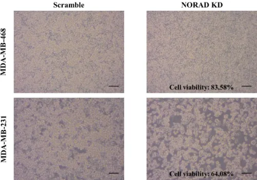
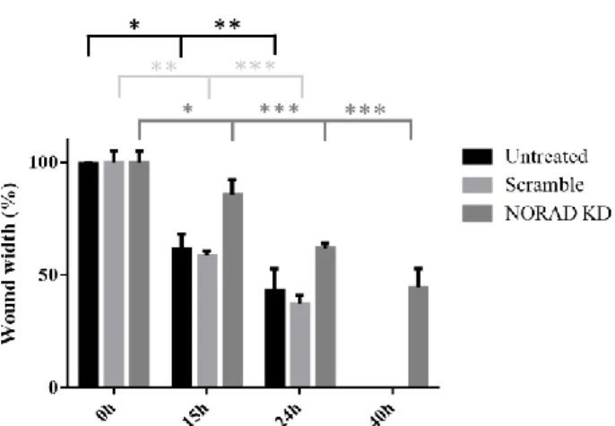
In the present study was found that NORAD expression is upregulated in a set of human epithelial breast cancer cell lines (MDA-MB-231, 436 and 468), which belong to the most aggressive triple negative subtype, in comparison to a non-malignant human mammary epithelial cell line (MCF-10A) by RT-qPCR. In agreement, was found that high NORAD expression levels in basal-like tumors are associated with poor prognosis, through Kaplan-Meier Plotter tool. In order to unravel the role of this lncRNA, tumor-relevant phenotypes were analyzed after its knockdown, using LNATM GapmeRs
and siRNAs, in the MDA-MB-231 cell line. Cell proliferation inhibition determined by alamarBlue®
reduction assay and cell migration inhibition determined by wound healing assay, suggests that NORAD acts as an oncogene. In addition, upon NORAD knockdown, was identified a shift in doxorubicin IC50
from 0.3779 μM to 0.05680 μM by alamarBlue® reduction assay caused by an increase in cell death,
indicating sensitization to chemotherapy. In fact, chromosomal instability generated upon NORAD knockdown, in combination with DNA damage induced by doxorubicin results in severe chromosome mis-segregations, incompatible with cell viability. In contrast, no cell cycle change was identified by flow cytometry because this lncRNA is not required for these aspects of the DNA damage response. This study also highlights the role of p53, guardian of the genome, in the response to these stress conditions, since its expression levels increase (RT-qPCR and western blot), inducing cell cycle arrest or apoptosis.
These results provide evidences that the lncRNA NORAD represents a tumor marker for disease diagnosis, patient prognosis or therapy response, and represents a therapeutic target in breast cancer.
IV
Resumo
O projeto ENCODE (Enciclopédia de Elementos do DNA) estima que apenas 2% do genoma humano codifica proteínas, enquanto 75% é transcrito em RNAs não codificantes de pequenas e de grandes dimensões. Cerca de 16.000 genes codificam mais de 28.000 RNAs longos não codificantes (lncRNAs), os quais têm sido alvo de interesse por participarem em diferentes processos biológicos (desenvolvimento, doenças, etc.). Geralmente, os lncRNAs são constituídos por mais de 200 nucleótidos, pouco conservados evolutivamente, pouco abundantes e heterogéneos. Recentemente foi descoberto um transcrito com 5,3 kb (LINC00657) que se destaca por ser muito conservado evolutivamente, muito abundante e constitutivamente expresso. Localiza-se predominantemente no citoplasma onde pode regular a estabilidade e tradução de RNAs mensageiros (mRNAs) e influenciar a sinalização celular. No núcleo pode organizar a estrutura subnuclear, regular a transcrição e interações cromossómicas. Após danos no DNA, este lncRNA é ativado de modo dependente de p53, sendo por isso denominado NORAD (RNA não codificante ativado por danos no DNA). NORAD, que contém 17 regiões de reconhecimento de proteínas de ligação ao RNA designadas Pumilio (sequência específica: UGUANAUA), captura uma quantidade significativa destas proteínas, impedindo que inibam a tradução de mRNAs alvo com os quais interagem através da sua região 3’, onde se encontra a sequência específica. Esses transcritos atuam ao nível da homeostasia da linha germinal, atividade neuronal, mitose, replicação e reparação do DNA. É importante referir que a interação entre o lncRNA NORAD e as proteínas Pumilio é induzida por outra proteína de ligação ao RNA, SAM68, que se liga a jusante das regiões de reconhecimento das proteínas Pumilio. Assim, na ausência de NORAD, as proteínas Pumilio são ativadas e inibem a tradução de mRNAs alvo, o que perturba a segregação dos cromossomas durante a mitose e provoca aneuploidia. Por sua vez, a aneuploidia pode contribuir para o ganho de funções por parte de oncogenes e a perda de funções por parte de genes supressores de tumores, originando tumores. Neste caso, NORAD atua como um gene supressor de tumores. Contudo, quando a acumulação de erros durante a mitose se torna demasiado elevada, as células não têm capacidade de resposta e morrem. Neste caso, NORAD atua como um oncogene. Estudos demonstraram a expressão de NORAD em vários tipos de cancro, no entanto, alguns resultados são contraditórios no que respeita à sua função. Nas células e tecidos dos cancros da mama, do esófago e do pâncreas detetaram-se níveis de expressão mais elevados em comparação com as respetivas células e tecidos normais, bem como uma correlação entre os seus níveis e o pior prognóstico dos pacientes. Além disso, depois da redução da expressão de NORAD, analisaram-se os efeitos nas propriedades das células e verificou-se redução da viabilidade, proliferação, migração e invasão in vitro e in vivo. Pelo contrário, nas células e tecidos do cancro do fígado detetaram-se níveis de expressão mais baixos em comparação com as respetivas células e tecidos normais, bem como uma correlação entre os seus níveis e o melhor prognóstico dos pacientes. Além disso, depois da redução da expressão de NORAD, analisaram-se os efeitos nas propriedades das células e verificou-se aumento da proliferação, ciclo celular, migração e invasão in vitro e in vivo.
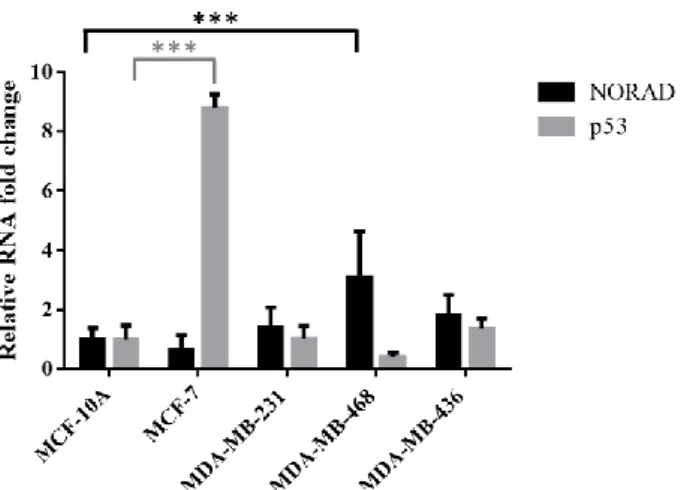
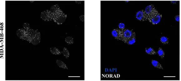
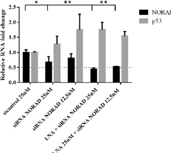
O presente estudo centra-se no cancro da mama por ser o mais frequentemente diagnosticado e o segundo mais mortífero em mulheres. É também uma doença complexa pois apresenta diferentes subtipos (luminal, HER2 positivo e basal) definidos de acordo com a presença de três recetores (ER, PR e HER2) e que apresentam diferentes características morfológicas e genéticas, resultando em diferentes tratamentos e prognósticos. Em primeiro lugar, determinaram-se os níveis basais de expressão de NORAD em células de cancro da mama (MCF-7, MDA-MB-231, 468 e 436) em comparação com células normais (MCF-10A) por RT-qPCR, tendo sido registados níveis de expressão mais elevados nas células de cancro da mama mais agressivas, isto é, negativas para os três recetores (MDA-MB-231, 468 e 436). A predominante localização citoplasmática deste lncRNA foi confirmada por smRNA FISH. Em segundo lugar, efetuou-se a redução da expressão de NORAD usando LNATM GapmeRs que atuam
V
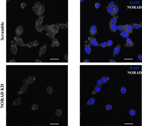
resultado foi confirmado por RT-qPCR e por smRNA FISH. Em terceiro lugar, analisaram-se os efeitos da redução da expressão de NORAD nas propriedades tumorais da linha celular MDA-MB-231, e verificou-se inibição da proliferação através do ensaio da redução de alamarBlue® e inibição da
migração através do ensaio de migração in vitro. A capacidade de proliferação e de metastização são características fundamentais do cancro, logo, estes resultados indicam que este lncRNA desempenha um papel importante na tumorigénese. De seguida, avaliou-se o efeito da combinação da redução da expressão de NORAD com quimioterapia. Para isso, utilizou-se a doxorrubicina que pertence à classe das antraciclinas, intercala-se entre pares de bases no DNA e inibe a topoisomerase II, provocando danos no DNA. Nestas condições, identificou-se uma redução do IC50 da doxorrubicina de 0,3779 μM para
0,05680 μM através do ensaio da redução de alamarBlue®, o que sugere que a redução da expressão de
NORAD sensibiliza as células do cancro da mama à quimioterapia. Isto assume principal relevância em casos de resistência à quimioterapia. De acordo com este resultado, nas mesmas condições verificou-se um aumento da apoptose celular através de citometria de fluxo. De facto, a instabilidade cromossómica originada pela redução da expressão de NORAD e os danos no DNA originados pela doxorrubicina, levam à acumulação de demasiados erros genéticos que resultam na morte celular. Por outro lado, não se registaram alterações no ciclo celular para além da acumulação das células na fase G2 induzida pela
doxorrubicina, porque este lncRNA não participa nesse tipo de resposta aos danos no DNA. No presente estudo é ainda destacado o gene supressor de tumores p53 como o guardião do genoma por ser ativado em condições de stress, ligar-se a proteínas envolvidas na reparação do DNA (XPB, RPA e rad51) e promover a transcrição de p21 que inibe cinases dependentes de ciclinas que regulam as transições G1
-S e G2-M, induzindo arrastamento do ciclo celular. Em alternativa, quando os danos no DNA são
demasiado elevados, ocorre apoptose celular através da libertação do citocromo c, da expressão de proteínas pro-apoptóticas da família BCL-2 (por exemplo, puma, noxa, bid e bax), de componentes da sinalização do recetor de morte celular (por exemplo, DR5 e Fas/CD95) ou da maquinaria efetora de morte celular (por exemplo, caspase-6, Apaf-1 e PIDD). De facto, perante a instabilidade cromossómica gerada pela redução da expressão de NORAD e os danos no DNA gerados pela doxorrubicina, detetou-se um aumento da expressão de p53 por RT-qPCR e western blot. Por fim, recorreu-detetou-se ao teste de Kaplan Meier para avaliar o efeito da expressão de NORAD na sobrevivência de pacientes com cancro da mama. Apenas para o subtipo basal que é o mais agressivo, se verificou uma correlação entre níveis de expressão de NORAD elevados e menor tempo de sobrevivência livre de recidiva.
Os resultados obtidos permitem inferir que o lncRNA NORAD pode representar um biomarcador importante para o diagnóstico de cancro, o prognóstico do paciente e a resposta a terapias, e pode representar um alvo terapêutico. No futuro, aumentar a expressão de NORAD em células da mama normais (MCF-10A) e avaliar se existe aquisição de fenótipo tumoral, seria uma boa estratégia para esclarecer se este lncRNA é fundamental na tumorigénese.
VI
Index
Agradecimentos………II Abstract………...………III Resumo………...…IV List of figures, tables and equations………..………....VII List of abbreviations…….………..……….VIII
1. Introduction………...1
1.1 Hallmarks of cancer………1
1.2 Tumor microenvironment ………..1
1.3 Breast cancer………...2
1.4 Molecular classification of breast cancer………2
1.5 Cancer and DNA damage: an intimate relationship………...4
1.6 p53, guardian of the genome……….…………..5
1.7 NORAD, a lncRNA induced by DNA damage………..6
1.8 Interaction with Pumilio proteins………...6
1.9 NORAD expression profiles predicts clinical outcome in multiple human cancers………..8
1.10 Long non-coding RNAs………8
1.11 Mechanisms of action………...9
1.12 LncRNA-based diagnostics and therapies in cancer………...10
1.13 Chemotherapy……….11
1.14 Combinatorial approaches………..11
2. Objectives………...12
3. Materials and methods………12
3.1 Cell culture………12
3.2 RNA isolation, cDNA synthesis and RT-qPCR………...13
3.3 Single-molecule RNA fluorescence in situ hybridization (smRNA FISH)………..14
3.4 NORAD knockdown……….14
3.5 Cell viability……….14
3.6 Cell migration………...15
3.7 Assessment of doxorubicin IC50………...15
3.8 Combination studies with NORAD knockdown and doxorubicin………...15
3.9 Protein isolation and western blot……….15
3.10 Cell apoptosis………..16
3.11 Cell cycle………16
3.12 Statistical analysis………...17
4. Results and discussion………17
4.1 NORAD and p53 expression profiles in human epithelial breast cancer cell lines………..17
4.2 NORAD is predominantly cytoplasmic………18
4.3 NORAD knockdown optimization protocol……….19
4.4 NORAD knockdown inhibits cell proliferation..………..21
4.5 NORAD knockdown inhibits cell migration………22
4.6 NORAD knockdown sensitizes breast cancer cells to chemotherapy………..23
4.7 NORAD knockdown promotes cell death………25
4.8 NORAD knockdown does not affect cell cycle………26
4.9 Correlation between NORAD expression and survival of breast cancer patients ………...27
5. Conclusions and future perspectives………...28
VII
7. Supplementary figures………33
List of figures, tables and equations Table 1.1 Therapeutic agents and corresponding mechanisms of action, specific to each breast cancer subtype………...…3,4 Figure 1.1 NORAD activation or inactivation and subsequent events……….7
Table 3.1 Characterization of the human epithelial breast cancer cell lines used in this study………..13
Table 3.2 Gene-specific primer pairs and sequences used in RT-qPCR………13
Equation 3.1 Cell viability (%)………...15
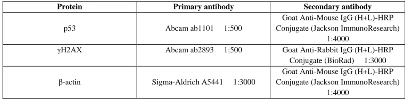
Table 3.3 Protein-specific antibodies used in western blot………16
Figure 4.1 NORAD and p53 mRNA basal levels in human epithelial breast cancer cells……….18
Figure 4.2 NORAD subcellular localization………...18,19 Figure 4.3 NORAD and p53 mRNA levels after NORAD knockdown, using LNATM GapmeRs and siRNAs, with 48h interval between transfections………...20
Figure 4.4 NORAD transcripts after knockdown I..………...21
Figure 4.5 NORAD knockdown effect on cell viability……….22
Figure 4.6 NORAD knockdown effect on cell migration………..22,23 Figure 4.7 Effect of NORAD knockdown combination with chemotherapy……….23
Figure 4.8 NORAD and p53 mRNA levels after NORAD knockdown and/or incubation with doxorubicin……….24
Figure 4.9 p53 and γH2AX protein levels after NORAD knockdown and/or incubation with doxorubicin……….25
Figure 4.10 NORAD knockdown and doxorubicin effects on cell apoptosis………26
Figure 4.11 NORAD knockdown and doxorubicin effects on cell cycle………...27
Figure 4.12 Breast cancer patients overall survival and relapse-free survival according to NORAD expression levels……….28
Figure 7.1 NORAD and p53 mRNA levels after NORAD knockdown, using LNATM GapmeRs, with 24h interval between transfections……….33
Figure 7.2 NORAD and p53 mRNA levels after NORAD knockdown, using a combination of LNATM GapmeRs, with 24h interval between transfections………33
Figure 7.3 NORAD and p53 mRNA levels after NORAD knockdown, using a combination of LNATM GapmeRs, with 48h interval between transfections………33
Figure 7.4 NORAD and p53 mRNA levels after NORAD knockdown, using a combination of LNATM GapmeRs, with 24/48h interval between transfections………..34
Figure 7.5 NORAD transcripts after knockdown II………...34
Figure 7.6 Doxorubicin IC50……….………..35
VIII
List of abbreviations
RAS – rat sarcoma gene TP53 – tumor protein p53
BRCA 1/2 – breast cancer susceptibility 1/2 gene RB1 – retinoblastoma 1 gene
ER – estrogen receptor PR – progesterone receptor
HER2 – human epidermal growth factor receptor 2 PARP – poly (ADP-ribose) polymerase
Mdm2 – murine double minute 2 gene ARF – ADP-ribosylation factor CDK – cyclin-dependent kinase BCL-2 – B-cell lymphoma 2 gene
Puma – p53 upregulated modulator of apoptosis Bid – BH3 interacting domain death agonist Bax – Bcl-2-associated X protein
oxLDL – oxidized low-density lipoprotein HIF-1α – hypoxia-inducible factor 1α VEGF – vascular endothelial growth factor MMP – matrix metalloproteinase
PCA3 – prostate cancer associated 3 PCGEM1 – prostate-specific transcript 1
CREB3L1 – cAMP response element-binding protein 3-like 1 PTEN – phosphatase and tensin homolog
ATM – ataxia telangiectasia mutated
MDC1 – mediator of DNA damage checkpoint 1 Mre11 – meiotic recombination 11 homolog Nbs1 – Nijmegen breakage syndrome 1 gene 53BP1 – TP53-binding protein 1
PP2A – protein phosphatase 2A
RNF 2/8/168 – ring finger protein 2/8/168
BMI1 – B lymphoma Mo-MLV insertion region 1 homolog HCC – hepatocellular carcinoma
TNM – tumor, node and metastasis
UICC – Union for International Cancer Control OS – overall survival
RFS – relapse-free survival sncRNA – short non-coding RNA lncRNA – long non-coding RNA
NORAD – non-coding RNA activated by DNA damage PRE – PUMILIO recognition element
PUM – Pumilio protein
PUF – Pumilio-Fem3-binding factor RNP – ribonucleoprotein
ND – NORAD domain
CIN – chromosomal instability RBP – RNA-binding protein
IX SAM68 – SRC associated in mitosis
ceRNA – competing endogenous RNA ACTB – β-actin
RT-qPCR – real-time quantitative reverse transcriptase polymerase chain reaction Ct – threshold cycle
smRNA FISH – single-molecule RNA fluorescence in situ hybridization DAPI – 4’, 6-diamidino-2-phenylindole
CRISPR-Cas9 – clustered regulatory interspaced short palindromic repeats-associated endonuclease 9 TALEN – transcription activator-like effector nucleases
RNAi – RNA interference shRNA – short hairpin RNA siRNA – small interfering RNA LNA – locked nucleic acid Scr – scramble
KD – knockdown
IC50 – half maximal inhibitory concentration
DMEM – Dulbecco’s Modified Eagle Medium FBS – fetal bovine serum
PBS – phosphate-buffered saline PFA – paraformaldehyde FA – formamide
SSC – saline sodium citrate
SDS-PAGE – sodium dodecyl sulfate-polyacrylamide gel electrophoresis APS – ammonium persulfate
TEMED – tetramethylethylenediamine HRP – horseradish peroxidase
BCA – bicinchoninic acid
FACS – fluorescence activated cell sorting 7-AAD – 7-aminoactinomycin D
PS – phosphatidylserine RT – room temperature
1
1. Introduction
1.1 Hallmarks of cancer
In 2000, Hanahan and Weinberg proposed six hallmarks of cancer, which are distinctive and complementary capabilities that enable tumor growth and metastatic dissemination: (1) sustaining proliferative signaling; (2) evading growth suppressors; (3) enabling replicative immortality; (4) activating invasion and metastasis; (5) inducing angiogenesis; and (6) resisting cell death (1).
Their acquisition is possible by two enabling characteristics. Most prominent is genomic instability, which consists in the accumulation of alterations in the genome, some that confer selective advantage on subclones of cells, enabling their outgrowth and eventual dominance in a local tissue environment (2). These alterations in the genome include gain-of-function mutations in oncogenes (e.g. RAS) (is sufficient to have a mutation in one of the two alleles) that normally stimulate cell growth, division and survival, and loss-of-function mutations in tumor suppressor genes (e.g. TP53) (is necessary to have a mutation in both alleles) that normally prevent unrestrained cellular growth, promote DNA repair and cell cycle checkpoints activation (3). This process can be accelerated by an increasing sensitivity to mutagenic agents, breaking down components of the genomic maintenance machinery, compromising surveillance systems that force genetically damaged cells into either senescence or apoptosis, and losing the telomeric DNA. Regarding the other enabling characteristic, some tumors are densely infiltrated by cells of the innate and adaptive immune system, which reflect an attempt of eradication or contribute to multiple hallmarks by supplying bioactive molecules to the tumor microenvironment, including growth factors that sustain proliferative signaling, survival factors that limit cell death, pro-angiogenic factors and extracellular matrix-modifying enzymes that facilitate angiogenesis and metastasis, and inductive signals of epithelial-mesenchymal transition and of other hallmark-facilitating programs. In addition, inflammatory cells can release chemicals, notably reactive oxygen species, that are actively mutagenic for nearby cancer cells (2).
Yet, other attributes of cancer cells have been proposed to be functionally important for the development of cancer: reprogramming of cellular energy metabolism and active evasion from attack and elimination by immune cells. Otto Warburg observed that even in the presence of oxygen, cancer cells can reprogram their glucose metabolism, almost limiting it to glycolysis. They compensate for the approximately 18-fold lower efficiency of ATP production in comparison to mitochondrial oxidative phosphorylation, by upregulating glucose transporters, notably GLUT1, which substantially increase glucose import into the cytoplasm (2,4). Of note, glycolytic intermediates participate in various biosynthetic pathways, including those generating nucleosides and amino acids, which facilitate the formation of macromolecules and organelles required for the assembly of new cells. Moreover, tissues are monitored by an ever-alert immune system, responsible for recognizing and eliminating the majority of incipient cancer cells. Therefore, solid tumors that do appear, disable components of the immune system such as cytotoxic T lymphocytes and natural killer cells, through secretion of TGF-β or other immunosuppressive factors, and recruit inflammatory cells that are actively immunosuppressive such as regulatory T cells and myeloid-derived suppressor cells (2).
1.2 Tumor microenvironment
Initially, tumors were thought of as a collection of relatively homogeneous cells, whose entire biology could be understood by elucidating the cell-autonomous properties. However, the recognition of the tumor complexity changed this perspective. In fact, the biology of a tumor can only be understood by studying the individual specialized cell types within it, as well as the tumor microenvironment that they construct during the course of multistep tumorigenesis. These are: cancer cells, cancer stem cells,
2
endothelial cells, pericytes, immune inflammatory cells, cancer-associated fibroblasts, stem and progenitor cells of the stroma. The likelihood that signaling interactions between cancer cells and their supporting stroma evolve during the course of multistep tumorigenesis, complicates the goal of fully elucidating the mechanisms of cancer pathogenesis. Therefore, understanding these dynamic variations is crucial to the development of novel therapies designed to successfully target both primary and metastatic tumors (2).
1.3 Breast cancer
Breast cancer is the most frequently diagnosed cancer and the second-leading cause of cancer death in women (5,6). Although commonly thought of as a woman’s disease, it also occurs in men. In 2017, in situ breast carcinoma (no evidence of invasion) was diagnosed in about 63,410 women, while invasive breast carcinoma was diagnosed in about 252,710 women and 2,470 men in the United States of America. The number of breast cancer deaths was 40,610 women and 460 men. Despite the incidence of female breast cancer is rising and the incidence of male breast cancer is stable, the death rates declined due to improvements in early detection and treatment. Potential risk factors are: personal or family history of breast or ovarian cancer; inherited mutations in BRCA1, BRCA2 or other breast cancer susceptibility genes; benign breast conditions, such as atypical hyperplasia; high breast tissue density; high natural levels of sex hormones; use of oral contraceptives; never having children; having the first child after age 30; long menstrual history; and postmenopausal hormone use. The most common symptom is a lump in the breast. Women with 40 or more years of age should do annual mammography, which is a low-dose x-ray procedure used to detect breast cancer at an early stage. Treatment usually involves either breast-conserving surgery (surgical removal of the tumor and surrounding tissue) or mastectomy (surgical removal of the breast), depending on tumor characteristics and patient preference. Axillary lymph nodes are generally removed and evaluated during surgery to determine whether the tumor has spread beyond the breast. Treatment may also involve (instead, before and/or after surgery) radiotherapy, chemotherapy, hormonal therapy or targeted therapy (7). Breast cancer recurrence can occur in two forms: distant metastasis and locoregional relapse. The late dissemination of cells that seed metastasis or local relapse suggests that they have most of the mutational processes active in the primary tumor. However, distant metastasis acquire diver mutations not seen in the primary tumor. Therefore, genome sequencing may help to decide the therapy (8). The 5-year relative survival rate in women for
in situ and invasive breast carcinoma are 99% and 90%, respectively. However, men are more likely to
be diagnosed at an advanced stage because of lack of awareness and screening for this disease. Therefore, the 5-year relative survival rate in men is 84% (7).
1.4 Molecular classification of breast cancer
Long before the advent of modern molecular profiling techniques, histopathologists recognized the heterogeneity of breast cancer through morphological observations, which was later confirmed through DNA microarrays. The identification of three markers (ER, PR and HER2) by gene expression profiling and immunohistochemistry, led to the classification of breast cancer into at least five subtypes: luminal A, luminal B, HER2, basal and normal (9).
Luminal breast cancers are the most common subtype, divided into luminal A and B, and expressing components of the luminal epithelial layer of normal breast ducts, such as ESR1 (estrogen receptor), LIV1 and CCND1 (estrogen receptor activation), GATA3, FOXA1, XBP1 and cMYB (10–12). Luminal tumors are often low grade, with fewer than 30% having mutations in p53 gene, i.e. substitutions that originate p53 protein with potential new functions (10,13). Luminal A tumors have, usually, higher expression of ER-related genes and lower expression of proliferative genes than luminal B tumors, carrying a significantly better prognosis. Therefore, luminal A tumors benefit from endocrine therapy alone, whereas luminal B tumors benefit from chemotherapy added to endocrine therapy (Table
3
1.1). Endocrine therapy blocks the body’s ability to produce hormones or interferes with their activity (10,14).
The HER2-enriched subtype is characterized by low expression of ER and related genes, high expression of receptor tyrosine kinases, including FGFR4, EGFR, HER2 and genes within the HER2 amplicon (e.g. GRB7), and mutations in p53 gene (72%) (10–12). These tumors are high grade and poorly differentiated, with bad prognosis. Nevertheless, they are sensitive to HER2-molecular targeted agents and chemotherapy (Table 1.1) (10).
The basal-like subtype was so named because the expression pattern mimicked that of the normal basal/ myoepithelial breast cells (10). Of note, low-to-absent expression of HER2, ER, PR and p-cadherin/p63; high expression of HER1/EGFR, vimentin, c-Kit, cytokeratins 5/6, 14 and 17 (15). Tumors that are negative for HER2, ER and PR expression, are named triple negative. P53 mutations are the most frequent (80%), consisting of deletions/insertions that result in lack of p53 protein. Loss of p53 function through mutations in downstream effectors also occurs. In basal-like tumors, p53 regulates epithelial-mesenchymal transition and stem cell properties through upregulation of miR-200c. Loss of RB1 and BRCA1 functions are also common. Furthermore, many of the elements of the PI3K/AKT/mTOR and RAS/RAF/MEK/MAPK pathways are amplified (12,13). This subtype comprises high grade, ductal or metaplastic carcinomas that exhibit more marked nuclear pleomorphism, areas of central necrosis, high mitotic rates, a pushing border of invasion and a brisk lymphocytic infiltrate (15,16). The bad prognosis is due to inherent aggressiveness and poor therapy options in comparison to the other subtypes (10).
Unfortunately, a significant number of patients become resistant to the available treatments, so recent clinical trials are investigating molecular-targeted agents and combinatorial approaches (Table 1.1) (12).
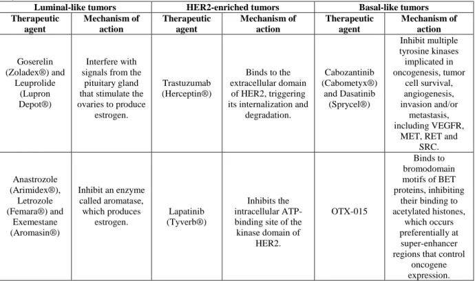
Table 1.1: Therapeutic agents and corresponding mechanisms of action, specific to each breast cancer subtype. These
therapeutic agents can be combined with conventional chemotherapy, especially in the case of basal-like tumors (12,14,15,17– 20).
Luminal-like tumors HER2-enriched tumors Basal-like tumors Therapeutic agent Mechanism of action Therapeutic agent Mechanism of action Therapeutic agent Mechanism of action Goserelin (Zoladex®) and Leuprolide (Lupron Depot®) Interfere with signals from the
pituitary gland that stimulate the ovaries to produce estrogen. Trastuzumab (Herceptin®) Binds to the extracellular domain of HER2, triggering its internalization and
degradation. Cabozantinib (Cabometyx®) and Dasatinib (Sprycel®) Inhibit multiple tyrosine kinases implicated in oncogenesis, tumor cell survival, angiogenesis, invasion and/or metastasis, including VEGFR,
MET, RET and SRC. Anastrozole (Arimidex®), Letrozole (Femara®) and Exemestane (Aromasin®) Inhibit an enzyme called aromatase, which produces estrogen. Lapatinib (Tyverb®) Inhibits the intracellular ATP-binding site of the kinase domain of HER2. OTX-015 Binds to bromodomain motifs of BET proteins, inhibiting their binding to acetylated histones, which occurs preferentially at super-enhancer regions that control
oncogene expression.
4
Table 1.1 (continuation): Therapeutic agents and corresponding mechanisms of action, specific to each breast cancer subtype. These therapeutic agents can be combined with conventional chemotherapy, especially in the case of basal-like tumors
(12,14,15,17–20).
Luminal-like tumors HER2-enriched tumors Basal-like tumors Therapeutic agent Mechanism of action Therapeutic agent Mechanism of action Therapeutic agent Mechanism of action Tamoxifen (Soltamox®), Toremifene (Fareston®) and Fulvestrant (Faslodex®) Attach to ER, preventing estrogen from binding. Pertuzumab (Perjeta®) Binds to the extracellular dimerization domain of HER2, blocking its heterodimerization
with other HER family. Rucaparib (Rubraca®) and Olaparib (Lynparza®) Inhibit PARP enzymes, which are
involved in DNA repair, resulting in the formation of toxic PARP-DNA complexes, DNA damage and, ultimately, cell death. Everolimus (Afinitor®) Inhibits mTOR, since the PI3K/AKT/mTO R pathway promotes ER transcriptional activity. Trastuzumab-DM1 (Kadcyla®) Trastuzumab binds to HER2 and, upon internalization, DM1 is released and binds to tubulin, disrupting microtubule dynamics and inhibiting cell division. Cetuximab (Erbitux®) Attaches to EGFR, inducing its internalization and preventing its ligands from binding. EGFR is involved in cell survival, cycle, migration, invasion and angiogenesis. Valproic acid (Depakene®) Inhibits HDAC, which downregulates ER, epigenetically. Tanespimycin and Ganetespib Inhibit Hsp90, a ubiquitous molecular chaperone fundamental for correct folding and
maturation of numerous cellular proteins, including HER2. In the absence
of Hsp90 activity, HER2 is subject to proteolysis. Scriptaid, Vorinostat (Zolinza®) Inhibit HDAC, which downregulates tumor suppressor genes, as well as genes involved in cell cycle, proliferation, differentiation and apoptosis.
1.5 Cancer and DNA damage: an intimate relationship
Each of the approximately 1013 cells in the human body receives tens of thousands of DNA
lesions per day (21). DNA damage can be generated spontaneously: dNTP misincorporation during DNA replication, interconversion between DNA bases caused by deamination, loss of DNA bases following depurination, modification of DNA bases by alkylation, oxidation of DNA bases and DNA breaks caused by reactive oxygen species derived from cellular metabolism. DNA damage can also be generated by environmental physical agents: ionizing radiation and ultraviolet light from the sun, clinical diagnosis employing X-rays or radiotherapy, which can originate pyrimidine dimers, photoproducts, oxidation of DNA bases, single-strand and double-strand breaks. Regarding environmental chemical agents: chemotherapeutic alkylating agents attach alkyl groups to DNA bases, crosslinking agents introduce covalent links between bases of the same or different DNA strands, and topoisomerase inhibitors induce the formation of single-strand and double-strand breaks by trapping topoisomerase-DNA complexes (22). However, cells have evolved mechanisms – collectively termed the DNA damage response – to detect DNA lesions, signal their presence and promote their repair (21). Normal cells harbor surveillance mechanisms, known as checkpoint pathways that control the entry and progression through the cell cycle, consisting of five phases: interphase, G1, S, G2 and mitosis (23).
DNA-damage response factors arrest the cell cycle to provide ample time for the repair of DNA lesions before S-phase (G1 arrest) and/or before mitosis (G2 arrest) (24). The repair mechanisms employed
single-5
strand break repair, non-homologous end joining or homologous recombination (22). Faulty DNA damage response confers sensitivity to DNA damaging agents, which can be counteracted by the accumulation of mutations in genes implicated in cancer development and therapy resistance. However, extensive DNA damage ends up with cell death (25,26).
1.6 p53, guardian of the genome
P53 is a central player in tumorigenesis (27). Normally, p53 is bound to Mdm2, which inhibits its transcription-activating ability. In addition, Mdm2 has E3 ubiquitin ligase activity, catalyzing the ubiquitinylation of p53 and targeting it for proteasomal degradation (23). Upon DNA damage, p53 becomes phosphorylated on a serine residue in the N-terminus, which displaces Mdm2 and increases p53 levels (23,26). On the other hand, in the presence of aberrantly high (not persistent) oncogenic signaling (e.g. Myc, Ras), E2F transcription factor 1 induces ARF expression, which binds to and sequesters Mdm2 in the nucleolus (23,28). Finally, stabilized p53 binds to proteins involved in DNA repair (XPB, RPA and rad51) and activates transcription of p21, a cyclin-dependent kinase inhibitor, which binds to and inhibits various cyclin-cdk complexes that regulate mainly G1-S but also G2-M
transition, arresting cells in G1 phase before DNA replication and/or in G2 phase before mitosis, for
DNA repair (29). Of note, Mdm2 is transcriptionally activated by p53. Alternatively, cells become permanently arrested (senescence) or, when exposed to extensive DNA damage, are directed to apoptosis (23). Apoptosis occurs through the p53-induced release of cytochrome c, expression of pro-apoptotic BCL-2 family members (e.g. puma, noxa, bid and bax), components of death-receptor signaling (e.g. DR5 and Fas/CD95), the apoptotic effector machinery (e.g. caspase-6, Apaf-1 and PIDD) or others with less well-defined roles (e.g. PERP, PML and p53AIP) (28). The p53-mediated DNA damage response does not discriminate between damaged cells that are potential tumor cells and those that are not, causing the widespread pathologies following radiotherapy or chemotherapy (e.g. breathing problems, infertility, bowel changes, bladder inflammation, cognitive dysfunction and second cancer) (18,30). Inactivation of p53 leads to genomic instability since damaged DNA can replicate, accumulating mutations and rearrangements that are passed on to daughter cells at an increased rate, which contributes to tumorigenesis (23). Interestingly, prolonged metaphase arrest causes de-condensation of chromosomes and entry to “pseudo G1” phase at the tetraploid DNA content, followed by aberrant DNA replication, resulting in the formation of polyploid giant cells. The prevention of this phenomenon is mediated by p21, which acts in a similar way as in normal G1 phase to prevent replication of damaged DNA (29). Moreover, p53 can restrict chromosomal instability through its ability to cull cells at risk of aberrant mitosis, particularly following centrosome amplification and telomere dysfunction. Therefore, inactivation of p53 also deregulates the spindle assembly checkpoint, leading to chromosome mis-segregation and tetraploidization; allows cells to replicate and initiate chromosome breakage-fusion-bridge cycles, upon telomere dysfunction (31).
In a study performed by Silwal-Pandit and colleagues, approximately 28.3% of breast tumors presented p53 mutations. The majority of mutations were non-synonymous single base substitutions (73.4%) that result in full-length proteins with altered biological activity. Small deletions (18.7%) and insertions (5.2%) were predicted to encode truncated proteins. Complex mutations, comprising both deletions and insertions (2.0%), and tandem mutations (0.7%) were uncommon and of unclear biological significance. In addition, the p53 pathway may also be disrupted by allelic deletions (loss of heterozygosity) of p53 (32). Surprisingly, cancer cells drive selection for partial loss-of-function p53 mutations that retain pro-survival properties (30). Besides, certain p53 mutants bind p63 or p73, leading to changes in transcriptional profiles that alter receptor tyrosine kinase signaling to promote invasion and metastasis. Others cooperate with the SWI/SNF complex to upregulate the angiogenesis regulator VEGFR2 (31). Thus, the evolutionarily accreted multifunctionality of p53 has compromised its efficacy as a tumor suppressor (30).
6
1.7 NORAD, a lncRNA induced by DNA damage
To discover lncRNAs involved in the DNA damage response, Lee and colleagues analyzed a published dataset of murine lncRNAs induced by doxorubicin in a p53-dependent manner. A 4.9 kb transcript, annotated as 2900097C17Rik, stood out because of its high conservation (approximately 65% nucleotides identity with its human ortholog, a 5.3 kb transcript, annotated as LINC00657 and referred to as NORAD, which stands for non-coding RNA activated by DNA damage); high abundance (in humans approximately 300 to 1,400 copies per cell, like abundant housekeeping transcripts such as
ACTB); ubiquitous expression across tissues and cell lines, at comparable levels, except for neuronal
tissues and cell lines, where is more expressed; and predominant location in the cytoplasm (33,34). This lncRNA is located in the chromosome 20q11.23, is intergenic, starts from a strong promoter overlapping a CpG island, the transcription start site is rich in H3K4me3-modified histones, the central 3.5 kb can be decomposed into 12 repeating units of approximately 300 nt each, terminates with a canonical poly(A) site and is unspliced. Some of these features indicate that NORAD is transcribed by RNA polymerase II. Within the repeats 1-10 are combinations of four sequence and structure conserved motifs: (i) one or two Pumilio recognition elements (PREs); (ii) a short-predicted stem-loop with four paired bases and a variable loop sequence; (iii) a U-rich stretch of 2-5 bases; and (iv) a stem-loop with eight or nine base pairs. Despite its p53-dependent induction, were not identified p53 binding sites in NORAD, suggesting indirect regulation (33,34).
1.8 Interaction with Pumilio proteins
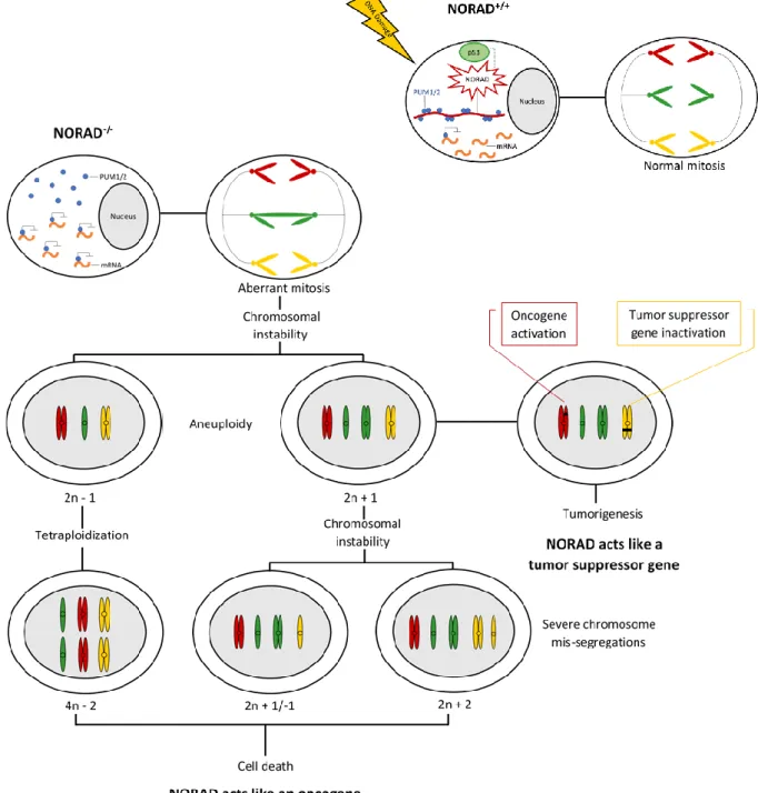
PUMILIO 1 (PUM1) and PUMILIO 2 (PUM2) are highly conserved RNA-binding proteins, mainly located in the cytoplasm, that belong to the Pumilio-Fem3-binding factor (PUF) family, exhibiting 91% similarity in their RNA-binding domains and regulating almost the same set of targets. Pumilio proteins bind to an 8 nt specific sequence (UGUANAUA), named Pumilio recognition element (PRE), in the 3’ untranslated region of target mRNAs, forming a multivalent cytoplasmic ribonucleoprotein (RNP) complex. Then, recruit 3’ de-adenylation factors and antagonize translation induction by the poly(A) binding protein, or destabilize the 5’ cap-binding complex, repressing gene expression. NORAD appears to be the preferred PUM2 target transcript and binds to it with great affinity because is composed of at least 17 Pumilio-binding motifs, mostly located in or near the conserved positions of the five repeated 400 nt Norad Domains (ND1 to ND5). The overall number of binding sites present in NORAD for Pumilio proteins (approximately 1,200) is comparable to their abundance in U2OS cells (approximately 200 and 550 copies per cell for PUM1 and PUM2, respectively). Upon DNA damage and in a p53-dependent manner, this lncRNA is activated and sequesters a significant fraction of the total cellular pool of Pumilio proteins, limiting the repression of target mRNAs involved in germline homeostasis, neuronal activity, mitosis, DNA repair and replication (Figure 1.1). Therefore, even small changes in NORAD levels can severely influence PUMILIO availability (33,34). Inactivation of NORAD results in PUMILIO hyperactivation and excessive repression of its targets, which perturb accurate chromosome segregation during mitosis, causing aneuploidy. Aneuploidy may contribute to gain of function of oncogenes and loss of function of tumor suppressor genes, leading to tumorigenesis. In this case, NORAD acts like a tumor suppressor gene. However, severe chromosome mis-segregations end up with cell death. In this case, NORAD acts like an oncogene (Figure 1.1). On the other hand, the canonical p53-dependent response is not affected (25,33). PUM1/2 overexpression not only results in changes in gene expression like those observed upon NORAD inactivation but also is sufficient to induce chromosomal instability (CIN) in NORAD+/+ cells. CIN is the most common form
of genomic instability, and comprises changes in chromosome number and structure. Conversely, PUM1/2 inactivation, significantly but not entirely, rescues chromosomal instability in NORAD-/- cells
(33,35). Of note, Pumilio proteins were already detected in myeloid leukemia, non-small cell lung cancer, ovarian cancer, and their levels were positively associated with tumor stage. After interfering
7
with their expression, cell proliferation, clone formation, migration and invasion abilities were inhibited, while cell apoptosis was increased significantly. These results suggest that Pumilio proteins play an important role in tumorigenesis and progression (36). 90% of other PUM-bound transcripts have two or fewer PREs. Furthermore, the human genome contains at least 43 loci with NORAD-like sequences, which exhibit 84% - 98% identity to NORAD over approximately 500 bp. These sequences may serve as binding sites for other RBPs that may either facilitate the binding of PUM1/2 or affect its stability and activity. Therefore, NORAD also regulates the expression of genes that are not PUMILIO targets (33,34). In fact, an RNA-binding protein, SAM68 was found to bind to conserved secondary structures immediately downstream from the PREs in NORAD repeat units. It was also found to be required for efficient recruitment of PUM2 to NORAD, regulation of PUM activity by NORAD and proper chromosome segregation in mammalian cells (Figure 1.1). Interestingly, SAM68 may have an additional, NORAD-independent effect on PUM activity (37).
Figure 1.1: NORAD activation or inactivation and subsequent events. Upon DNA damage and in a p53-dependent manner,
8
and replication. NORAD inactivation and subsequent repression of Pumilio proteins’ target mRNAs, results in aneuploidy and tumorigenesis or severe chromosome mis-segregations and cell death (25,33,34,37).
1.9 NORAD expression profiles predicts clinical outcome in multiple human cancers
Liu and colleagues profiled cDNA arrays from OriGene consisting of 43 breast tumors and 5 normal breasts, and observed that in 9 of 43 samples (21%), LINC00657 expression level was above a 2-fold of the mean expression level, which was associated with low overall survival (Kaplan-Meier test). They also found that expression of LINC00657 was upregulated in breast cancer cell lines MCF-7 and MDA-MB-231, as compared to non-malignant human mammary epithelial cells (RT-qPCR). Therefore,
LINC00657 knockout by CRISPR/Cas9 in LM-4142, a derivative cell line from MDA-MB-231, caused
significant reduction of cell viability (MTT assay) and number of colonies (clonogenic assay)(38). In a study performed by Wu and colleagues, expression of NORAD was significantly upregulated in esophageal squamous cell carcinoma tissues (larger size and higher UICC stage) compared to adjacent normal tissues from 106 patients (RT-qPCR). Besides, patients with higher expression of NORAD had poorer overall survival and disease free survival (5-year follow-up, Kaplan-Meier test) (39).
Li and colleagues found that NORAD expression was significantly higher in 75 pancreatic cancer samples than in 55 normal pancreas tissues (GEO database), which was confirmed in 33 pancreatic cancer samples with complete clinicopathological information along with the corresponding normal pancreas tissues (RT-qPCR). Moreover, patients with higher NORAD expression had shorter overall survival and recurrence-free survival rates (Kaplan-Meier test). Therefore, NORAD knockdown using shRNAs markedly suppressed cell migration and invasion in vitro (wound healing, transwell migration and invasion assays), and in vivo since mice with NORAD expression presented more metastasis, including more distant metastasis (dual-luciferase reporter assay, hematoxylin and eosin staining) (40).
Furthermore, in a study performed by Bao and colleagues in endothelial cells, low concentration of oxLDL (oxidized low-density lipoprotein) induced oxidative stress and, consequently, expression of
LINC00657. This lncRNA acted as a competing endogenous RNA inhibiting expression of
miR-590-3p, which in turn suppressed HIF-1α. The upregulation of HIF-1α promoted expression of many growth factors and cytokines, including VEGF, MMP-2 and MMP-9, involved in cell survival (MTS assay), migration (wound healing and transwell assays) and tube formation (tube formation assay), ultimately leading to angiogenesis in atherosclerosis. miR590-3p was also found to participate in the carcinogenesis of different types of cancer such as prostate, liver and lung. Therefore, LINC00657 might also induce angiogenesis in these tumors (41).
However, Hu and colleagues discovered that LINC00657 was downregulated in hepatocellular carcinoma (HCC) tissues and cell lines (RT-qPCR), which was associated with high tumor size, vascular invasion, TNM stage and poor overall survival (tumor characterization, Kaplan-Meier test). Therefore, NORAD knockdown using shRNAs and siRNAs, promoted cell cycle (flow cytometry), proliferation (CCK-8 and colony-formation assays), migration and invasion (transwell chamber detection) in vitro, as well as tumor growth in vivo (tumor monitorization, H&E staining and immunohistochemistry) (42). These results suggest that lncRNA NORAD expression or downregulation accounts for tumorigenesis and progression. Thus, it may represent a tumor marker for disease diagnosis, patient prognosis or therapy response, and it may represent a therapeutic target.
1.10 Long non-coding RNAs
Less than 2% of the genome encodes proteins, while at least 75% is transcribed into non-coding RNAs: short (sncRNAs) and long non-coding RNAs (lncRNAs) (43). Based on genomic organization and relationship to protein-coding genes, lncRNAs can be classified into: (1) sense or (2) antisense,
9
when overlapping one or more exons of another transcript on the same or opposite strand, respectively; (3) bidirectional, when its expression and of a neighboring coding transcript on the opposite strand are initiated in close genomic proximity; (4) intronic, when derived from an intron of a second transcript; or (5) intergenic, when it is an independent unit within the genomic interval between two genes (38,44). Generally, lncRNAs are defined by a length of >200 nucleotides and lack an open reading frame of significant length (less than 100 amino acids), are poorly conserved, less abundant and heterogeneous (33,45). However, the emerging evidence that some putative lncRNAs may encode short, translated open reading frames, and that coding RNAs can exert translation-independent functions, indicates that distinction between mRNA and lncRNA may be less absolute than once thought (46). Similarly to protein-coding genes, conservation is usually higher at the promoter regions of lncRNAs, which are enriched for A/T nucleotides and diminished in CpG patterns, and contain distinct transcription factor binding sequences and a unique pattern of chromatin marks, i.e., higher density of methylation at transcriptional start sites, regardless of expression levels (46). The lower levels of gene expression may not necessarily be the result of low RNA copy number in all cells, but may be the result from restricted expression in only a subpopulation of cells. Indeed, in a study performed by Djebali and colleagues, a considerable fraction (29%) of all expressed lncRNAs was detected in only one of the cell lines studied, whereas 10% were detected in all cell lines. Conversely, a considerable fraction (53%) of all expressed protein-coding genes were constitutive, whereas only 7% were cell-line specific (47). LncRNAs may be located in the nucleus, where regulate transcription, organize subnuclear structure and mediate chromosomal interactions, or in the cytoplasm, where modulate mRNA stability and translation, and influence cellular signaling cascades (33,39). The number of lncRNAs encoded by the genome has increased with animal evolution, suggesting that the presence of lncRNAs is linked to organismal complexity. Still, many human lncRNAs have syntenic orthologs in lower organisms (homologous genes present on the same chromosome, in different but related species). When performing comparative studies to analyze lncRNA conservation, should be taken into account that not just the sequence but also the structure, might determine function (43,48).
1.11 Mechanisms of action
LncRNAs function in the recruitment of protein factors for regulation of chromatin states. They may act as a scaffold onto which multiple protein complexes can assemble, recruit factors involved in gene activation or act as cis-acting molecular tethers. In fact, they are inherently attached to chromatin via DNA:RNA hybridization during transcription, are generally transcribed from a single locus in the genome, their length allows them to reach out and capture epigenetic complexes while tethered (either by Pol II or by bridging factors like YY1), and the unmasking of 3’ degradation signals upon transcriptional termination would limit the RNA’s half-life and prevent diffusion and action at ectopic sites. For example, HOTAIR interacts with the LSD1/CoREST/REST complex that demethylates histone H3K4 to prevent gene activation, synergizing the repressive effects of the PRC2 complex that also interacts with this lncRNA (49).
LncRNAs also directly affect the process of transcription, acting as decoys for transcription factors, transcriptional coregulators or inhibitors of Pol II activity. For example, lncRNAs transcribed from SINEs, an abundant class of retrotransposons, may block transcription of heat-shock genes by binding Pol II to prevent formation of preinitiation complexes (49).
In addition, they act in mRNA processing, stability and translation. For example, natural antisense transcripts may affect the alternative splicing of their overlapping transcripts by masking splice sites through base complementarity. For example, the α-thyroid hormone receptor gene erbAα produces two mRNA isoforms and has a downstream translated antisense transcript called RevErb, whose 3’ untranslated region overlaps the last splice acceptor site of the long erbAα isoform, tilting the balance in favor of the short erbAα isoform (49).
10
Moreover, they are key regulators of nuclear compartments. For example, MALAT1 interacts with the PRC1 subunit Cbx4/Pc2 and participates in the shuttling of genes between nuclear compartments for silencing and activation. In the presence of extracellular growth signals, unmethylated Cbx4 binds MALAT1 and localizes its target genes, along with coactivating factors such as LSD1, to interchromatin granules that usually cluster around nuclear speckles, for transcription; in the absence of signal, Cbx4 becomes methylated, binds another lncRNA, TUG1, associates with corepressors such as Ezh2, and translocates to silencing compartments called Polycomb bodies (49).
Interestingly, the long and small ncRNA worlds intertwine. LncRNAs may interfere with miRNA-mediated mRNA destabilization by masking the binding sites for miRNAs or by sequestering miRNAs in their 3’ untranslated regions that contain miRNA-binding sites. Moreover, lncRNAs can themselves be host genes for small RNAs. For example, in X chromosome inactivation, Tsix might repress Xist partly through an RNAi-related pathway, by base pairing to form an RNA duplex that yields Dicer-dependent small RNAs (siRNAs) (49).
However, in some cases it may be the act of transcription, and not the transcript itself, that is important. For example, in the control region of the β-globin locus, polyadenylated transcripts of varying lengths are generated from the HS2 enhancer site. No biological function is ascribed to these transcripts, but because HS2 is unable to activate globin expression when a transcriptional terminator is placed between it and the globin promoter, the phenomenon is interpreted to be transcription rather than transcript dependent. Indeed, intergenic transcription occurs throughout the globin locus and plays a role in establishing open chromatin domains marked by permissive histone modifications such as H3K4me2/3 and H3 acetylation (49).
1.12 LncRNA-based diagnostics and therapies in cancer
LncRNAs have key roles in gene regulation and, consequently, affect various aspects of cellular homeostasis, including proliferation, survival, migration or genomic stability. Cancer is primarily caused by genetic alterations that result in the deregulation of the gene networks that are responsible for the maintenance of cellular homeostasis. In addition, tumor suppressor genes and oncogenes may regulate the transcription of lncRNAs. For their application as biomarkers, lncRNAs should present tissue- and cell type-specific expression and, should be stable and easily detectable in body fluids to permit noninvasive diagnosis (43). For example, the lncRNA PCA3 is a specific and sensitive marker of prostate cancer in patient urine samples, which was the first Food and Drug Administration-approved test based on a lncRNA (46). Similarly, the lncRNA HULC is highly expressed in patients with hepatocellular carcinoma and can be detected in the blood by conventional PCR methods. Since lncRNAs regulate specific facets of protein activity, they also represent potential therapeutic targets. Of note, drugs that target lncRNAs can be more refined and less toxic than conventional protein-targeting drugs (43). Some therapeutic strategies utilize oligonucleotides for the knockdown of lncRNAs. siRNAs consist of an antisense (or guide) strand and a sense (or passenger) strand, forming a duplex 19 to 25 bp in length with 3' dinucleotide overhangs, and are mainly located in the cytoplasm. When bound by Argonaut 2 in the RNA-induced silencing complex, the passenger strand is discarded, leaving the guide strand free to bind an RNA target that is subsequently degraded (50,51). Alternatively, LNATM GapmeRs
are antisense oligonucleotides enriched with locked nucleic acids in the flanking regions and DNA in a central region, and are mainly located in the nucleus. The locked nucleic acids-containing flanking regions confer nuclease resistance and increase target binding affinity; the central DNA activates RNase H-cleavage of the target RNA upon binding (51,52). Still, LNATM GapmeRs share some limitations with
siRNAs: inefficient delivery, incomplete knockdown, off-target effects and temporary inhibition. On the other hand, genome-editing methods, such as transcription activator-like effector nucleases (TALENs) and clustered regulatory interspaced short palindromic repeats (CRISPR) – associated endonuclease 9 (Cas9), could be effective for stable knockdown of lncRNAs. Moreover, features of
11
lncRNAs can be exploited for other therapeutic strategies that do not necessarily involve targeting them. The plasmid BC-819 (DTA-H19) was developed to carry the gene for the A subunit of diphtheria toxin under the regulation of the promoter of the H19 lncRNA with tumor-specific expression. Intratumoral injection induces the expression of high levels of diphtheria toxin, resulting in a reduction of tumor size in human trials in a broad range of carcinomas and reduction of the risk of affecting normal tissues (43).
1.13 Chemotherapy
Chemotherapy is the use of drugs to treat cancer. Selectivity towards cancer cells occurs because of their higher proliferation relative to normal cells, with the exception of cells from the bone marrow, gastrointestinal mucosa and hair follicles. However, since drugs cannot distinguish between normal and cancer cells, common side-effects include myelosuppression, nausea, vomiting and hair loss. Moreover, cancer cells are often defective in their ability to repair DNA damage (53). Once chemotherapeutic agents are rapidly eliminated from the systemic circulation, high doses are required for antitumor efficacy. Only a small number of cancer types can be cured with chemotherapeutic agents (e.g. childhood blastomas) (54). Tumors can be intrinsically resistant to chemotherapy before treatment or acquire resistance during treatment (35). Chemotherapeutic agents are divided into: alkylating agents, platinum compounds, antimetabolites, anthracyclines, topoisomerase inhibitors, tubulin-binding drugs and tyrosine kinase inhibitors (55).
Adriamycin (generic name doxorubicin) is an anthracycline isolated from the bacterium
Streptomyces peucetius var. caesius, and is the hydroxylated congener of daunorubicin (56). This drug
is used for treatment of solid tumors arising in the breast, bile ducts, endometrial tissue, esophagus and liver, osteosarcomas, soft-tissue sarcomas and non-Hodgkin’s lymphoma (57). It is known that doxorubicin intercalates between base pairs in DNA, preventing DNA replication and protein synthesis; inhibits topoisomerase II, resulting in an increased and stabilized cleavable enzyme-DNA linked complex during DNA replication and in the non-repair of double-strand breaks (56). However, a new mechanism of action has been recently revealed, which depends on a transmembrane precursor, cAMP response element-binding protein 3-like 1 (CREB3L1), with a transcription factor domain located in the N terminus that projects into the cytosol, while the C terminus projects into the lumen of the endoplasmic reticulum. Doxorubicin induces the synthesis of ceramide that triggers the transportation of CREB3L1 from the ER to the Golgi complex, where occurs the regulated intramembrane proteolysis of CREB3L1, which releases the N terminus, allowing translocation to the nucleus and transcription of genes that suppress cell proliferation (58,59). These genes encode cyclin inhibitors p21 and p18, c-myc antagonists Mxi-1, GADD45γ, SPARC and RGC32 (60). The major side effect is cardiotoxicity associated with oxygen free radical formation from reduced doxorubicin intermediates (61).
1.14 Combinatorial approaches
The discoveries on the molecular mechanisms of cancer development have prompted the search for agents that interfere with pathways controlling cancer cell survival, proliferation and invasion, reducing the toxicity to normal cells (62). However, considering the heterogeneity of tumors, acting in only one pathway is not an effective treatment (63). Therefore, the combination of conventional chemotherapeutic agents with novel molecular-targeted agents might be a promising therapeutic approach. First, since the targets and mechanisms of action of these agents are different, there is no cross-resistance. Second, since in combinatorial approaches, the concentrations are low, chemotherapeutic agents side effects and molecular-targeted agents off-target effects are reduced. Third, alterations in expression and/or activity of genes that regulate mitogenic signals caused by molecular-targeted agents, not only may perturb cell growth but also may reduce resistance and sensitize cancer cells to chemotherapeutic agents (54,62). For example, the chemotherapeutic agent 5-fluorouracil (5FU) creates fluorinated nucleotides that are incorporated into DNA in place of thymidine, inhibiting DNA
12
replication and causing cell death. The lncRNA HOTAIR contributes to colorectal cancer and 5FU resistance through the recruitment of EZH2, epigenetic silence of miR-218, upregulation of VOPP1 expression and subsequent activation of the NF-kB/TS pathway. Thus, suppression of HOTAIR results in sensitivity to 5FU (64).
2. Objectives
The recently discovered human lncRNA NORAD is induced after DNA damage in a p53-dependent manner and plays a critical role in the maintenance of genomic stability. NORAD inactivation results in chromosomal instability and aneuploidy, which contributes to tumorigenesis or severe chromosome mis-segregations and cell death. Moreover, it has been detected in several types of cancer. In breast cancer, pancreatic cancer and esophageal squamous cell carcinoma, the lncRNA expression is upregulated and its knockdown inhibits cell viability, proliferation, migration and invasion. However, in hepatocellular carcinoma the lncRNA expression is downregulated and its knockdown promotes cell cycle, proliferation, migration and invasion. Having this in mind (NORAD action as an oncogene or tumor suppressor gene and contradictory results in different types of cancer), the main aim of the present study was to unravel the role of this lncRNA in breast cancer, which is the most frequently diagnosed and the second-leading cause of cancer death in women, in order to ascertain if it represents a potential therapeutic target.
Hence, mRNA basal levels of NORAD were determined in a set of human epithelial breast cancer cell lines (MCF-7, MDA-MB-231, 468 and 436) in comparison to a non-malignant human mammary epithelial cell line (MCF-10A). Correlation between the lncRNA expression levels and patients prognosis was also established. Then, its knockdown was performed and tumor-relevant phenotypes (cell viability, cycle, migration and apoptosis) were analyzed.
Another goal of this project was to explore the p53 pathway since it is a tumor suppressor gene, considered as the guardian of the genome, which also plays an important role in response to DNA damage. Understanding how it works may help to explain some of the effects observed upon NORAD knockdown. Therefore, mRNA and proteins levels were determined in different conditions.
Finally, due to the increasing number of resistance cases to chemotherapy, it was also important to evaluate, for the first time, the outcome of combining NORAD knockdown with chemotherapeutic agents i.e. if NORAD knockdown increases the sensitivity of human epithelial breast cancer cell lines to chemotherapeutic agents, in this case doxorubicin which is used in breast cancer management in the clinical setting.
3. Materials and methods
3.1 Cell culture
The non-malignant human mammary epithelial cell line MCF-10A was generously offered by Doctor Sérgio de Almeida (Instituto de Medicina Molecular/João Lobo Antunes, Lisbon). The human epithelial breast cancer cell lines MDA-MB-231, 436, 468 and MCF-7 were generously offered by Doctor Sérgio Dias (Instituto de Medicina Molecular/João Lobo Antunes, Lisbon) and are characterized in Table 3.1. MCF-10A cells were cultured in DMEM/F-12 (Dulbecco’s Modified Eagle Medium/Nutrient Mixture F-12, Gibco by Life Technologies), supplemented with 5% v/v horse serum (Gibco by Life Technologies), epidermal growth factor (20 ng/mL) (Millipore), hydrocortisone (0.5 µg/mL), cholera toxin (100 ng/mL), insulin (10 µg/mL) and 1% v/v penicillin-streptomycin (Gibco by Life Technologies). MDA-MB-231, 436, 468 and MCF-7 cells were cultured in DMEM (Gibco by Life Technologies), supplemented with 10% v/v heat inactivated fetal bovine serum (FBS), 1% v/v L-glutamine and 1% v/v penicillin-streptomycin (Gibco by Life Technologies). All cells were maintained at 37°C in a humidified atmosphere (90%) containing 5% CO2, at the exponential growth phase under
13
adherent conditions. All cells were visualized and photographed in a Primo Vert inverted microscope (Carl Zeiss) incorporated with an AxioCam ER c5s.
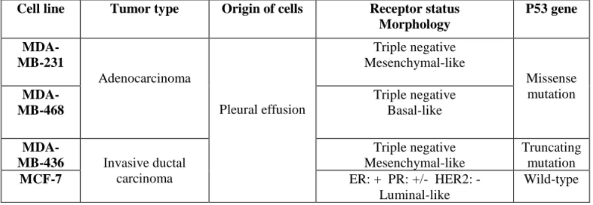
Table 3.1: Characterization of the human epithelial breast cancer cell lines used in this study. MCF-7 cells belong to the
luminal subtype and present a wild-type p53 gene sequence, while MDA-MB-231, 468 and 436 cells belong to the tripe negative subtype, which is the most aggressive, and present p53 mutations (5,9,65,66).
3.2 RNA isolation, cDNA synthesis and RT-qPCR
Total RNA was isolated using NZYol, following manufacturer’s instructions (NZYTech). RNA quantity and quality was verified by measuring the absorbance at 230nm, 260nm and 280nm using NanoDrop 2,000 Spectrophotometer (Thermo Fisher Scientific). Ideally, the A260/280 and A260/230 ratios
should be between 1.8 and 2.0. cDNA synthesis was performed with 200ng - 1µg of RNA and random hexamers using the Roche Transcriptor High Fidelity cDNA Synthesis Kit. The program on thermocycler consists of 10 minutes at 65°C (annealing of primers), 10 minutes at 29°C (extension of primers), 60 minutes at 48°C (cDNA synthesis) and 5 minutes at 85°C (degradation of other RNA and DNA molecules). Finally, real-time quantitative reverse transcriptase polymerase chain reaction (RT-qPCR) was performed in the ViiA 7 Real-Time PCR System (Thermo Fisher Scientific) using SYBR Green PCR master mix (Thermo Fisher Scientific). The program consists of 40 cycles: 2 minutes at 50°C (stage 1 – initial denaturation), 10 minutes at 95°C (stage 2 – dissociation of dsDNA into ssDNA), 15 seconds at 95°C and 1 minute at 60°C (stage 3 – annealing and extension of primers), 15 seconds at 95°C, 1 minute at 60°C and 15 seconds at 95°C (stage 4 – dissociation of dsDNA with incorporated dye molecules into ssDNA, melting curve). For results analysis, a comparative quantification between the threshold cycle (Ct) values of the genes of interest (NORAD and p53) in both the test sample and
calibrator sample (e.g. untreated), and the Ct values of the housekeeping genes (β-actin and 18S) in the
same two samples, was performed using the 2-∆∆Ct method. Gene-specific primer pairs (Sigma-Aldrich)
(10 µM) and sequences are presented in Table 3.2. Primer pairs efficiency was tested.
Table 3.2: Gene-specific primer pairs and sequences used in RT-qPCR. The genes of interest are NORAD and p53, while
the housekeeping genes are β-actin and 18S.
Cell line Tumor type Origin of cells Receptor status Morphology P53 gene MDA-MB-231 Adenocarcinoma Pleural effusion Triple negative Mesenchymal-like Missense mutation MDA-MB-468 Triple negative Basal-like MDA-MB-436 Invasive ductal carcinoma Triple negative Mesenchymal-like Truncating mutation MCF-7 ER: + PR: +/- HER2: - Luminal-like Wild-type
NORAD 1 Forward: 5’ – TGTTTGTGCAGTGGTTCAGG – 3’ Reverse: 5’ – TCTTGCCTCGCTGTAAACAG – 3’
NORAD 2 Forward: 5’ – AAAGTGTACAACGGCCTGTC – 3’ Reverse: 5’ – ATGGGGTTTCACCATGTTGG – 3’ NORAD 3 Forward: 5’ – AGCGAAGTCCCGAACGACGA – 3’
Reverse: 5’ – TGGGCATTTCCAACGGGCCAA – 3’ p53 Forward: 5’ – CCCCTCCTGGCCCCTGTCATCTTC – 3’
Reverse: 5’ – GCAGCGCCTCACAACCTCCGTCAT – 3’ β-actin Forward: 5’ – TGACGTGGACATCCGCAAAG – 3’
Reverse: 5’ – CTGGAAGGTGGACAGCGAG – 3’ 18S Forward: 5´ – GGATGTAAAGGATGGAAAATACA – 3’