0103 - 5053 $6.00+0.00
Article
* e-mail: [email protected]
Isoprenoid Compounds from
Euphorbia portlandica.
X-ray Structure of Lupeportlandol,
a New Lupane Triterpene
Ana M. Madureiraa, Maria Teresa Duarteb, Maria Fátima M. Piedadeb,c José R. Ascensob and Maria-José U. Ferreira*,a
a
Centro de Estudos de Ciências Farmacêuticas, Faculdade de Farmácia da Universidade de Lisboa, Av. das Forças Armadas, 1600-083 Lisboa, Portugal
b
Centro de Química Estrutural, Instituto Superior Técnico, Av. Rovisco Pais, 1096 Lisboa, Portugal
c
Departamento de Química e Bioquímica, Faculdade de Ciências da Universidade de Lisboa, Campo Grande, 1749-016 Lisboa, Portugal
O estudo fitoquímico dos extratos de Me2CO da planta inteira e seca de Euphorbiaportlandica levou ao isolamento de um novo álcool triterpênico pentacíclico, com o esqueleto do lupano, designado por lupeportlandol. A sua estrutura foi estabelecida como 3α-hidroxi-19αH-lup-20(29)-eno. Foram também isolados os já conhecidos triterpeno pentacíclico glutinol e o esteróide β-sitostenona. A caracterização do novo composto e do seu derivado acetilado foi baseada em métodos espectroscópicos e numa análise de difração de raio-X. O acetato de lupeportlandol mostrou-se inativo em ensaios de citotoxicidade in vitro contra três linhagens de células tumorais humanas: MCF-7 (câncer da mama), NCI-H460 (câncer do pulmão) e SF-268 (câncer do SNC).
Phytochemical survey of the Me2CO extracts of the whole dried plant Euphorbiaportlandica led to the isolation of a new pentacyclic triterpene alcohol, with the lupane skeleton, named lupeportlandol. Its structure was established as 3α-hydroxy-19αH-lup-20(29)-ene. The known pentacyclic triterpene glutinol and the steroid β-sitostenone were also isolated. The characterization of the new compound and its acetylated derivative was based on spectroscopic methods and an X-ray diffraction analysis. Lupeportlandol acetate was inactive in cytotoxicity assays in vitro against three human tumor cell lines: MCF-7 (breast cancer), NCI-H460 (non-small cell lung cancer) and SF-268 (CNS cancer).
Keywords:Euphorbia portlandica, triterpenes, steroids, lupane, X-ray diffraction
Introduction
Euphorbia portlandica L., from Euphorbiaceae family, is frequently found in the coast of Portugal, mainly in sand and rocks near the sea. Euphorbia species have been used in the traditional medicine for treatment of cancers, tumors and warts for hundred of years.1 They are characterized by
the existence of a latex very rich in isoprenic compounds, whose major constituents are tetra and pentacyclic triterpenes that revealed a wide spectrum of biological activities.2-5
Among pentacyclic triterpenes, lupane derivatives have demonstrated antiviral activity 6-8 as well as anti-proliferative
activity against various tumor cell lines.9-11
Previous studies on this species have afforded rearranged jatrophane-type diterpenes, which have been
found to be effective modulators of multidrug resistance in tumor cells.12 The present paper reports the isolation
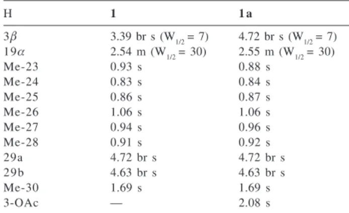
and structure determination of a new pentacyclic triterpene alcohol with the lupane skeleton (1), as well as the isolation
of the known compounds glutinol (2) and β-sitostenone (3) from the Me2CO extracts of the whole dried plant
Euphorbia portlandica. The evaluation of the acetylated derivative of compound 1 (1a) for its in vitro effect on the
growth of three human cancer cell lines is also reported.
Experimental
General experimental procedures
determined on a Perkin-Elmer 1310 instrument. The NMR spectra were recorded on a Varian Unity-300 NMR spectrometer (1H 300 MHz; 13C 75.4 MHz), with TMS as
internal standard and CDCl3 as solvent. MS were taken on a Kratos MS25RF spectrometer (70 eV) and HRMS on a Finnigan FT/MS 2001-DT. Column chromatography was carried out on SiO2 (Merck 9385). TLC was performed on precoated SiO2 F254 plates (Merck 5554 and 5744) and visualized under UV light and by spraying with sulphuric acid-acetic acid-water (1:20:4) followed by heating. HPLC was carried out on a Merck-Hitachi instrument, with UV detection, using a Merck LiChrospher 100 RP-18 (10 µm, 250 x 10 mm) column. The purity of the isolated compounds was monitored by means of analytical TLC and HPLC or GC being the latter analyses performed on a Hewlet Packard-5890 with a HP-17 column (10 m x 0.53 mm x 2.0 µm, 50 % PhMe silicone), isothermally, at 270 ºC, with He as carrier gas (20 mL min-1) and injection
and detection temperature 300 ºC; cholesterol acetate was used as an internal standard.
Plant material
Euphorbia portlandica was collected in the west coast of Portugal near the beach of Vale Furado, Nazaré, in September 1997 and identified by Dr. Teresa Vasconcelos of Instituto Superior de Agronomia, University of Lisboa. A voucher specimen (nº 248) has been deposited at the herbarium (LISI) of Instituto Superior de Agronomia.
Extraction and isolation
The air-dried whole powdered plant (4.8 Kg) of Euphorbia portlandica was extracted, by maceration, with acetone (7 x 8 L) at room temperature. Evaporation of the solvent (under vacuum, 40 ºC) from the crude extract gave a residue (367 g), which was suspended on a MeOH/H2O solution (1:1, 2 L) and extracted with n-hexane (3 x 1 L). The n-hexane extract was dried (Na2SO4) and evaporated (40 ºC), yielding a residue (170 g) that was submitted to column chromatography on SiO2 (1.2 Kg), using mixtures of n-hexane/EtOAc of increasing polarity (100:0 to 0:100) and EtOAc/MeOH (75:25) as eluting solvents yielding eight crude fractions after TLC and GC control. The more apolar crude frations (n-hexane/EtOAc; 100:0 to 92.5:7.5) contained mainly waxes and were discarded. The residue (27 g) of the crude fraction A (n-hexane/EtOAc; 92.5:7.5) was subjected to column chromatography on SiO2 (1 Kg) using n-hexane/EtOAc (100:0 to 75:25); eleven fractions eluted with n-hexane/EtOAc(96:4 to 95:5), after combined (1.5 g) and concentrated, crystallized from the elution
solvent affording 80 mg of pure compound 2 (Rf: 0.36,
CH2Cl2; GC Rt: 1.73). The mother liquor (1.3 g) were subjected to column chromatography (130 g) with n-hexane/CH2Cl2 (100:0 to 0:100) and CH2Cl2/EtOAc (50:50 and 0:100); the residue (120 mg) of fractions eluted with n-hexane/CH2Cl2, 80:20 to 77.5:22.5, after being subjected to crystallization with acetone, yielded respectively 10 mg of 1 (Rf: 0.48, CH2Cl2; GC Rt: 1.91)
and 180 mg of 2.Purification of the mother liquor residue
of 1, by preparative TLC (2 x n-hexane/CH2Cl2, 50:50),
yielded 26 mg of 1. The crude fraction B (n-hexane/EtOAc,
92.5:7.5 to 90:10) was submitted to three successive column chromatographies, both with mixtures of n-hexane/CH2Cl2 as above and with n-hexane/Me2CO (0:100 to 90:10), furnishing a product (65 mg; n-hexane/ Me2CO; 96:4), that was further purified by preparative TLC (2 x CH2Cl2), affording 40 mg of a product that was pooled to the fractions eluted with n-hexane/Me2CO, 95:5 (20
mg) and subjected to reverse phase HPLC (MeOH/H2O,
95:5; 5 mL min-1) yielding 10 mg of 3 (HPLC R
t: 51 min;
GC Rt: 1.86; Rf: 0.26, CH2Cl2).
Lupeportlandol (3α-hydroxy-19αH-lup-20(29)-ene) (1). White needles, mp 212-214 ºC (Me2CO); [α]D25 =+ 44°
(CHCl3, c 0.10); HRMS, m/z: 426.38552 [M]+,(426.38561
Calc. for C30H50O); IR (KBr), νmax/cm-1: 3405, 3104, 2944,
2919, 2866, 1640, 1456, 1381, 1082, 996, 884; 1H NMR
and 13C NMR see Tables 1 and 2;EIMS, m/z (rel. int.): 426
[M]+ (6), 411[M - CH
3]+ (2), 408 [M - H20]+ (1), 393
[M-CH3 - H20]+ (2), 315 (7), 218 (23), 207 (87), 189 (100), 175
(24), 161 (24), 149 (18), 147 (24), 135 (47), 121 (34), 107 (39), 95 (41), 81 (48), 69 (48), 55 (42), 43 (45).
Lupeportlandol acetate (3α-acetoxy-19α
H-lup-20(29)-ene (1a). Compound 1 (26 mg) was acetylated with
Ac2O-pyridine 1:1 at room temperature, overnight. The usual workup gave 25 mg of 1a (Rf: 0.77, CH2Cl2): White
needles, mp 230-232 ºC (Me2CO); [α]D25= + 25° (CHCl 3,
c 0.40); IR (KBr), νmax/cm-1: 2939, 2858, 1733, 1645, 1453,
1373, 1243, 1180, 1140, 1103, 1056, 1036, 1017, 983, 965, 941, 883; 1H NMR and 13C NMR see Tables 1 and 2;
EIMS, m/z (rel. int.): 408 [M - HOAc]+ (2), 393 [M - HOAc
- CH3]+ (2), 218 (5), 202 (8), 189 (88), 136 (42), 135 (55),
121 (100), 119 (49), 109 (90), 107 (70), 55 (43), 43 (25).
X-ray crystallographic analysis
Appropriate crystals of 1 for X-ray diffraction analysis
were obtained by recrystallization from Me2CO. The
the intensity of three standard reflections was monitored every hour of X-ray exposure time showing no significant decay. The structure was solved and refined using the WinGX package.13 The program used to solve the structure
was SIR97, 14 and to refine was SHELXL 97.15 All hydrogens
were found except the O-H hydrogen that was placed in calculated position riding on a carrier atom with isotropic displacement parameter amounting 1.5 times the value of the equivalent isotropic displacement parameter of the carrier atom. Absolute structure configuration was confirmed by the Flack parameter (see Table 3). Graphics
were done with ORTEP-3 for Windows version 1.076,16
included in WinGX system.
Cancer cell growth assay
The in vitro effect of compound 1a on the growth of
human tumor cell lines MCF-7 (breast cancer), NCI-H460 (non-small cell lung cancer) and SF-268 (CNS cancer) was
performed according to established protocols.17-19
Doxorubicin was used as the positive control.
Results and Discussion
The acetone extract of the whole dried plant Euphorbia portlandica was partitioned between a MeOH/H2O solution and hexane. Fractionation of the hexane extract yielded a new pentacyclic triterpene alcohol with a lupane skeleton, named lupeportlandol, which structure was established as
3α-hydroxy-19αH-lup-20(29)-ene (1). The known
pentacyclic triterpene glutinol (2) and the steroid β-sitostenone (3) were also isolated and identified.
Compound 1 showed a molecular ion, in the HREIMS,
at m/z 426.38552 indicating the molecular formula C30H50O (calc. 426.38561). The IR spectrum of 1 showed absorption
bands for a hydroxyl group (3405 cm-1) and an exocyclic
methylene group (3104, 1640, 884 cm-1). The EI-MS
spectrum showed the molecular ion peak at m/z 426, the common fragments of pentacyclic triterpenes and a base peak at m/z 189 (Figure 1). This fragment may arise from the cleavage between C-8/C-14 and C-12/C-13 bonds with H-transfer and suggests the lupane or hopane skeleton for
1.20, 21 The 1H NMR spectrum of 1 (Table 1) showed the
presence of six tertiary methyl groups (δ 0.83, 0.86, 0.91, 0.93, 0.94, and 1.06) and one methyl group attached to an sp2 carbon at δ 1.69. The latter methyl group and the
Table 3. Crystal data and structure refinement for lupeportlandol
(1)
Empirical formula C30H50O
Formula weight 426.70
Temperature 293 (2) K
Wavelength 1.54180 Å
Crystal system Monoclinic
Space group P21
Unit cell dimensions a = 7.574 (2) Å b = 16.563 (3) Å c = 10.139 (2) Å β = 98.15 (2) deg.
Volume 1259.1(5) Å3
Z 2
Calculated density 1.126 Mg/m3 Absorption coefficient 0.482 mm-1
F(000) 476
Crystal size 0.5 x 0.3 x 0.2 mm
Theta range for data collection 4.41 to 66.89 deg. Limiting indices -1 < = h < =9; -1 < = < = 19;
-12 < = l < = 12
Reflections collected / unique 3079 / 2486 [R (int) = 0.0272] Completeness to theta = 66.89 99.9 %
Absorption correction None
Refinement method Full-matrix least-squares on F2 Data / restraints / parameters 2486 / 1 / 477
Goodness-of-fit on F2 1.053
Final R indices [I > 2 sigma (I)] R1 = 0.0318; wR2 = 0.0845 R indices (all data) R1 = 0.0333; wR2 = 0.0862 Absolute structure parameter -0.2 (4)
Extinction coefficient 0.0222(14)
Largest diff. peak and hole 0.144 and - 0.126 e. Å-3
Table 2. 13C NMR data of compounds 1 and 1a (CDCl
3, δ in ppm)
Carbon 1 (δC) 1a (δC) DEPT Carbon 1(δC) 1a (δC) DEPT
1 33.3 33.9 CH2 1 6 36.9 36.9 CH2
2 25.3 27.8 CH2 1 7 41.8 41.8 C
3 76.3 78.4 CH 1 8 50.6 50.7 CH
4 37.5 37.2 C 1 9 44.8 44.8 CH
5 49.1 50.3 CH 2 0 150.6 150.6 C
6 18.3 18.2 CH2 2 1 30.6 30.6 CH2
7 34.0 34.0 CH2 2 2 41.4 41.4 CH2
8 41.1 41.2 C 2 3 28.2 27.9 CH3
9 50.4 50.3 CH 2 4 22.1 21.8 CH3
1 0 37.3 37.2 C 2 5 15.9 16.0 CH3
1 1 20.7 20.7 CH2 2 6 15.9 15.9 CH3
1 2 25.2 25.2 CH2 2 7 14.5 14.6 CH3
1 3 34.7 34.8 CH 2 8 20.6 20.6 CH3
1 4 43.1 43.2 C 2 9 108.9 108.9 CH2
1 5 27.5 27.6 CH2 3 0 25.1 25.2 CH3
Table 1.1H NMR data of 1 and 1a (CDCl
3, δ in ppm, J in Hz)
H 1 1 a
3β 3.39 br s (W1/2 = 7) 4.72 br s (W1/2 = 7) 19α 2.54 m (W1/2 = 30) 2.55 m (W1/2 = 30)
Me-23 0.93 s 0.88 s
Me-24 0.83 s 0.84 s
Me-25 0.86 s 0.87 s
Me-26 1.06 s 1.06 s
Me-27 0.94 s 0.96 s
Me-28 0.91 s 0.92 s
29a 4.72 br s 4.72 br s
29b 4.63 br s 4.63 br s
Me-30 1.69 s 1.69 s
exocyclic methylene protons at δ 4.72 and 4.63, support the existence of an isopropenyl group in the molecule. The proton at δ 3.39 was ascribed to H-3 and must be equatorially (3α-OH) oriented since it appears as a broad singlet (W1/2 = 7 Hz), resulting from small couplings to the protons H-2 (J 3eq,2ax ≅ J 3eq, 2eq) and is slightly downfield shifted (∆δ≅ 0.20 ppm) relatively to the normal chemical shift of H-3, axially oriented in common triterpenes, that appears as a double doublet near δ 3.20. 22, 23 The multiplet
appearing at δ 2.54 was assigned to H-19.
The 13C NMR spectrum showed signals for 30 carbon
atoms with multiplicities assigned by DEPT experiment The low field region of this spectrum shows the olefinic
carbons at δ 108.9 (CH2) and 150.6 (C) assigned to the terminal double bond of the isopropenyl group and one methine carbon at δ 76.3. In the high-field region seven CH3, ten CH2, five methines, and five quaternary carbons were identified. Acetylation of 1, with Ac2O-pyridine,
yielded a monoacetyl derivative 1a that showed essentially
the same 1H NMR spectrum, except for the signal due to
H-3 shifted to lower field (δ 4.72 brs) and the acetyl methyl resonance (δ 2.08.)
The above results agree with a lupane skeleton for 1
having an unusual configuration for the hydroxyl group at C-3 (3α-OH) that was further confirmed by the carbon resonances of ring A which were shifted upfield (except C-10), when compared to those reported for lupeol (4), whereas
paramagnetic shielding is observed for Me-24. 23 However,
by comparing the NMR data of 1 with those reported for
epilupeol (5) significant differences are observed for proton
and carbon resonances of rings D and E. 24, 25 A literature
survey revealed that the chemical shift of the angular methyl group Me-28 of 1 (δ 0.91), closely resembled those found for lupane derivatives (δ≅≅≅≅≅0.90) with a cis D/E ring junction nepehinol (6) (H-18β, Me-28β and H-19α)22,26 and
17-epi-lupenyl acetate (7) (H-18α, Me-28α and H-19β) 27 as well as
19αH-lupeol (8) 23 but different from lupeol (H-19β; δ 0.79) despite the same trans D/E ring junction (H-18α and Me-28β) in both compounds.23 The stereochemistry of 1 was
partially solved by through space proton-proton correlations observed in a NOESY spectrum. Indeed, the correlations between Me-25/Me-26, Me-26/Me-28 and Me-28/ isopropenyl (Figure 2) and the assumption of the same β configuration for both methyl groups at C-10 and C-8, as found in lupeol, provided evidence for a β configuration for both the isopropenylgroup and Me-28.
The less clear stereochemical aspects of 1, as the
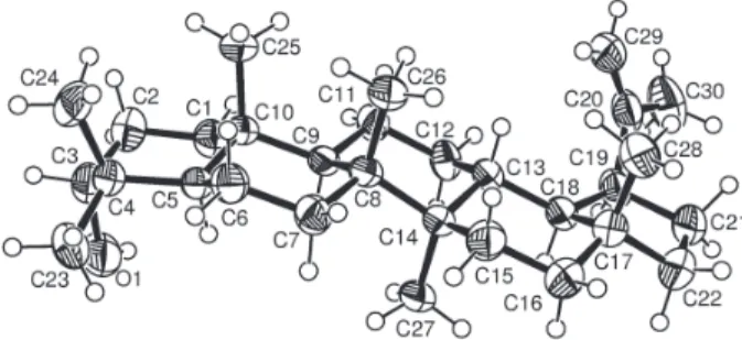
ambiguity in the stereochemistry of C-18, led to an X-ray diffraction analysis of compound 1. Figure 3 shows the
ORTEP diagram of the molecule establishing that the structure of lupeportlandol is 3α-hydroxy-19α H-lup-20(29)-ene, a new epimer of both compounds 5 (at C-19) and 8 (at
C-3). The distances and angles within the molecule, as well
Figure 1. Diagnostic fragment for lupane (R2 = H; R3 = CH
3) and hopane (R2= CH
3; R3 = H) triterpenes.
as the determined conformation of the rings, agree with the data from other triterpenic derivatives.28-31 This data also
agree with the conformation found both in 3α,4α -epoxy-D:A-friedo-18β,19αH-lupane30 and 3β
-hydroxy-20-oxo-29(20→19)abeolupane31 where no double bonds were
found in the carbon skeleton of the molecule.
Compounds 2 and 3, respectively identified as glutinol
and β-sitostenone, were also isolated. Their physical and spectral data were in agreement with those reported in the literature. 22, 32-34
The ability of compound 1a to inhibit the in vitro
growth of MCF-7, NCI-H460 and SF-268 cell lines was evaluated. It showed to be inactive (GI50 > 100 µM) against
the three cell lines.
Acknowledgments
The authors are grateful to Prof. Maria São José and Dr. Madalena Pedro from Faculdade de Farmácia, University of Porto, Portugal, for the tumor cell growth assays. The authors thank also Dr. Teresa Vasconcelos (ISA, University of Lisbon, Portugal) for identification of the plant and Mr. I. Marques (IST) for low resolution mass spectra.
This work was supported by FCT (POCTI, Quadro Comunitário de Apoio III) and PRODEP.
Supplementary Information
Supplementary crystallographic data for the structure have been deposited at the Cambridge Crystallographic Data Centre no. CCDC 225799. Copies of the data can be obtained free of charge via www.ccdc.cam.ac.uk/conts/ retrieving.html (or from Cambridge Crystallographic Data Centre, CCDC, 12 Union Road, Cambridge CB2 1EZ, UK; fax +44 1223 336033; or e-mail: [email protected]).
References
1. Hartwell, J. L.; Lloydia1969, 62, 153.
2. Huang, L.; Chen, C. H.; Curr. Drug Targets Infect. Disord.
2002, 2, 33.
3. Novotny, L.; Vachalkova, A.; Biggs, D.; Neoplasma2001, 48, 241.
4. Tanaka, R.; Kinouchi, Y.; Tokuda, H.; Nishino, H.; Matsunaga, S.; Planta Med. 2000, 66, 630.
5. Madureira, A. M.; Ascenso, J. R.; Valdeira, L.; Duarte, A.; Frade, J. P.; Freitas, G.; Ferreira, M. J. U.; Nat. Prod. Res.
2003, 17, 375.
6. Sun, I. C.; Kashiwada, Y.; Morris-Natschke, S. L.; Lee, K. H.; Curr. Top. Med. Chem. 2003, 3, 155.
7. Hashimoto, F.; Kashiwada, Y.; Cosentino, L. M.; Chen, C. H.; Garrett, P. E.; Lee, K. H.; Bioorg. Med. Chem.1997, 5, 2133. 8. Baltina, L. A.; Flekhter, O. B.; Nigmatullina, L. R.; Boreko, E. I.; Pavlova, N. I.; Nikolaeva, S. N.; Savinova, O. V., Tolstikov, G. A.; Bioorg. Med. Chem. Lett. 2003, 13, 3549.
9. Hata, K.; Hori, K.; Ogasawara, H.; Takahashi, S.; Toxicol. Lett.
2003, 143, 1.
10. Setzer, W. N.; Setzer, M. C.; Mini Rev. Med. Chem. 2003, 3, 540.
11. Chaturvedula, V. S. C.; Schilling, J. K.; Johnson, R. K.; Kingston, D. G.; J. Nat. Prod.2003, 66, 419.
12. Madureira, A. M.; Gyémánt, N.; Ugocsai, K.; Ascenso, J. R.; Abreu, P. M.; Hohmann, J.; Molnár, J.; Ferreira. M. J.; Planta Med. 2004, 70, 45-49
13. Farrugia, L. J.; J. Appl. Crystallogr. 1999, 32, 837.
14. Altomare, A.; Burla, M. C.; Camalli, M; Cascarano, G.; Giacovazzo, C.; Guagliardi, A.; Moliterni, A. G. G.; Polidori, G.; Spagna, R.; J. Appl. Crystallogr. 1998, 32, 115. 15. Sheldrick, G. M.; SHELXL97- Program for Refining Crystal
Structures, Univ. of Göttingen, Germany, 1993. 16. Farrugia, L. J.; J. Appl. Crystallogr. 1997, 30, 565.
17. Monks, A.; Scudiero, D.; Skehan, P.; Shoemaker, R.; Paull, K.; Vistica, D.; Hose, C.; Langley, J.; Cronise, P.; Vaigro-Wolff, A.; Gray-Goodrich, M.; Campbell, H.; Mayo, J.; Boyd, M.; J. Nalt. Cancer Inst.1991, 83, 757.
18. Skehan, P.; Storeng, R.; Scudiero, D.; Monks, A.; McMahon, J.; Vistica, D.; Warren, J. T.; Bokesch, H.; Kenney, S.; Boyd, M. R.; J. Natl. Cancer Inst.1990, 82, 1107.
19. Valente,C.; Ferreira, M. J. U.; Abreu, P. M.; Pedro, M.; Cerqueira,F.;Nascimento, M. S. J.;Planta Med. 2003, 69, 361.
20. Budzikiewick, H.; Wilson, J. M.; Djerassi, C.; J. Org. Chem.
1963, 85, 3688.
21. Ogunkoya, L.; Phytochemistry1981, 20, 121.
22. Akihisa, T.; Shimizu, N.; Kawaguchi, R.; Tamura, T.; Matsumoto, T.; J. Jpn. Oil Chem. Soc.1986, 35, 17. 23. Nazir, M.; Ahmad, W.; Kreiser, W.; Pak. J. Sci. Ind. Res. 1998,
41, 6.
24. Waterman, P. G.; Ampofo, S.; Phytochemistry 1985, 12, 2925. 25. Carvalho, L.; Seita, J.; Fitoterapia1995, 66, 273.
Figure 3. ORTEP diagram of compound 1 with numbering scheme
26. Ahmad, V. U.; Bano, S.; Mohammad, F. V.; Planta Med. 1985,
6. 521.
27. Shiojima, K.; Suzuki, H.; Kodera, N.; Kubota, K.; Tsushima, S.; Ageta, H.; Chand, H. C.; Chen, Y. P.; Chem. Pharm. Bull.
1994, 42, 2193.
28. Ferreira, M. J. U.; Lobo, A. M.; O’Mahoney, C. A.; Williams, D. J.; Wyler, H.; Helv. Chim. Acta1991, 74, 1329.
29. Konda, Y.; Iguchi, M.; Harigaya, Y.; Takayanagi, H.; Ogura, H.; Li, X.; Lou, H.; Onda, M.; Tetrahedron Lett. 1990, 37, 5315.
30. Yokoyama Y.; Moriyama, Y.; Tsuyuki, T.; Takahasi, T.; Iitaka, Y.; Bull. Chem. Soc. Jpn. 1980, 53, 2971.
31. Chiang, Y.; Kuo, Y.; J. Org. Chem.2002,67, 7656. 32. Tori, M.; Torh, T.; Tachibana, K.; Yamada S.; Tsuyuki, T.;
Takahashi, T.; Bull. Chem. Soc. Jpn. 1977, 50, 469. 33. Oleo, R. S. G.; Torres, L. M. B.; Roque, R. C.; Roque, N. F.;
Magn. Reson. Chem. 1994, 32, 378.
34. Greca, M. D.; Monaco, P.; Previtera, L.; J. Nat. Prod. 1990, 53, 1430.
Received: January 5, 2004