Authors
David Carvalho Fiel1 Iolanda Santos1 Joana Eugénio Santos1 Rita Vicente1
Susana Ribeiro1 Artur Silva1 Beatriz Malvar1 Carlos Pires1
1 Hospital do Espírito Santo de
Évora, Largo Senhor da Pobreza, Évora, Portugal.
Submitted on: 07/21/2018. Approved on: 09/23/2018.
Correspondence to: David Carvalho Fiel.
E-mail: davidcarvalhofiel@gmail.com
sulfonate bezoar - a rare entity
Perfuração do ceco associada a bezoar de poliestirenossulfonato de
cálcio - uma entidade rara
A hipercalemia é um dos distúrbios eletrolíti-cos mais comuns, responsável por um grande número de desfechos adversos, incluindo ar-ritmias potencialmente fatais. Quelantes de potássio são amplamente prescritos para o tratamento da hipercalemia, mas infeliz-mente são muitos os eventos adversos asso-ciados ao seu uso, em particular os gastro-intestinais. A identificação de pacientes com risco mais elevado para complicações graves associadas aos quelantes de potássio atual-mente em uso, como necrose e perfuração do cólon, pode evitar desfechos fatais. O presen-te artigo descreve o caso de um homem de 56 anos com diabetes secundário e doença re-nal crônica em tratamento por hipercalemia com poliestirenossulfonato de cálcio (PSC). Posteriormente o paciente apresentou abdô-men agudo devido a perfuração do ceco e foi submetido a uma ressecção ileocecal, mas acabou indo a óbito por choque séptico uma semana mais tarde. Durante a cirurgia, uma massa branca sólida foi isolada no lúmen do cólon. A massa foi identificada como um be-zoar de PSC, uma massa de fármaco de rara ocorrência formada no trato gastrointestinal que contribuiu para a perfuração. História pregressa de gastrectomia parcial e vagoto-mia foi identificada como provável fator de risco para o desenvolvimento do bezoar de PSC. Espera-se que os dois novos quelantes de potássio - patiromer e ciclossilicato de zir-cônio sódico - ajudem a tratar pacientes de alto risco em um futuro próximo.
R
ESUMOPalavras-chave: Hiperpotassemia; Potás-sio; Cálcio; Bezoares.
Hyperkalemia is one of the most com-mon electrolyte disorders, responsible for a high number of adverse outcomes, including life-threatening arrhythmias. Potassium binders are largely prescribed drugs used for hyperkalemia treatment but unfortunately, there are many adverse events associated with its use, mostly gas-trointestinal. Identification of patients at highest risk for the serious complications associated with the current potassium binders, such as colon necrosis and per-foration, could prevent fatal outcomes. The authors present a case of a 56-year--old man with secondary diabetes and chronic renal disease that was treated for hyperkalemia with Calcium Polystyrene Sulfonate (CPS). He later presented with acute abdomen due to cecum perforation and underwent ileocecal resection but ul-timately died from septic shock a week later. During surgery, a solid white mass was isolated in the lumen of the colon. The mass was identified as a CPS bezoar, a rare drug-mass formed in the gastroin-testinal tract that contributed to the per-foration. A previous history of partial gastrectomy and vagothomy was identi-fied as a probable risk factor for the CPS bezoar development. Hopefully, the two new potassium binders patiromer and (ZS-9) Sodium Zirconium Cyclosilicate will help treat such high-risk patients, in the near future.
A
BSTRACTKeywords: Hyperkalemia; Potassium; Calcium; Bezoars.
DOI: 10.1590/2175-8239-JBN-2018-0158
I
NTRODUCTIONHyperkalemia is one of the most com-mon electrolyte disorders, responsible for a high number of adverse outcomes, including life-threatening arrhythmias1.
Although little is known about the true
(ESRD)1. A correlation has also been established
be-tween hyperkalemia and other risk factors such as older age, Caucasian race, diabetes mellitus (DM) and renin-angiotensin-aldosterone system inhibitors (RAASi) use1. Treating such patients can be rather
challenging, as they are the ones that benefit the most from the inhibition of the renin-angiotensin-aldos-terone system but also are the most at risk of life--threatening hyperkalemia. As shown in various re-trospective and observational studies, several patients who should be medicated with RAASi according to guidelines have been prescribed with lower-than-the-rapeutic doses or have discontinued this medication due to hyperkalemia, with consequent worse outco-mes and mortality2. This emphasizes the importance
of strategies that can lower serum potassium levels and maintain levels in the normal range, such as po-tassium binders (PB). PB are artificial resins that bind potassium ions in the gastrointestinal tract (GIT), exchanging these ions for calcium (Ca2+) or sodium
(Na+) and hydrogen (H+) cations, therefore preventing
potassium from being absorbed.
There are two classes of PB widely commerciali-zed in most countries: calcium polystyrene sulfonate (CPS) and sodium polystyrene sulfonate (SPS), diffe-ring in the cation attached to the resin that is exchan-ged with potassium in the intestinal lumen. However, these drugs have poor digestive tolerability and cause adverse events, which commonly lead to the disconti-nuation of the drug by patients themselves: constipa-tion, diarrhea, and abdominal pain.
Patiromer and ZS-9 (sodium zirconium cyclosili-cate) are new PB not yet available in some countries, with good tolerability and promising results regar-ding the treatment of patients with hyperkalemia3.
In this article, the authors describe a case of a seve-re adverse event associated with PB, namely a cecum perforation associated with a PB bezoar (Table 1).
C
ASEPRESENTATIONA 56-year-old Caucasian man presented to the Emergency Department (ER) with a two-month--lasting painful lesion in his right foot. The patient had a history of chronic alcoholic pancreatitis and se-condary DM at young age, which later culminated in diabetic kidney disease. At hospital admission, he had stage 4 CKD with renal tubular acidosis type 4 (ATR 4). Other significant conditions were hypertension, history of duodenal ulcer with stenosis resolved by partial gastrectomy (with Bilroth II and vagothomy) at the age of 45, ischemic stroke at the age of 52, hypothyroidism, and major depressive disorder. He was chronically medicated with insulin, enalapril, ni-fedipine, darbapoetin alfa, sodium bicarbonate, clo-pidogrel, rosuvastatin, levothyroxine, escitalopram, and pantoprazole.He had also been medicated with a PB (calcium polystyrene sulfonate) in the past, during episodes of severe hyperkalemia, but it had been dis-continued a few weeks before the ER visit due to an excessive reduction in potassium levels. No history of allergies was reported.
On clinical observation in the ER, the patient pre-sented a deep ulcer with tendon exposure and perile-sional swelling - cellulitis - in the right foot, associa-ted with necrosis of the ipsilateral second and third toes. Abdomen examination was unremarkable. The patient had to be admitted for intravenous (IV) an-tibiotics and surgical debridement of the ulcer, with amputation of the second and third toes of the right foot. Despite long-term Piperacillin/tazobactam IV
PRESENTATION 22ADMITTANCEnd DAY OF 25ADMITTANCEth-30th DAY OF 33ADMITTANCErd DAY OF
Observation Right foot ulcer and cellulitis.
Hyperkalemia (K+ 6.0 mmol/L).
Hypokalemia (K+ 2 mmol/L).
Cecum perforation with peritonitis.
Management
• Long-term Piperacilin/ Tazobactan iv.
• Amputation of the 2nd-3rd right toes.
• Dietary potassium restriction.
• Insulin dose increase.
• CPS prescription.
• CPS suspension.
• Spironolactone prescription.
• Large amounts of K+ iv.
• Ileocecal resection (bezoar removed).
•Imipenem/Cilastatin initiation.
Outcome Amputation of the right leg.
Hypokalemia (2,0 mmol/L).
Refractory Hypokalemia.
CPS bezoar formation
Death due to septic shock.
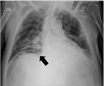
Figure 1. Patient’s thoracic radiography showing a pneumoperitoneum (black arrow).
Figure 2. Histopathological findings of the resected fragment showing serositis (black arrow), and transmural ischemia.
and local surgical intervention, the foot lesion conti-nued to worsen and the patient had to endure ampu-tation of the right leg on the 22nd day of admittance.
After amputation, he developed hyperkalemia (K+
6.0 mmol/L), which did not respond to dietary potas-sium restriction and insulin dose increase, and CPS was therefore prescribed. Potassium levels decreased steadily but more extensively than expected, and on the seventh day of treatment, resins were suspended. Despite this, his hypokalemia continued to worsen in the following days to levels as low as 2.0 mmol/L, and iv potassium supplementation was required in large amounts. Spironolactone was also prescribed. The patient complained of constipation and slight ab-dominal discomfort that could be solely attributed to hypokalemia, but was able to maintain a stool output every other day, so a major complication was unsus-pected at this time. The consulting nephrologists sug-gested that a rare event - the development of a bezoar of ion-exchange resin - was a likely explanation for an unresponsive hypokalemia in this setting.
On the 33th day of hospitalization, the patient
com-plained of diffuse abdominal pain and general weak-ness. His abdomen was distended and painful, with peritoneal reaction. The blood tests showed an eleva-tion of infeceleva-tion parameters (leucocytes 10.6x10³µL
with 89.6% neutrophil count; C-reactive protein of 25.6 mg/dL) that seemed to have no association with the initial clinical picture, as the amputation stump was clean with no signs of infection. Radiography re-vealed a pneumoperitoneum (Figure 1) and the patient was immediately transferred to the operation room for an exploratory laparotomy: a cecum perforation with peritonitis was diagnosed. This prompted an ileocecal resection with ileostomy and iv broad-spec-trum antibiotics prescription (Imipenem/Cilastatin). During the surgical procedure, a solid white mass was removed from the lumen of the resected cecum, inter-preted as a CPS bezoar.
The resected colon presented greyish external surface, hemorrhagic foci, and whitened plaques. Histologic examination (Figure 2) showed serositis and transmural ischemia. Whether cecum perforation was favored by mucosal ulceration from exposure to the resin or from lumen obstruction by the CPS bezo-ar was not completely established by the pathologist.
Enterococcus faecium was latter isolated in the pe-ritoneal effusion and blood cultures. Unfortunately, despite every support measures taken in the Intensive
Care Unit, the patient died one week after the colec-tomy due to septic shock.
D
ISCUSSIONPB are associated with many adverse events, mostly gastrointestinal, ranging from mild constipation to rare life-threatening complications such as the one described in this case report. Severe gastrointestinal adverse effects including colonic perforation have been documented in both type of resins, sodium and calcium polystyrene sulfonate, either with sorbitol or alone4. Although the colon is the most common
loca-tion of injury, it is increasingly recognized that injury may occur in more proximal sections of the GIT: in 30% of the cases there is an injury in the esophagus, stomach, or small intestine5.
have demonstrated that inoculation of tissue with SPS can lead to an inflammatory reaction within 24 hours and the release of cytokines and prostaglandins result in further impairment in local hemodynamic mechanisms, that culminate in vascular injury and mucosal lesion7. Ziv Harel et al. identified 58 cases of
severe gastrointestinal adverse events associated with SPS use in a review of case series and case reports including frank necrosis and perforation5. Therefore,
resins use alone can frequently induce GIT lesion, re-gardless of forming a drug bezoar and inducing bo-wel obstruction. In some cases, crystals of the drug can be found when assessing the pathologic sample, therefore corroborating the presence of the resin as the etiological agent of the lesion. However, despite existing in vivo, these crystals can often be lost during the physical preparation of the sample thus remaining undetectable.
In the present case, the onset of severe hypoka-lemia, despite discontinuation of CPS, enduring for days and requiring iv potassium supplementation, was highly suggestive of an unremitting PB influen-ce, best explained by the sustained presence of the drug inside the GI tract. The best assumption was that a CPS bezoar had formed inside the intestinal lumen.
A bezoar is a stiff, solid, and persistent foreign bo-dy that is located in the GIT. The majority of bezoars are located in the stomach; however, they may be encounte-red in the whole GIT, including the esophagus and colon. Depending on the material of origin, four different types of bezoars have been described: phytobezoars (hortobe-zoars), trichobezoars (pilobezoars, hairball), stone-like foreign bodies, and pharmacobezoars (drug-induced)8.
There is little knowledge on pharmacobezoars, as there are only nearly 30 published articles on the subject. The majority of pharmacobezoars develop in the stomach8,
formed by the anomalous binding of drugs due to altera-tions in GIT anatomical structure, motility, or secretion. It is thus expected that the most common risk factor is a history of previous gastric surgery9. DM and antacid
drug use are other recognized predisposing factors. The clinical diagnosis is usually difficult; therefore, pharma-cobezoars are usually diagnosed during an operation or endoscopy8.
Concerning PB use, numerous risk factors ha-ve been identified as contributors to the gastroin-testinal injury induced by these drugs: CKD and ESRD (elevated renin levels predispose patients
to non-occlusive mesenteric ischemia through an-giotensin II-mediated vasoconstriction); postope-rative status (hypotension, ileus-induced colonic distension with consequent reduced colonic blood flow, and decreased gut motility as a result of opioids; constipation increases the risk of injury); and solid organ transplantation (there might be an increased risk associated with immunosuppressi-ve medications that impair normal protectiimmunosuppressi-ve and reparative mechanisms of gastrointestinal cells)5.
PB should be avoided in these patients, or at least prescribed in a small dosage, with frequent moni-toring. In view of the present case of a PB bezoar development in a patient with history of partial gastrectomy and vagothomy, the authors believe that there should be a warning for PB prescription in such patients.
There is no specific treatment guidelines for PB bezoars, only for their complications. Neither en-doscopy nor laparotomy are advocated in an early stage to remove the resin mass. The use of osmo-tic catharosmo-tics should also be avoided. The current recommendation is drug interruption, along with hemodynamic improvement to prevent gastroin-testinal hypoperfusion that could lead to transmu-ral necrosis, which was the conduct taken in this case10. Unfortunately, cecum perforation occurred
a few days later. In the setting of free perforation to abdomino-pelvic cavity, the surgeon must seek the removal of PB crystals from the peritoneum. On-table colonic lavage can be used to make su-re that all the su-resin is su-removed from the lumen and the creation of a primary anastomosis is ill--advised10. There is frequent need for surgical
re--exploration due to new intestinal perforations that can occur from PB intraluminal or peritoneal remnants10.
The future of acute and chronic hyperkale-mia treatment is likely to be altered by two emer-ging and promising therapies: patiromer and so-dium zirconium cyclosilicate (Table 2). Patiromer (Veltassa®
), approved by the Food and Drug Administration (FDA) in October 2015 and by the European Medicines Agency (EMA) in July 2017, is a non-absorbable organic polymer that binds po-tassium in exchange for calcium, mostly in distal colon, where the concentration of free potassium is highest3. It is a non-selective cation-exchanger,
TABLE 2 COMPARISONBETWEENTHETWONEWHYPERKALEMIATHERAPIESRECENTLYINTRODUCEDINTHEMARKET
POLYSTYRENE
SULFONATE (PS) PATIROMER
ZS-9: SODIUM ZIRCONIUM CYCLOSILICATE
STRUCTURE Sulphonated cross-linked polystyrene copolymer.
Spherical organic polymer (oral suspension).
Microporous crystalline spherical inorganic polymer (powder).
MECHANISM
Not absorbed.
Exchanges K+ for Ca2+ or Na+.
Non-selective binding.
Not absorbed.
Exchanges K+ for Ca2+.
Non-selective binding.
Not absorbed.
Exchanges K+ for Na+.
Highly selective for K+
(125x superior than PS).
ACTION Stomach and mainly colon. Distal colon.
Sustained effects for 24-48h.
Duodenum, jejunum, Ileum and colon.
CKD POPULATION Tested in CKD population. Tested in CKD population. Tested in CKD population.
MAIN
ADVERSE
EVENTS
Abdominal pain.
Diarrhea or constipation.
Intestinal ulceration/ perforation.
Hypercalcemia, hypokalemia.
Hypomagnesemia.
Hypokalemia.
Constipation.
Edema.
Hypokalemia.
MAIN
ADVANTAGES Potassium binding.
Better tolerated.
Calcemia control in CKD.
Compatible with RAASi.
Better tolerated.
Powerful K+ binding effect.
Compatible with RAASi.
through 48 hours11. There are cases of
hypomagne-semia and constipation associated with patiromer, however it seems to be better tolerated when com-pared to other available PBs3.
Sodium zirconium cyclosilicate, also known as ZS-9 (Lokelma®), is a 2018 FDA/EMA appro-ved highly selective inorganic microporous cation exchanger that entraps potassium in the intesti-nal tract in exchange for sodium and hydrogen. Its great advantage is that it has 9.3 times more potassium-binding capacity than SPS and is more than 125 times more selective for potassium than the former. Although some GI adverse events have been described associated with ZS-9, such as edema and hypokalemia, several studies have shown that the drug has good safety profile, ca-pable of consistently reducing serum potassium levels3.
In conclusion, CPS and SPS administration can lead to severe gastrointestinal adverse events. Lack of an alternative drug for the treatment of chronic hyperkalemia makes the use of these drugs com-mon and render their side effects rather frequent. The authors describe a rare case of a PB pharma-cobezoar, seldom diagnosed, that contributed to
cecum perforation. They believe that the partial gastrectomy and vagothomy (performed several years before for treatment of duodenal ulcer wi-th stenosis) was an important risk factor for PB bezoar development, and suggest that in such pa-tients an alternative treatment option for hyperka-lemia should be sought. The recent development of Patiromer and ZS-9 as new PBs might change the paradigm of hyperkalemia therapy.
R
EFERENCES1. Kovesdy CP. Epidemiology of hyperkalemia: an update. Kidney Int Suppl 2016;6:3-6.
2. Epstein M. Hyperkalemia constitutes a constraint for im-plementing renin-angiotensin-aldosterone innhbition: the widening gap between mandated treatment guidelines and the real-world clinical arena. Kidney Int Suppl 2016;6:20-8.
3. Weir MR. Current and future treatment options for managing hyperkalemia. Kidney Int Suppl 2016;6:29-34.
4. Kamel KS, Schreiber M. Asking the question again: are cation exchange resins effective for the treatment oh hyperkalemia? Nephrol Dial Transplant 2012;27:4294-7.
5. Harel Z, Harel S, Shah PS, Wald R, Perl J, Bell CM. Gas-trointestinal adverse events with sodium polystyrene sul-fonate (Kayexalate) use: a systematic review. Am J Med 2013;126:264e9-24.
7. Haupt HM, Hutchins GM. Sodium polystyrene sulfonate pneu-monitis. Arch Intern Med 1982;142:379-81.
8. Erdemir A, Ağalar F, Çakmakçı M, Ramadan S, Baloğlu H. A rare cause of mechanical intestinal obstruction: Pharmacobe-zoar. Ulus Cerrahi Derg 2015;31:92-3.
9. Kement M, Ozlem N, Colak E, Kesmer S, Gezen C, Vu-ral S. Synergistic effect of multiple predisposing risk fac-tors on the development of bezoars. World J Gastroenterol 2012;18:960-4.
10. Rodríguez-Luna MR, Fernández-Rivera E, Guarneros-Zárate JE, Tueme-Izaguirre J, Hernández-Méndez JR. Cation Ex-change Resins and colonic perforation. What surgeons need to know. Int J Surg Case Rep 2015;16:102-5.
