Article
J. Braz. Chem. Soc., Vol. 26, No. 3, 537-544, 2015. Printed in Brazil - ©2015 Sociedade Brasileira de Química 0103 - 5053 $6.00+0.00
A
*e-mail: [email protected]
An Ion-Pair Reagent Incorporated Polystyrene Nanofiber Applied to Solid Phase
Extraction of 5-Hydroxytryptamine in Human Plasma
Yu Wang,a Xiaoling Zhou,b Yuqin Maa and Xuejun Kang*,a
aKey Laboratory of Child Development and Learning Science, Southeast University, 210096 Nanjing, China
bSuzhou Xianwei Nano Techonlogy Co., Ltd, 215123 Suzhou, China
Polystyrene (PS) nanofibers incorporated with ion-pair (IP) reagent, e.g., sodium dodecyl sulfonate (SDSn), were developed as functional adsorbents for the solid-phase extraction (SPE) of ionic neurotransmitter such as 5-hydroxytryptamine (5-HT), prior to the determination by high performance liquid chromatography-fluorescence detection (HPLC-FLD). A comprehensive study was initiated to optimize the preconcentration step by exploring the main factors that affect the extraction/preconcentration efficiency of 5-HT, such as the composition of nanofibers, eluent and its volume, amount of adsorbent, pH and ionic strength. The validity of this method was investigated and optimal analytical performance was achieved including a wide dynamic range of 0.50-200 ng mL−1, detection limits of 0.50 ng mL–1 and precision (as RSD%) lower than 4% for both intra-day and inter-day assays. This method was then applied to the determination of 5-HT in human plasma with satisfactory results.
Keywords: solid phase extraction, ion-pair reagent incorporated nanofiber, 5-hydroxytryptamine, human plasma
Introduction
Solid-phase extraction (SPE) is regarded as one of the most popular sample preparation methods. It has been used extensively to extract, purify and concentrate analytes from complex matrices, such as environmental and biological samples, attributed to its high preconcentration factors, low consumption of organic solvents, simplicity, relatively
high selectivity and the ease of automation and operation.1
Electrospinning, an electrostatic fiber fabrication technique, can be used to spin ultra-thin fibers from polymers. The nanoscale fibers are generated by the application of strong electric field, which imposes a uniaxial stretching of a viscoelastic jet derived from the polymer solution to continuously reduce the diameter and leads to formatting nanofibres. It is a simple and cost-effectiveness
technique for preparation of nanofibers.2 The ultra-thin
fibers produced by this process offer various advantages like high surface area to volume ratio, tunable porosity and the ability to manipulate nanofiber composition in order to get desired properties and function. Various applications have been reported, such as heavy metal removal, drug loading
and release, chronic wound care.3-5
Electrospun nanofibers were employed as adsorbent for SPE to extract and concentrate target analytes from a complex matrix, due to its pronounced micro and nano characteristics such as high surface area integrated with porous structure, and fiber diameter at nano range. Solid phase micro-extraction fibers are generated by electrospinning SU-8 (an epoxy-based negative photoresist), onto stainless-steel wires, which has been successfully used for the extraction of both nonpolar
and polar compounds.6 An µ-SPE device, employing
electrospun polypyrrole-polyamide nanofibers sheet as the extracting medium, has been applied to extract trace
amounts of malathion from aqueous samples.7 Moreover,
previously, our group has reported a packed-fiber solid-phase extraction (PFSPE), in which electrospun polystyrene (PS) nanofibers are packed into a small cartridge to extract
drugs, hormoneand vitamins in biological matrices,8-10
and even trace pollutants in environmental samples.11
One solution to the problem of adsorbing polar molecules is to introduce polar components into the solid phase, thus favoring the polar interaction between sorbent and analytes, and also enhancing the recoveries. Using hydrophilic sorbents and ion-pair (IP) reagent have been long known to alter selectivity and increase
retention of highly polar compounds on solid phase.12 In
the past, hydrophilic polymeric and ion-exchange sorbents were prepared by either copolymerizing monomers with desirable functional groups or by chemical modification with appropriate reagent, involving complicated multi-step process. In contrast, electrospinning can incorporate IP reagents directly into the nanofibers matrices, via a single step solution pre-mixing method. This, in fact, can be much convenient to enhance the overall performance of nanofibers for enrichment of polar target compounds. Basing on this hypothesis, a novel PS nanofiber integrated with counter ions was generated by incorporation of anionic surfactants (sodium dodecyl sulfonate, SDSn) into the spinning solution of PS polymer for the first time.
5-hydroxytryptamine (5-HT or serotonin) is a monoamine neurotransmitter widely distributed in biological systems and plays an important role in a number of pharmacological,
physical and fundamental biological processes.13,14
Consequently, to quantitatively analyze 5-HT is the key step for its extensive studies. However, the detection method of 5-HT was limited because of its very low concentration in human plasma and the presence of many native interfering. Therefore, it is necessary to pretreat samples such as clean-up and enrichment prior to analysis.
For this purpose, a solid phase extraction method, employing nanofibers adsorbent containing IP reagent, has been developed within our research group. 5-HT was selected as the model analyte to demonstrate the potential applications and advantages of this method. A special handheld device has been invented for biological sample pretreatment by PFSPE, allowing fine adjustment and optimization of factors influencing the extraction efficiency, such as the composition of nanofibers, eluent and its volume, amount of adsorbent, pH and salts. With sample pretreatment in place, the extraction method was further validated by successful determination of 5-HT level in human plasma samples.
Experimental
Chemicals and reagents
5-HT was purchased from Sigma-Aldrich (Shanghai, China), liquid chromatography grade methanol was obtained from Shandong Yuwang Chemical (Shangdong, China). Potassium dihydrogen phosphate, sodium lauryl
sulfate (SLS), sodium heptane sulfonate, sodium hexadecyl sulfonate, phosphoric acid were purchased from Xilong Chemical Co., Ltd and were of analytical reagent grade. Ethylene diamine tetraacetic acid (EDTA), trichloroacetic acid (TCA), sodium dodecyl sulfonate (SDSn), sodium dodecyl benzene sulfonate (SDBS) were purchased from Sinopharm Chemical Reagent Co.,Ltd and were also of analytical reagent grade. Double distilled water was used throughout the experiments. PS (Mw = 185,000), tetrahydrofuran (THF) and dimethylformamide (DMF) of analytical grade were purchased from Shanghai Chemical Agents Institute (Shanghai, China). The pipette tip (200 µL) was from Yonghua glasswork (Haimen, Jiangsu, China). Heparinized spiked human plasma was provided by Nanjing Blood Donor Service (Nanjing, China) and was stored
at −20 °C. Blank plasma sample was obtained from the
irradiation under ultraviolet light (6 W, 365 nm) for 2 h.15
This research had been approved by the Ethic Committee of Southeast University, Nanjing, China.
Instrumentation
The nanofibers were fabricated with electrospinning at the voltage of 17.5 kV supplied by a Dongwen high-voltage generator (DW-P403-1AC, Tianjin, China). The flow rate of the syringe was controlled by a SLGO syringe pump (TCI-I, Beijing, China). The morphology images of SDSn incorporated PS nanofibers were obtained by using a Hitachi S-3000N scanning electron microscope (SEM, Tokyo, Japan), at an acceleration voltage of 10 kV. ImageJ software 1.48v was used for the fiber diameter calculation. Fiber pore diameter, pore volume, and surface area were analysised by Micromeritics analyzer (ASAP 2020, Norcross, U.S.A).
High performance liquid chromatography (HPLC) was carried out on a Shimadzu LC serious system (Tokyo, Japan) equipped with a LC-20A pump, a Shimadzu RF-10AXL fluorescence detector and a HPLC software package. The separation was performed using a Shimadzu C18 column (150 mm × 4.6 mm, 5 µm) from Dikma (Dikma Techologies, Beijing, China), which was kept in an oven at 30 °C. The
mobile phase was composed of 50 mmol L–1 phosphoric acid
buffer solution (pH 4.5), 0.1 mmol L–1 EDTA and 5.8% (v/v)
methanol at a flow rate of 1.0 mL min–1. The injection volume
was 20 µL. The wavelengths of excitation and emission were 280 nm and 340 nm, respectively.
Fabrication of IP reagents incorporated PS nanofibers and PFSPE handheld device
prepared by dissolving an appropriate amount of PS into a mixture of DMF and THF (4/6, v/v), with addition of 5% m/v IP reagents (SDSn, SLS, SDBS, sodium heptane sulfonate and sodium hexadecyl sulfonate). This solution was loaded into a glass syringe (5 mL). The glass syringe was fitted to a steel needle with a tip diameter of 0.5 mm and a flat tip, which was connected to the anode. The aluminum foil collecting equipment was connected to the cathode. A voltage of 17.5 kV was supplied by a high-voltage generator. The distance between the needle tip and the collector was 20 cm. The feeding rate of the precursor
solution was fixed at 2.0 mL h–1. The morphology and size
of nanofibers were examined using SEM.
The PFSPE columns were developed according to the
procedure described in a previous article.11 3 mg of SDSn
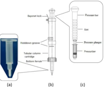
incorporated PS nanofibers were divided into four or five parts, and packed into the tip end of the solid-phase column. Then, the column was inserted into the handheld device as plotted in Figure 1. The SPE column could be inserted into the tubular column cartridge and fixed by holddown groove and bottom ferrule. Pushing the pressure bar, the sample solution could be pushed through the sorbent by the pressure of air forced by a gas tight plastic syringe (pressurizer). A steady flow speed could be maintained by adjusting bayonet lock and the slot on the pressure bar. Such design enabled a massive saving on the manual-work required for biological sample preparation, such as plasma, saliva and urine, which is usually volume-limited and highly viscous.
Recommend SPE procedure
The procedure recommended for the nanofiber solid phase extraction was as follow: plasma sample (500 µL)
was mixed with 60 µL (30%, m/v) of TCA solution by vortexing for 30 s, then centrifuged at 12,750 × g for 5 min. The supernatant was separated and adjusted to pH 6.0
with 50 µL of sodium hydroxide (0.3 mol L–1), before
injected through the nanofiber column by the handheld device carefully controlled in a drop wise manner (about
0.5 mL min–1). The column was preconditioned with 100 µL
of methanol and 200 µL of water. The target analyte retained on the PS-SDSn nanofibers was eluted with 100 µL of
methanol and 1 mol L–1 ammonium formate (1/1, v/v) into
the collecting tubes, then 20 µL of the eluent was injected into chromatographic system.
Conventional ion-pair solid-phase extraction (IP SPE)
PS nanofibers and the PFSPE columns were fabricated
as described in a previous paper.16 The PFSPE columns with
3 mg of PS nanofibers were activated by 100 µL of methanol
and 100 µL of water, followed with 100 µL of 0.05 mol L–1
SDSn solution. Meanwhile, 100 µL of 0.05 mol L–1 SDSn
solution was added to the 500 µL of prepared human plasma sample before loading to the preconditioned PFSPE columns. The flow rate was controlled in a drop wise
manner (about 0.5 mL min–1) and 5-HT was eluted with
100 µL of methanol.
Results and Discussion
Fabrication of SDSn incorporated PS nanofibers
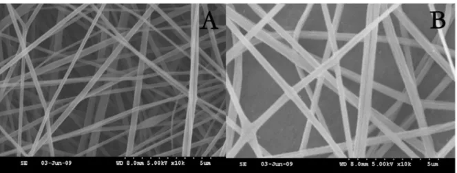
The SEM images of SDSn incorporated PS nanofibers and PS nanofibers are shown in Figure 2. The average fiber diameter has been calculated by use of free and open source ImageJ software based on the images of 20 nanofibers shown in Figure 2. The result was that the diameter of the PS nanofibers was 381 ± 94 nm (n = 20) and SDSn incorporated PS nanofibers was 232 ± 63 nm (n = 20). The pore diameter, pore volume, and surface area of PS nanofibers and SDSn incorporated PS nanofibers were
respectively 3.25 nm, 0.02 cm3 g−1, 9.65 m2 g−1 and 5.04 nm,
0.02 cm3 g−1, 19.52 m2 g−1.
The performance of IP reagents incorporated nanofibers as sorbents for SPE strongly relied on the composition of nanofibers. In this study, IP reagents of different hydrophilic groups, hydrocarbon length, and the concentration of them were investigated to elucidate interaction between 5-HT and the solid phase. The effect of nanofiber composition was investigated by using spiked plasma sample at the 5-HT
concentrations of 5, 50 and 200 ng mL–1.
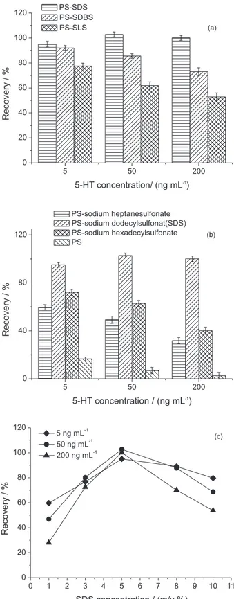
Three IP reagents (SDSn, SLS, and SDBS) with different hydrophilic group, i.e., sulfonic acid, benzene Figure 1. Schematic representation of handheld solid-phase extraction
sulfonic acid, and ester sulfate group were tested to seek optimal performance by adding an equal amount
(0.18 mol L–1) of them into the PS solution to prepare the
nanofibers. As shown in Figure 3a, the extract recoveries showed no difference among three nanofibers when the
spiked 5-HT concentration was 5 ng mL–1, but PS-SDSn
nanofibers performed better than the other two when the
spiked 5-HT concentration was 50 and 200 ng mL–1, thus,
SDSn was selected as the modifier to prepare IP reagents incorporated nanofibers by electrospinning.
Besides, the effect of hydrocarbon chain length of the sulfonate on the extraction was studied. Sodium heptane sulfonate, sodium dodecyl sulfonate, and sodium hexadecyl sulfonate were used to prepare nanofibers from the
electrospinning solution containing 0.18 mol L–1 of each
sulfonate. The extraction efficiency of 5-HT was tested and PS-SDSn nanofibers performed better than others, as presented in Figure 3b.
The effect of SDSn concentration on extraction efficiency was also investigated. The concentration of SDSn was tested form 1% to 10% (m/v). As seen in Figure 3c, the extraction efficiency of 5-HT increased when the percents (m/v) of SDSn was in the range of 1% to 5%, while in the range of 5% to 10%, the extraction efficiency decreased. Therefore, 5% was chosen as the optimum concentration of SDSn.
Optimization of the SPE procedure
In order to achieve high extraction efficiency for the target analyte, several important parameters, such as amount of adsorbent, eluent composition and its volume, pH and salt concentration in sample solution should be considered cautiously. A spiked plasma sample containing
50 ng mL–1 of 5-HT was used as model sample in the
optimized experiments.
Effect of packed nanofibers amount
The amount of nanofibers plays an important role in the solid-phase extraction process. It was found that extraction efficiency increased with the increasing fiber quantity up to 3.0 mg of packing quantity, and over 3.0 mg variations of 5-HT recoveries were not remarkable. Therefore, 3 mg was selected as the appropriate amount to achieve effective pretreatment.
Effect of eluent
The effect of eluent and its volume on extraction efficiency were tested for eluting 5-HT by using methanol and acetonitrile as solvent. It was found that methanol performed better than acetonitrile. This may be attributed to the difference in dielectric constant for methanol (33.7) and acetonitrile (38.8). Compared with acetonitrile, methanol has a lower dielectric constant; the molecules of this solvent exert a weaker ability to produce solvation to stabilize cations like ammonium ions in the eluent, which promotes the replacement of 5-HT by formation of ion-pair between ammonium and SDSn ions, and so methanol had the higher efficiency as the eluting solvent for 5-HT. To ensure maximum elution of 5-HT from the PFSPE columns, an eluent experiment was carried out by changing the volume of methanol from 50 to 200 µL. The results indicated that the recovery of 5-HT increased with the increasing of the methanol volume from 50 to 100 µL. Once above 100 µL, the recovery reached a plateau. Consequently, 100 µL of methanol was chosen as the eluent.
Effect of pH
degree of ionization of the 5-HT and the ability to form IP with counter ion(s). The effect of pH on the extraction of
5-HT was examined in the range of pH 3.0 to 10.0. The maximum extraction efficiency of 5-HT was achieved at pH 6.0, while the lowest was obtained at pH 3.0 or 10.0.
This was attributed to the alkaline nature of 5-HT (pKa 9.8)17
and the pka of alkyl sulfonates. When sample solution is at weak acidic pH, 5-HT can combine with protons to form cation, and it mainly exists in its unionized form or even in negative ion form at basic pH, which could not be attracted by conter ions. The pKa of alkyl sulfonates ranges in values between 2.0 and 3.0, depending on the substituents on the
alkyl group.18 Thus, at pH less than 3, SDSn exists mainly in
the neutral form. The IP complex between SDSn molecular and positive charged 5-HT is hardly formed. When the pH is higher than 3.0, SDSn hydrolyzes to form sulfonate, which interacts with 5-HT by electrostatic force. Thus, the pH of sample solution was adjusted to pH 6.0.
Effect of salt concentration
The effect of salt concentration on the extraction efficiency was tested with sodium chloride ranging from
0 to 500 mmol L–1. Results shown in Figure 4 clearly
suggested that the addition of sodium chloride decreased retention of 5-HT on PS-SDSn nanofibers. Generally, addition of NaCl to sample solution increases the ionic strength as well as changes the physical properties of Nernst
diffusion layer.19 It is well known that adding salt to solution
would decrease the solubility of organic compounds
in water phase,10 resulting in an increase in the amount
of the analytes extracted by the nanofibers. However, IP incorporated nanofibers adsorbing 5-HT primarily was based on electrostatic interaction, thus causing the competition mechanism. This means that ions with the same charge compete with the target analytes, thereby decreasing the extent of IP formation and the extraction
efficiency.14 The addition of NaCl also reduces mass
transfer by changing the Nernst diffusion layer, probably leading to the decreased rate of the formation of the ion
pairs,18 and therefore the decreased extraction efficiency.
Consequently, salt should be eliminated from the system.
Reusability of SDSn incorporated PS nanofibers
Commonly SPE cartridges are for single-use only. Considering the adsorbing site was introduced onto the fiber surface by incorporated electrospinning rather than chemically synthesis, the potential to reuse SDSn incorporated nanofibers should be investigated to indicate
the stability of adsorbing sites existed in the fibers. After per
circle of samples treatment as shown in recommend SPE procedure section, 100 µL of methanol were employed to
5 50 200
0 20 40 60 80 100 120
R
e
c
o
v
e
ry
/
%
5-HT concentration/ (ng mL-1
)
PS-SDS PS-SDBS
PS-SLS (a)
5 50 200
0 40 80 120
Recovery
/
%
5-HT concentration /(ng mL-1)
PS-sodium heptanesulfonate PS-sodium dodecylsulfonat(SDS) PS-sodium hexadecylsulfonate
PS (b)
0 1 2 3 4 5 6 7 8 9 10 11
0 20 40 60 80 100 120
R
e
c
o
v
e
ry
/
%
SDS concentration / (m/v %)
5 ng mL-1
50 ng mL-1
200 ng mL-1
(c)
wash the column followed by 200 µL of water to eliminate any possible residue of 5-HT. The column was then reused to investigate the recovery of 5-HT. The results are shown in Figure 5. It is clear that the variation of recoveries were not obvious for the former four times, however, the recoveries of 5-HT were reduced when the column was reused for the fifth time. Therefore, these adsorbing sites were stable enough to support multiple uses up to 4 times.
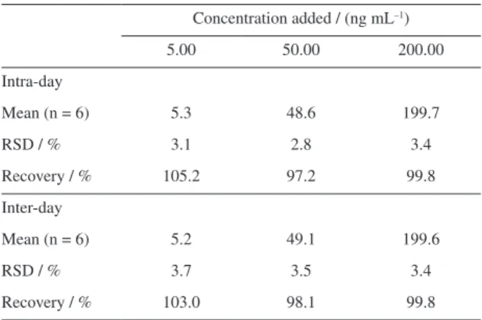
Reproducibility, linearity, and sensitivity
Quantitative analysis using nanofiber-packed SPE was evaluated by determination of 5-HT in spiked plasma
sample under optimized conditions. The calibration curves, prepared on six consecutive days, showed to be linear
in the concentration range of 0.50-200 ng mL–1 (with
r2 value of 0.999). The detection limit, calculated at a
signal-to-noise ratio of 3 (S/N = 3), was 0.5 ng mL–1. The
precision and accuracy of the method were determined by consecutively analyzing six replicates of spiked plasma at
the concentrations of 5, 50 and 200 ng mL–1 as shown in
Table 1. The average coefficients of variation (RSD / %) of intra-day and inter-day assays were found to be below
4%. The average recovery at 5, 50, 200 ng mL–1 for 5-HT
in spiked plasma samples was 105.2%, 97.2% and 99.8%, respectively (n = 6).
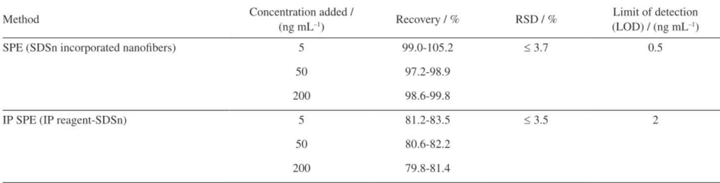
Comparison with conventional IP SPE
In this section, the comparative studies were performed between the recommended SPE procedure and conventional
IP SPE, in terms of extraction recovery,reproducibility and
sensitivity. The comparisons were investigated by using spiked plasma sample at 5-HT concentrations of 5, 50 and
200 ng mL–1. The results were summarized in Table 2.
Compared with IP SPE, SPE with SDSn incorporated nanofibers showed higher recoveries and low detection limit. Another advantage of the new method was the fact that no IP reagent was added into the sample, which would be preferable for mass spectrometry analysis.
Application
To evaluate the applicability of the developed method in real clinical samples (Figure 6), 12 human plasma samples from 6 Tourette children before and after treatment
were prepared and analyzed.19 Under the optimized
conditions, 500 µL of each sample was extracted by the SDSn incorporated nanofibers. The results revealed that
0 1 2,5 5 10 50 500
0 20 40 60 80 100 120
R
e
c
o
v
e
ry
/
%
Sodium chloride concentration / (mmol L-1
)
Figure 4. Effect of salt addition to the sample. Nanofibers: 3 mg; SDSn concentration: 5% (m/v); 5-HT concentration: 50 ng mL–1; sample solution volume: 500 µL (pH 6.0); sample flow rate: 0.5 mL min–1; eluent: methanol and 1 mol L–1 ammonium formate (1/1, v/v). Error bars illustrate the relative standard deviation from six replicates determinations.
5 50 200
0 20 40 60 80 100 120
Recovery
/
%
5-HT concentration / (ng mL-1)
First time Second time Third time Forth time Fifth time
Figure 5. Reusability of SDSn incorporated nanofibers. Nanofibers: 3 mg; SDSn concentration: 5% (m/v); sample solution volume: 500 µL (pH 6.0); sample flow rate: 0.5 mL min–1; eluent: methanol and 1 mol L–1 ammonium formate (1/1, v/v). Error bars indicate the relative standard deviation from six replicates determinations.
Table.1. The intra- and inter-day validation method for the determination of 5-HT in human plasma
Concentration added / (ng mL–1)
5.00 50.00 200.00
Intra-day
Mean (n = 6) 5.3 48.6 199.7
RSD / % 3.1 2.8 3.4
Recovery / % 105.2 97.2 99.8
Inter-day
Mean (n = 6) 5.2 49.1 199.6
RSD / % 3.7 3.5 3.4
the plasma concentration of 5-HT was decreased in most children after treatment (Figure 7).
Table 2. Comparison of PFSPE with IP SPE
Method Concentration added /
(ng mL–1) Recovery / % RSD / %
Limit of detection (LOD) / (ng mL–1)
SPE (SDSn incorporated nanofibers) 5 99.0-105.2 ≤ 3.7 0.5
50 97.2-98.9
200 98.6-99.8
IP SPE (IP reagent-SDSn) 5 81.2-83.5 ≤ 3.5 2
50 80.6-82.2
200 79.8-81.4
Figure 7. The change of concentration of 5-HT in Tourette children’s plasma.
Figure 6. HPLC-FLD chromatograms. (a) 200 ng mL–1 5-HT standard
solution; (b) separation of human plasma sample without nanofiber-packed solid phase extraction; (c) extraction of 500 µL of human plasma sample with 3 mg of SDSn incorporated nanofibers. The mobile phases were composed of 50 mmol L–1 phosphoric acid buffer solution (pH 4.5), 0.1 mmol L–1 EDTA and 5.8% (v/v) methanol, which was used at a flow rate of 1.0 mL min–1. The wavelengths of excitation and emission were 280 nm and 340 nm and injection volume was 20 µL. Other conditions were as stated in recommend SPE procedure.
Conclusion
A new functional SPE adsorbent based on IP reagent incorporated PS nanofibers was prepared and successfully applied to the extraction and preconcentration of 5-HT in human plasma samples. Several factors affecting the PFSPE procedure including the composition of nanofibers, eluent compostion and its volume, amount of adsorbent, pH and salt concentration were optimized systematically. The LOD, recovery, and precision of proposed method meet the requirements of 5-HT detection. Thus, an accurate, simple, and sensitive method based on HPLC-FLD was developed for the determination of 5-HT in human plasma, which can be used in physiological and psychological studies.
Acknowledgments
Nanjing Municipal Hospital of Chinese Medicine for kindly providing clinical plasma samples.
References
1. Zhao, X. L.; Li, J. D.; Shi, Y. L.; Cai, Y. Q.; Mou, S. F.; Jiang, G. B.; J. Chromatogr. A 2007, 1154, 52.
2. Alireza, V.; Samad, M. F.; IET Nanobiotechnol. 2014, 8, 83. 3. Yunpeng, H.; Yue-E, M.; Tianxi, L.; J. Appl. Polym. Sci.2014,
131, 40864.
4. Goonoo, N.; Bhaw-luximon, A.; Jhurry, D.; J. Biomed. Nanotechnol. 2014, 10, 2173.
5. Martina, A.; Sally, L. M.; Peter, K.; Macromol. Biosci. 2014,
14, 772.
6. Zewe, J. W.; Steach, J. K.; Olesik, S. V.; Anal. Chem. 2010, 82, 5341.
7. Bagheri, H.; Aghakhani, A.; Akbari, M.; Ayazi, Z.; Anal. Bioanal. Chem. 2011, 400, 3607.
8. Kang, X. J.; Chen, L. Q.; Zhang, Y. Y.; Liu, W.; Gu, Z. Z.; J. Sep. Sci. 2008, 31, 3272.
9. Chen, L. Q.; Kang, X. J.; Sun, J.; Deng, J. J.; Gu, Z. Z.; Lu, Z. H.; J. Sep. Sci. 2010, 33, 2369.
10. Liu, Z. Y.; Kang, X. J.; Fang, F.; Microchim. Acta 2010, 168, 59.
11. Qi, D. J.; Kang, X. J.; Chen, L. Q.; Zhang, Y. Y.; Wei, H. M.; Gu, Z. Z.; Anal. Bioanal. Chem. 2008, 390, 929.
12. Marce, R. M.; Borrull, F.; J. Chromatogr. A 2008, 885, 273. 13. Goyal, R. N.; Gupta, V. K.; Oyama, M.; Bachheti, N.; Talanta
2007, 72, 976.
14. Carson, M. C.; J. Chromatogr. A 2000, 885, 343.
15. Masanori, Y.; Yutaka, K.; Zenzo, T.; Takao, K.; Chem. Pharm. Bull. 1977, 25, 75.
16. Kang, X. J.; Pan, C.; Xu, X.; Yao, Y. F.; Wang, Y. Y.; Qi, D. J.; Gu, Z. Z.; Anal. Chim. Acta 2007, 587, 75.
17. Huang, Y. F.; Chiang, C. K.; Lin, Y. W.; Liu, K.; Hu, C. C.; Bair, M. J.; Chang, H. T.; Electrophoresis 2008, 29, 1942. 18. Jingming, W.; Hian, L. L.; Anal. Chem. 2006, 78, 7292. 19. Long, H. Y.; Zhang, J. C.; Chin. J. Integr. Tradit. West. Med.
(Chin. Ed.) 2012, 32, 926.
Submitted: July 22, 2014