Case Report
Sergi Bermúdez i Badia*, Ela Lewis and Scott Bleakley
Combining virtual reality and a myoelectric limb
orthosis to restore active movement after stroke:
a pilot study
Abstract: We introduce a novel rehabilitation technology
for upper limb rehabilitation after stroke that combines a
virtual reality (VR) training paradigm with a myoelectric
robotic limb orthosis. Our rehabilitation system is based
on clinical guidelines and is designed to recruit specific
motor networks to promote neuronal reorganization. The
main hypothesis is that the restoration of active
move-ment facilitates the full engagemove-ment of motor control
networks during motor training. By using a robotic limb
orthosis, we are able to restore active arm movement in
severely affected stroke patients. In a pilot evaluation, we
have successfully deployed and assessed our system with
three chronic stroke patients by means of behavioral data
and self-report questionnaires. The results show that our
system is able to restore up to 60% of the active movement
capability of patients. Further, we show that we can assess
the specific contribution of the biceps/triceps movement
of the paretic arm in a VR bilateral training task.
Question-naire data show enjoyment and acceptance of the
devel-oped rehabilitation system and its VR training task.
Keywords: electromyogram (EMG); motor rehabilitation;
personalization; robotic orthosis; stroke; virtual reality
(VR).
DOI 10.1515/ijdhd-2014-0333
Received April 8, 2013; accepted May 23, 2013; previously published online August 5, 2014
Introduction
Currently, stroke is one of the main causes of adult
dis-ability, and by 2030 it is expected to be one of the main
contributors to the burden of disease worldwide [1]. An
important goal in the management of stroke patients,
and in particular in patients with spasticity, involves
res-toration of normal limb position and ease of passive and
active movement execution with the aim of improving
functional outcomes such as the ability to carry out
activi-ties of daily living [2]. This is a very demanding task for
trained therapists and especially problematic in patients
with low level of motor control and yet aggravated in the
presence of spasticity. In fact, 85% of stroke survivors
will present a motor deficit contralateral to the location
of the brain lesion [3]. Additionally, 20%–40% will also
suffer from increased muscle tone or spasticity, which will
further limit their level of independence in the activities of
daily living [4, 5]. The large economical and psychological
impacts of stroke on our society, in particular on relatives
and public health systems, make it necessary to find
alter-native and novel approaches to address these issues.
Nowadays, it is well understood that recovery after
stroke depends on brain mechanisms that allow
undam-aged brain areas, such as contralateral or secondary
net-works, to take over the functions of the damaged areas
[6–8]. In the chronic stage of stroke, neuronal plasticity is
the main contributor to true recovery, being dependent on
the size, severity, and location of the lesion [9, 10].
There-fore, modern rehabilitation approaches should aim at
pro-viding an effective way of driving cortical plasticity and
recruiting alternative motor areas to achieve functional
brain reorganization, while being accessible to the widest
range of patients, in particular to those with the poorest
prognostic. During the intent to perform a motor action,
cortical areas devoted to motor control generate
particu-lar activity patterns – reflecting the synchronization and
desynchronization of neural activity – known as Sensory
Motor Rhythms [11]. These activity patterns encode motor
control signals that can reach the paretic arm as long as
there are remaining cortico-spinal tracts after stroke [12].
Control commands effectively transmitted to the limbs
can be assessed by measuring electric potentials at the
muscles (electromyogram, EMG). Depending on the brain
*Corresponding author: Sergi Bermúdez i Badia, PhD, Universidade da Madeira, Campus Universitário da Penteada, 9020-105 Funchal, Portugal, E-mail: [email protected]
Ela Lewis: Myomo Inc., One Broadway, Cambridge, MA 02142, USA Scott Bleakley: Department of Occupational Therapy, University of Pittsburgh, Pittsburgh, PA, USA
lesion, the amount of motor control and, therefore, of
active movement is compromised.
To overcome this limitation, we propose a hybrid
virtual reality (VR) and robotic approach for the restoration
of correct limb pose and active movement. The objective of
our hybrid system is to restore motor control of the upper
limbs when active movement is compromised but weak
EMG responses are still present. Our technology is able to
detect residual EMG activation and, by means of a robotic
orthosis, enable motor-impaired patients to exercise
movement even when active movement is severely
com-promised. There are data that suggest that the restoration
of active movement may play a crucial role in mobilizing
cortical plasticity, and, therefore, in accelerating recovery
after stroke. First, although passive movement exercising
is able to engage motor networks by means of
propriocep-tive feedback [13], it has been shown not to be the most
effective way of engaging overt execution motor areas [14].
Second, the activation of motor-related networks does not
only depend on the action intent, but also on the type of
actions and their completion. It has been shown that both
the observation and performance of meaningful
goal-oriented actions can engage additional networks such as
the mirror neuron system (MNS), which is also known as
the action recognition system [15–17]. The discovery of the
MNS has allowed the emergence of novel stroke
rehabilita-tion approaches based on clear neuroscientific hypotheses
on brain recovery mechanisms [18–22]. In this project, we
propose restoration of active movement as a crucial step
to fully engage both the motor control networks and the
MNS. Therefore, by restoring active movement and
engag-ing patients in physical trainengag-ing with meanengag-ingful
goal-ori-ented actions, our hybrid VR robotic system is designed to
facilitate true recovery by means of cortical plasticity.
Previous myoelectric driven robotic interventions [23]
have been shown to lead to improved Fugl-Meyer scores
of the upper extremities [24] and reduced spasticity as
assessed by the modified Ashworth score [25]. Many control
techniques have been explored for myoelectric driven
movement assistance such as fuzzy controllers [26] or
compliant systems [27, 28]. In this project, we use a unique
wearable and portable orthosis with integrated myoelectric
measurement capabilities that restore correct limb position
(mPower1000, Myomo Inc., Cambridge, MA, USA). Further,
we believe the combination of the myoelectric orthosis
approach with a VR training paradigm is appropriate
because of the inherent properties of VR systems for motor
rehabilitation. The VR approaches allow for a combination
of features including: low cost; personalization of
train-ing; unsupervised traintrain-ing; goal-oriented actions;
adapt-ability to a broad range of patients; quantifiable outcome
measures; extended feedback; and motivation thanks to
the use of game elements [29]. Our VR training environment
builds on previous work [30, 31], on training principles
that we have shown to be effective in the chronic phase of
stroke [32], and on accelerating recovery in the acute phase
of stroke [33]. Thus, our hybrid system exploits a
state-of-the-art information and communication technologies, a
myoelectric robotic approach, and a neuroscience-based
rationale to provide a novel personalized rehabilitation
training system that addresses the physical sequels and
social impact of stroke. The approach presented here puts
special emphasis on patients without (or with minimal)
active movement capabilities and those with spasticity,
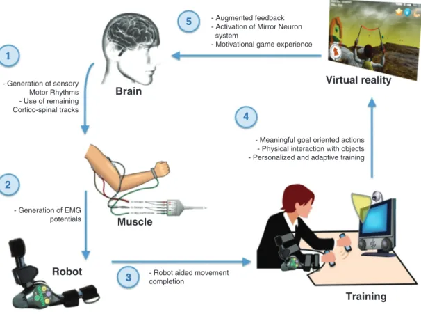
enabling them to train active movement (see Figure 1).
Methods
In our approach, we take advantage of the use of VR because it can support requirements for an effective training. VR allows creating fully controlled environments that define training tasks specifically designed to target the individual needs of patients. Additionally, intensive movement training can be supported through motivating tasks that use augmented feedback and reward [29]. VR allows not only for the individualization of training and monitoring by physi-cians, but also enables patients to play a more active role in their rehabilitation process and self-monitor their own improvements. Besides, our VR-based rehabilitation system has been integrated into a game-like interaction, capitalizing in motivational factors that are essential for recovery [34]. Nevertheless, the main novelty of our approach is the combination of an online adaptation during VR train-ing of the level of assistance provided by a robotic limb orthosis with EMG measurement capability (Figure 1). This technology is designed to restore active movement, compensate for fatigue, and optimize training duration, intensity and repetition.
Limb orthosis
The mPower 1000 robotic device (Myomo Inc., Cambridge, MA, USA) is a portable limb orthosis that is controlled through EMG signals that are measured by an on-board data sampler. Two EMG channels and one actuated joint are used to restore active movement based on biceps/triceps EMG activation or relaxation (see Figure 1, points 2 and 3). The mPower assists its user in the completion of arm move-ments by means of an embedded electric motor that is activated on the detection of biceps and/or triceps EMG activity. The EMG signals are compared to the baseline EMG activity level of the user and an assistive force is executed (either arm extension or contraction) when EMG changes (muscle contraction or relaxation) are detected. This approach makes therapy accessible to patients with residual EMG activation or involuntary and permanent EMG activation, correcting limb position and allowing them to train active contraction/relaxa-tion to gain movement control. The mPower connects to the virtual environment through a virtual serial port over Bluetooth, allowing its remote control from within the training environment. This wire-less connection provides information on the orthosis settings, arm
- Generation of sensory Motor Rhythms - Use of remaining Cortico-spinal tracks
1
5
4
2
3
- Generation of EMG potentials- Robot aided movement completion
- Augmented feedback - Activation of Mirror Neuron system
- Motivational game experience
- Meaningful goal oriented actions - Physical interaction with objects - Personalized and adaptive training
Brain
Muscle
Robot
Training
Virtual reality
Figure 1 Diagram of the developed virtual reality and robotic limb orthosis training paradigm showing the role of each technological com-ponent (numbered from 1 to 5).
position, and EMG readings, and it allows remote adjustment of the level of motor assistance during training from 0% to 100%.
Tracking
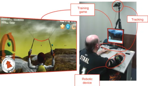
The tracking technology used in this project is the ARToolKit (ARTool-works, Inc., Seattle, WA, USA). The ARToolKit is an augmented reality software toolkit that enables tracking the position (x, y, z) and ori-entation in space of predefined unique markers by using a webcam as an input device. In our system, the ARToolKit was used to track two handles (7 cm diameter × 12 cm high) tagged with unique visual markers. Thus, an overhead webcam is used to track the position and orientation of the markers, providing the virtual environment with precise information about the position and movement trajectories of the user’s hands during training. Users of the system are instructed to grasp and move these handles around a table top in order to interact with the virtual environment (see Figure 2, right panel).
Virtual environment
The virtual environment and training task are based on the Neu-rorehabilitation Training Toolkit (NTT), which is freely accessible at http://neurorehabilitation.m-iti.org/NTT. The NTT is a virtual training environment developed with the open-source game engine Panda3D (www.panda.org) that was designed following neurosci-entific and therapeutic guidelines for stroke rehabilitation, such as
relevance of training to ADLs, neuronal mechanisms of recovery, nar-rative, personalization or individualization, augmented feedback, and engagement (See [30] for a detailed description of the training rational). In essence, the training task is a game experience consisting of a bimanual coordination task that uses upper limb motor actions as control signals. Bimanual upper limb training was selected because it has been shown to enhance excitability of cortical motor networks and lead to improved functional outcomes [35, 36]. The bimanual motor actions are mapped onto the actions of an avatar that controls a glider in the virtual environment, i.e., the physical arm movements of the user are used to control the steering direction of a virtual glider (see Figure 2, left panel). Feedback on performance and on-screen information is extensively used to inform the user on the immediate game goals and motor actions to be performed, as well as a reward mechanism is used. The goal of the game is to gather the largest possi-ble number of collectapossi-ble items in the virtual environment. Two types of collectable objects are present – easy (balloons) and difficult (stars) – that are accumulated to an on-screen score to provide feedback on performance. In addition, the amount of arm movement measured by the limb orthosis is also provided as a visual score. All tracking and training data are logged as a text file for later analysis.
Pilot evaluation
The objective of this pilot study was to assess the acceptance and usability of the system, as well as the impact of the level of assistance of the limb orthosis on task performance and overall arm movement.
Training game
Tracking
Robotic device
Figure 2 Prototype of the myoelectric-based interactive system for rehabilitation.
Left panel: an adaptive training in the form of a game defines the training parameters for a bimanual coordination motor task. The training offers augmented feedback on performance, sustains motivation, and automatically modifies the level of motor assistance offered by the limb orthosis. Right panel: the different components of the system (robotic device, tracking setup, and training game task) while being used by a stroke patient.
We evaluated the system with 3 chronic stroke survivors (47–63 years old; > 6 months poststroke) in a laboratory setting at the University of Pittsburgh (see Table 1). All subjects had a very low level of control of their paretic arm but were able to generate voluntary EMG activa-tion, and hence able to drive the robotic orthosis. All subjects used the robotic orthosis in biceps mode (only controlled by biceps EMG activ-ity) and used the system for a single training session of approximately 20 min. During the training session, the level of assistance of the ortho-sis was randomly changed between 40% and 90% every time a virtual item was collected. After the training session, subjects were asked to report on their experience by answering a questionnaire about enjoy-ment, engageenjoy-ment, and usability rated using a Likert scale from 1 to 5. All subjects gave their informed consent to participate in this study.
Results
This is a unique system that not only engages users in a
game-like VR training experience, but also makes use of a
myoelectric-capable orthosis to restore active movement.
However, the effectiveness of the orthosis assistance in
Table 1 Patients’ demographics.
Age, years 47 63 50
Sex Male Male Male
Stroke type Hemorrhagic Ischemic Ischemic Stroke
location Left Left Right
Handedness Right Right Right
movement restoration and how to optimally integrate it
in an interactive training experience need to be studied
before any longitudinal deployment. For these reasons, we
performed a number of experiments in which we exposed
three stroke survivors to single training sessions of the
combined VR and myoelectric limb orthosis paradigm. The
integrated system allows us to simultaneously measure
both the movement of the arm end effector (tracked by a
marker on the handle) and the specific movement of the
biceps as measured by the orthosis (see Figure 2, right
panel). Training data were recorded synchronously with
tracking data as well as the limb orthosis settings.
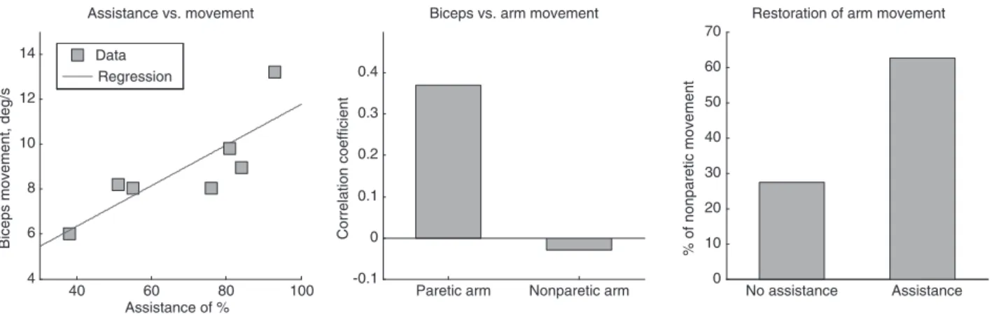
The analysis of the multimodal data revealed a linear
effect of the level of assistance of the limb orthosis with the
amount of biceps movement as measured by the system
in deg/s (see Figure 3, left panel). The existence of this
linear relationship between the level of assistance and
movement execution is an important feature of the system
because it will enable us to integrate, into the VR training
environment itself, straight forward statistical modeling
techniques to automatically adjust the level of assistance
depending on the characteristics of each user. This will
allow us to improve the level of motor control or
compen-sate for fatigue or loss of force during training. Moreover,
the system provides us with additional valuable data,
allowing us to quantify the particular contribution of the
biceps/triceps movement to overall movement (Figure 3,
middle panel). In our experiment we could assess that
the movement of the elbow joint (measured by the limb
orthosis) showed a low correlation coefficient with that of
the end effector (r = 0.37). This reveals a low contribution of
the elbow joint, and, therefore, a low biceps/triceps
contri-bution, to the bimanual control task defined in our
train-ing. This correlation indicates that possible compensatory
strategies were used during training. Further, our VR
train-ing system allows us to assess and compare differences
between paretic and nonparetic arms. This enables us to
monitor recovery over time using the nonparetic arm as
reference. Of particular interest is the comparison of
move-ment capability of the paretic and nonparetic arms when
the orthosis assistance is enabled. During our pilot
experi-ment we have quantified the impact of the active orthosis
on the overall movement of the arm and we verified that
the myoelectric orthosis was able to restore the paretic arm
movement to about 60% of the nonparetic arm in patient 3.
These results are yet more remarkable when compared to
the absence of assistance, in which case the overall
move-ment of the paretic arm was below 30% of that of the
non-paretic one (see Figure 3, right panel).
Questionnaire data revealed a good acceptance of the
system, the most positive aspects being: fun [4.3],
enter-taining [4], and willingness to use it as regular motor
training [4.6]. Subjects reported that the system was easy
to understand [3.6] but also considered it as a challenging
training task [1.6].
Discussion
Here we presented a novel hybrid system that integrates
VR training and a myoelectric limb orthosis. This system
is an extension of the NTT that aims at restoring arm
movement in severely affected stroke patients by
integrat-ing an EMG-driven portable robotic limb orthosis. This
system is specifically designed to be used by patients
without or with minimal active movement capabilities,
i.e., those with the poorest prognostic, enabling them to
train active movement. The robotic orthosis is combined
with a gaming training environment that is based on
clinical guidelines and designed to recruit the MNS and to
promote neuronal reorganization. By using a robotic limb
orthosis, we are able to restore active arm movement in
severely affected stroke patients. We hypothesize that the
restoration of active movement facilitates the full
engage-ment of motor control networks during motor training.
In this first pilot experiment, we have successfully
deployed and tested our biohybrid VR-interactive
reha-bilitation system with three chronic stroke patients. The
system was evaluated by means of quantitative behavioral
data – acquired by the system itself – and self-report
ques-tionnaires. Initial results show that our system is capable
of online adjusting the assistance level provided by the
orthosis, and that the level of assistance has a linear effect
on the overall arm movement. We showed that the
myo-electric orthosis is able to restore up to 60% of the active
movement capability. Although encouraging, these results
require further investigation to better understand how
the level of assistance relates to improvements in motor
control and also to the overall recovery process of the
paretic arm in a longitudinal intervention. Another of the
strengths of the presented approach is that our technology
allows assessing the individual contribution of the biceps/
triceps movement to the overall bilateral training task. We
40 60 80 100 4 6 8 10 12 14 Assistance of %
Biceps movement, deg/s
Assistance vs. movement No assistance Assistance 0 10 20 30 40 50 60 70
Restoration of arm movement
% of nonparetic movement
Paretic arm Nonparetic arm -0.1 0 0.1 0.2 0.3 0.4
Biceps vs. arm movement
Correlation coefficient
Data Regression
Figure 3 Effect of the myoelectric limb orthosis during the virtual training task.
Left panel: effect of the level of assistance of the limb orthosis on the amount of biceps movement. Middle panel: quantification of the con-tribution of the biceps movement to the overall arm movement, computed as the correlation value of the biceps and arm movements during training. Right panel: restoration of arm movement. Percentage of arm movement of the paretic arm as compared to the nonparetic arm in presence and absence of robotic assistance. Example data from patient 3.
have shown that we can objectively assess and monitor
the active contribution of the elbow joint to overall arm
movement as well as detect and quantify the presence of
compensatory strategies. Questionnaire data reveal a high
level of acceptance of the system and its VR training task,
although it was found to be challenging. This is an
expect-able result because in this experimental protocol patients
with no or very low active movement were exposed to
varying levels of orthosis assistance, including low levels
of assistance. This effect will be minimized in the future
by using an algorithmic solution to automatically adjust
the level of assistance to each patient, thus maximizing
the outcome of training. The long-term impact of these
technologies will be assessed in a randomized controlled
trial in the inpatient rehabilitation unit of the Hospital of
Funchal.
Acknowledgments: The authors would like to thank Prof.
Daniel P. Siewiorek (Carnegie Mellon University) and Steve
Kelly (Myomo Inc.) for their support and contribution to
this project. Support for this research was provided by the
Fundação para a Ciência e Tecnologia (Portuguese
Foun-dation for Science and Technology) through the Carnegie
Mellon Portugal Program under grant CMU-Pt/0004/2007,
and through the EC FP7 program under grant 303891
RehabNet FP7-PEOPLE-2011-CIG.
References
1. WHO. The global burden of disease: 2004 update. Geneva: World Health Organization, 2008.
2. Esquenazi A. Falls and fractures in older post-stroke patients with spasticity: consequences and drug treatment considerations. Clin Geriatrics 2004;12:27–35.
3. Lai SM, Studenski S, Duncan PW, Perera S. Persisting conse-quences of stroke measured by the Stroke Impact Scale. Stroke 2002;33:1840–4.
4. Watkins CL, Leathley MJ, Gregson JM, Moore AP, Smith TL, Sharma AK. Prevalence of spasticity post stroke. Clin Rehabil 2002;16:515–22.
5. Sommerfeld DK, Eek EU, Svensson AK, Holmqvist LW, von Arbin MH. Spasticity after stroke: its occurrence and association with motor impairments and activity limitations. Stroke 2004;35:134–9. 6. Seitz RJ, Butefisch CM, Kleiser R, Homberg V. Reorganisation of
cerebral circuits in human ischemic brain disease. Restor Neurol Neurosci 2004;22:207–29.
7. Sabatini U, Toni D, Pantano P, Brughitta G, Padovani A, Bozzao L, et al. Motor recovery after early brain damage. A case of brain plasticity. Stroke 1994;25:514–7.
8. Dobkin BH. Training and exercise to drive poststroke recovery. Nat Clin Pract Neurol 2008;4:76–85.
9. Nudo RJ. Postinfarct cortical plasticity and behavioral recovery. Stroke 2007;38:840–5.
10. Murphy TH, Corbett D. Plasticity during stroke recovery: from synapse to behaviour. Nat Rev Neurosci 2009;10:861–72. 11. Hatsopoulos N. Rhythms in motor processing: functional
implications for motor behavior. Short course organized by the Society for Neuroscience “Rhythms of the neocortex: Where do they come from and what are they good for. Washington, DC: Society for Neuroscience, 2009.
12. Butefisch CM, Kleiser R, Seitz RJ. Post-lesional cerebral reorgan-isation: evidence from functional neuroimaging and transcranial magnetic stimulation. J Physiol Paris 2006;99:437–54.
13. Carel C, Loubinoux I, Boulanouar K, Manelfe C, Rascol O, Celsis P, et al. Neural substrate for the effects of passive training on sensorimotor cortical representation and colon; a study with functional magnetic resonance imaging in healthy subjects. J Cerebral Blood Flow Metabol 2000;20:478–84. 14. Szameitat AJ, Shen S, Conforto A, Sterr A. Cortical activation
during executed, imagined, observed, and passive wrist move-ments in healthy volunteers and stroke patients. Neuroimage 2012;62:266–80.
15. Gallese V, Fadiga L, Fogassi L, Rizzolatti G. Action recognition in the premotor cortex. Brain 1996;119:593–609.
16. Keysers C, Kohler E, Umilta MA, Nanetti L, Fogassi L, Gallese V. Audiovisual mirror neurons and action recognition. Exp Brain Res 2003;153:628–36.
17. Kohler E, Keysers C, Umilta MA, Fogassi L, Gallese V, Rizzolatti G. Hearing sounds, understanding actions: action representation in mirror neurons. Science 2002;297:846–8. 18. Altschuler EL, Wisdom SB, Stone L, Foster C, Galasko D,
Llewellyn DM, et al. Rehabilitation of hemiparesis after stroke with a mirror. Lancet 1999;353:2035–6.
19. Ertelt D, Small S, Solodkin A, Dettmers C, McNamara A, Binkofski F, et al. Action observation has a positive impact on rehabilitation of motor deficits after stroke. Neuroimage 2007;36:T164–73.
20. Merians AS, Tunik E, Fluet GG, Qiu Q, Adamovich SV. Innovative approaches to the rehabilitation of upper extremity hemi-paresis using virtual environments. Eur J Phys Rehabil Med 2009;45:123–33.
21. Michielsen ME, Selles RW, van der Geest JN, Eckhardt M, Yavuzer G, Stam HJ, et al. Motor recovery and cortical reorgani-zation after mirror therapy in chronic stroke patients: a phase II randomized controlled trial. Neurorehabil Neural Repair 2011;25:223–33.
22. Rizzolatti G, Fabbri-Destro M, Cattaneo L. Mirror neurons and their clinical relevance. Nat Clin Pract Neurol 2009;5:24–34. 23. Hu XL, Tong K, Song R, Zheng XJ, Leung WW. A comparison
between electromyography-driven robot and passive motion device on wrist rehabilitation for chronic stroke. Neurorehabil Neural Repair 2009;23:837.
24. Fugl-Meyer AR, Jaasko L, Leyman I, Olsson S, Steglind S. The post-stroke hemiplegic patient. 1. a method for evaluation of physical performance. Scand J Rehabil Med 1975;7:13–31. 25. Bohannon RW, Smith MB. Interrater reliability of a modified
Ashworth scale of muscle spasticity. Phys Ther 1987;67:206–7. 26. Kiguchi K, Tanaka T, Fukuda T. Neuro-fuzzy control of a robotic
exoskeleton with EMG signals. Fuzzy Syst, IEEE Trans on 2004;12:481–90.
27. Tsagarakis NG, Caldwell DG. Development and control of a ‘soft-actuated’ exoskeleton for use in physiotherapy and training. Autonomous Robots 2003;15:21–33.
28. Andreasen DS, Alien SK, Backus DA. Exoskeleton with EMG based active assistance for rehabilitation. 9th International Conference on Rehabilitation Robotics, ICORR, IEEE, 2005. 29. Lucca LF. Virtual reality and motor rehabilitation of the upper
limb after stroke: a generation of progress? J Rehabil Med 2009;41:1003–100.
30. Bermúdez i Badia S, Cameirão MS. The Neurorehabilitation Training Toolkit (NTT): a novel worldwide accessible motor train-ing approach for at-home rehabilitation after stroke. Stroke Res Treat 2012;2012:802157.
31. Cameirão MS, Bermúdez i Badia S, Duarte E, Verschure P. Neurorehabilitation using the virtual reality based rehabilitation gaming system: methodology, design, psychometrics, usability and validation. J Neuroeng Rehabil 2010;7:48.
32. Cameirão MS, Bermúdez i Badia S, Duarte E, Frisoli A, Verschure PF. The combined impact of virtual reality neurorehabilitation
and its interfaces on upper extremity functional recovery in patients with chronic stroke. Stroke 2012;43:2720–8.
33. Cameirão MS, Bermúdez i Badia S, Duarte E, Verschure P. Virtual reality based rehabilitation speeds up functional recovery of the upper extremities after stroke: a randomized controlled pilot study in the acute phase of stroke using the Rehabilitation Gam-ing System. Restor Neurol Neurosci 2011;29:287–98.
34. Maclean N, Pound P, Wolfe C, Rudd A. Qualitative analysis of stroke patients’ motivation for rehabilitation. Br Med J 2000;321:1051–4.
35. Stoykov ME, Stinear JW. Active-passive bilateral therapy as a prim-ing mechanism for individuals in the subacute phase of post-stroke recovery: a feasibility study. Am J Phys Med Rehabil 2010;89:873. 36. Byblow WD, Stinear CM, Smith M-C, Bjerre L, Flaskager BK,
McCambridge AB. Mirror symmetric bimanual movement prim-ing can increase corticomotor excitability and enhance motor learning. PLoS One 2012;7:e33882.