DIAGNOSIS OF THE PULMONARY TUBERCULOSIS BY POLYMERASE CHAIN REACTION: A COMPARATIVE
STUDY BETWEEN HIV-POSITIVE AND -NEGATIVE INDIVIDUALS
Rosemeri Maurici da Silva1, Tairone Machado2, Maria Luiza Bazzo3
1
Programa de Mestrado em Ciências da Saúde, Universidade do Sul de Santa Catarina, Tubarão, SC, Brasil; 2Universidade do Sul de Santa Catarina, Tubarão, SC, Brasil; 3Departamento de Análises Clínicas, Universidade Federal de Santa Catarina,
Florianópolis, SC, Brasil.
Submitted: December 12, 2010; Approved: January 16, 2012.
ABSTRACT
This study was performed to assess the efficiency of polymerase chain reaction (PCR) directly from sputum
for the diagnosis of pulmonary tuberculosis by comparison between HIV-positive and HIV-negative
individuals. Sputum samples were collected from hospitalized patients admitted with a clinical diagnosis of
pulmonary tuberculosis, and subjected to smear microscopy, culture on LJ medium and detection of M.
tuberculosis by PCR. Sensitivity, specificity, and predictive values (positive and negative) were calculated
using smear and/or culture at day 42 as the gold standard, by comparing the yield in positive and
HIV-negative individuals. Regardless of serostatus, the technique’s yield had 62% sensitivity, 70% specificity,
79% positive predictive value, 50% negative predictive value, and 65% accuracy. HIV-negative had 64%
sensitivity, 74% specificity, 75% positive predictive value, 63% negative predictive value, and 68%
accuracy. HIV-positive had 59% sensitivity, 33% specificity, 87% positive predictive value, 10% negative
predictive value, and 56% accuracy. The PCR showed a higher yield in HIV-negative individuals compared
to HIV-positive individuals.
Key words: Tuberculosis. HIV. PCR. Sputum.
The special program of the World Health Organization for
research and training in tropical diseases expressed concern
regarding the diagnosis and treatment of tuberculosis in
developing countries, arguing that the primary impediment to
controlling the disease lies on inadequate case detection. The
pandemic of HIV, immunosuppression linked or not linked to
this virus, and the worldwide increase of TB cases with M.
tuberculosis drug resistant stress the need for better diagnostic
tools. Although the initial diagnosis of mycobacterial disease is
based on clinical data, the definitive diagnosis depends on
laboratory isolation and identification of the microorganism
(19).
Early diagnosis has a crucial role in TB control. However,
the bacilloscopy has a low sensitivity in paucibacillary clinical
samples and the culture in Löwenstein-Jensen medium is slow;
besides, the laboratory results may take several weeks (12, 13).
The reversal of this scenario will require the development
of new strategies to increase the quality and speed of TB
diagnosing. The estimate for the next 20 years is that the
increase in case detection will reduce the incidence by 41% and
new treatment regimens will control the disease and reduce its
transmission by up to 59%. The combination of new diagnostic
methods and new drugs may result in a decreased incidence of
around 76% during this same period of time (22).
Detection of mycobacterial DNA directly from sputum by
amplification of the 16S rDNA gene allows the rapid
identification of species (20). However, the amplification of
this gene in sputum has proven challenging because it presents
sensitivity values below those desired for diagnosis (2, 5, 7).
This study was conducted to assess the yield of PCR
directly from sputum, comparing the yielding capacity between
HIV-positive and HIV-negative individuals.
Sputum samples were obtained from in-patients with a
clinical diagnosis of TB, with a maximum of two days of
treatment, admitted to a TB reference hospital, from January to
November 2009, and processed within two hours after
collection. Each sample was homogenized and separated into
three parts: one for sputum smear microscopy according to
Ziehl-Nielsen staining, one for DNA extraction and subsequent
PCR detection, and the third part for the decontamination
procedure by the Petroff method and culturing in
Löwenstein-Jensen solid medium. Smear preparation, Ziehl-Nielsen
staining and slide reading followed the recommendations
outlined in the Manual of Tuberculosis Bacteriology (11).
DNA extraction from sputum was performed by alkaline
lysis; all reagents had molecular biology grade purchased from
Invitrogen® (Carlsbad, CA, USA) (17): sputum was resuspended in GET (50 mM glucose, 25 mM Tris-HCl, pH 8.0
and 10 mM of EDTA), followed by cell lysis solution 1% SDS,
0.2 M NaOH. The pH was neutralized with a solution of 3M
potassium acetate, pH 4.8 to 5.0. Then, the sample was treated
with proteinase K 20mg/ml. The extraction was performed
with phenol/chloroform/isoamyl alcohol (25:24:1) and the
DNA was precipitated in ethanol in the presence of salt and
resuspended in 20 µl of TE (10 mM Tris pH 7.4, 1 mM
EDTA). Quality control of the DNA extracted and verification
of inhibitors in PCR reaction were made with primers ZR-244
and F-285 that amplify a 350-bp fragment of 16S rRNA
conserved for eubacteria (16).Detection primers were obtained
from the rDNA sequence corresponding to nucleotides of the
16S rRNA gene (7). Antisense primer MYC-264 (4) nucleotide
1638 to 1657 (3'TGCACACA GGCCACAAGGGA-5') and
sense primer F-285 nucleotides 631 to 648 (5'-AGAGTTTG
ATCCTGGCTCAG -3') amplified a fragment of 1027 bp. The
PCR reaction was performed in a volume of 50 µl containing
dimethyl sulfoxide (DMSO) under the following conditions:
1.5 mM MgCl2, 1% DMSO, 0.8 mM dNTP (dATP, dCTP,
dGTP, dTTP), 10 ρmoles of each primer, 1X Taq polymerase
buffer and 1.25 U recombinant Taq polymerase (Invitrogen® Carlsbad, CA, USA) and 1 µl of DNA template. Amplification
condition used was 94°C for 1 minute, 60°C for 1 minute, 72°C
for 1 minute in 35 cycles and a final cycle of 72°C for 10
minutes. PCR was performed in a PCR thermocycler
Eppendorf® brand, Mastercycler Personal model. The electrophoretic separation of DNA extracted and PCR products
was performed with the application of 5 µl of these materials
with the addition of 1 µl of 6X sample buffer (30% glycerol,
0.25% Bromophenol blue, 0.25% xylene cyanol and 10% 10X
TAE - 40 mM Tris-acetate/1 mM EDTA) in 1% (w/v) agarose
gel, in 1X TAE buffer at 200V for ten minutes. Visualization
was achieved by ethidium bromide staining (1 µg/ml). Gels
were photographed under ultraviolet light at 320ηm
(HOEFER-MacroVue UV-20), using a gel
photo-documentation system (DOC-PRINT® Biosystems). The size of fragments was estimated by comparison with the molecular
size marker of 50pb (Invitrogen® Carlsbad, CA, USA). The pattern sample used as positive control of PCR amplification
was DNA extracted from the BCG vaccine which contains
attenuated bacillus Calmette-Guérin.
the Petroff technique (11). The cultures that showed no
bacterial growth up to 42 days were considered negative.
The PCR assay results were interpreted without
knowledge of the results of the reference standard. PCR
reactions were repeated if a false-negative result occurred.
The SPSS 16.0® software was used to calculate sensitivity, specificity, predictive values (positive and negative), taking the
culture on the 42nd day as the gold standard.
The research project was submitted to and approved by
the Ethics Committee in Human Research (CEPSH) of the
University of Southern Santa Catarina.
Eighty-five patients were consecutively evaluated; mean
age was 43.6 years (SD±11.7); 69 (81.2%) were male. With
regard to ethnicity, 71 (83.5%) were Caucasians. Of the
participants, 60 (70.6%) were HIV-negative and 25 (29.4%)
were HIV-positive.
Bacilloscopy and/or culture was positive in 55 (64.7%) of
the samples and negative in 30 (35.3%). PCR was positive in
43 (50.6%) of the samples and negative in 42 (49.4%).
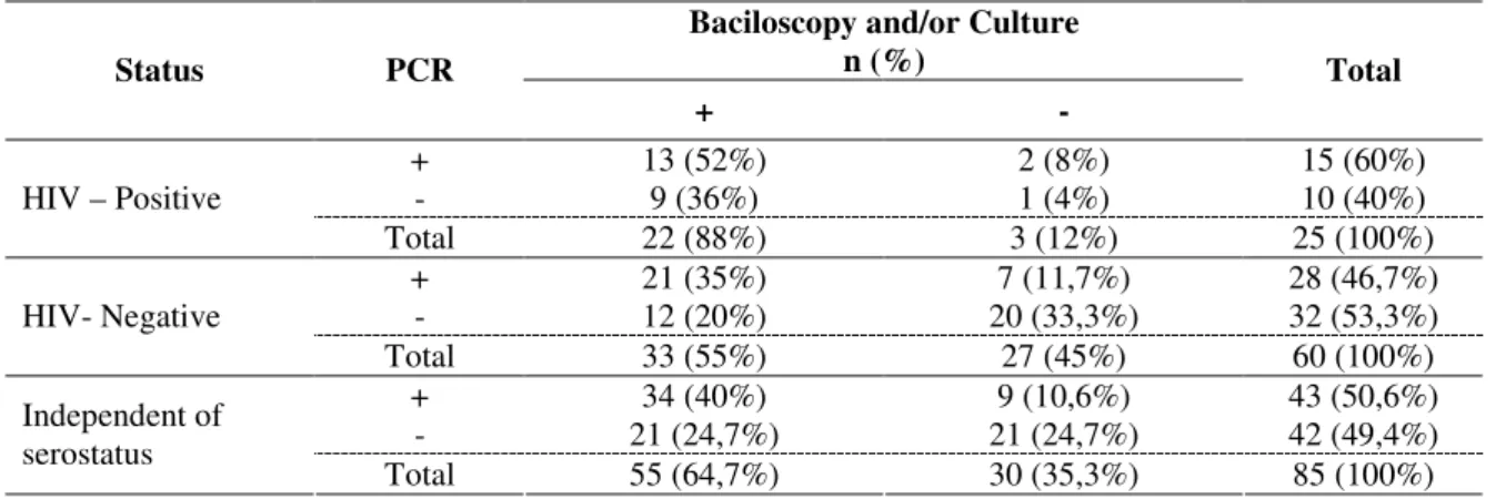
Table 1 shows the distribution of participants according to
the bacilloscopy and/or culture, PCR and HIV seropositivity.
Table 2 shows the yield of PCR using the result of
bacilloscopy and/or culture as the golden standard.
Table 1. Distribution of the participants according to the result of bacilloscopy and/or culture, PCR and HIV seropositivity.
Baciloscopy and/or Culture n (%)
Status PCR
+ -
Total
+ 13 (52%) 2 (8%) 15 (60%)
- 9 (36%) 1 (4%) 10 (40%)
HIV – Positive
Total 22 (88%) 3 (12%) 25 (100%)
+ 21 (35%) 7 (11,7%) 28 (46,7%)
- 12 (20%) 20 (33,3%) 32 (53,3%)
HIV- Negative
Total 33 (55%) 27 (45%) 60 (100%)
+ 34 (40%) 9 (10,6%) 43 (50,6%)
- 21 (24,7%) 21 (24,7%) 42 (49,4%)
Independent of serostatus
Total 55 (64,7%) 30 (35,3%) 85 (100%)
Table 2. PCR yield compared to the golden standard.
Statistical measures (CI 95%) Status
Sensitivity Specificity PPV PPN Accuracy
HIV – Positive 59
(39-80)
33 (-20-86)
87 (69-100)
10 (-8-25)
56 (37-75)
HIV- Negative 64
(47-80)
74 (57-90)
75 (59-91)
63 (46-80)
68 (57-80) Independent of
serostatus
62 (49-74)
70 (54-86)
79 (67-91)
50 (35-65)
65 (55-75)
Studies show that from colonies grown on LJ, PCR has
100% sensitivity in the amplification of the 16S rDNA gene,
while sputum sensitivity values range between 22% and
72.4%; these values are always associated with the quality of
sputum (2, 3). DNA extracted directly from sputum contains
bacteria of the upper respiratory tract and mouth, fungi,
leukocytes and other cells. However, a greater sensitivity of
PCR through the culture could not explain the discrepancy in
yield, whose values of specificity between HIV positive and
The intensity of amplification is different for each sample
and seems not to be related to the number of bacilli found in
bacilloscopy. Ievens and Goossens (8), in a meta-analysis
article, stated that some authors ascribe the different results in
different methodologies when using the same sample by
unequal distribution of the mycobacteria present in sputum to
the difficulty of perfect sample homogenization, mainly
because mycobacteria appear to be heavily clustered in some
samples, which makes their separation and an equal
distribution in sputum very difficult. This characteristic has
been observed by the authors, daily, in sputum smears that,
despite being prepared after proper sputum homogenization,
have mycobacteria grouped with irregular distribution in the
slide.
PCR is an alternative method for diagnosis of pulmonary
tuberculosis among HIV-positive and negative individuals,
besides the culture and/or bacilloscopy, with the advantage of a
rapid and simultaneous identification of M. tuberculosis, but
the disadvantage of a higher cost. It is said that, although the
PCR presents specificity and negative predictive value lower
than desirable in samples of HIV-positive individuals, the
technique can still be advantageous when compared with
conventional methods for the rapid diagnosis of paucibacillary
pulmonary tuberculosis. To date, there has been no other
method more effective when the combination of conventional
clinical, radiological and microbiological findings does not
establish the diagnosis.
The PCR technique can reduce the diagnosis time and
may increase the detection of mycobacteria in smear-negative
TB. However, variations in procedures for in-house PCR could
explain the wide variability of sensitivity and specificity
reported in several studies. None of them present a comparison
between HIV-positive and negative individuals (1, 5, 10, 18).
Other factors, such as the quantity of bacilli, can influence
the performance of PCR. Wu et al. (23), using nested-PRA for
the hsp65 gene, identified 100% of samples with 3+ bacilli,
95% of samples with 2+ bacilli, and only 53% of samples with
1+ or fewer bacilli.This could explain the lower yield in
HIV-positive patients often shown in paucibacillary samples.
Another possibility could be the presence of non-tuberculous
mycobacteria in this specific group of patients, which reduces
the yield.
Lima et al. (9) showed that the PCR, compared to other
methods (bacilloscopy and culture), showed 77.5% sensitivity,
slightly higher than that found in this study in HIV-negative
individuals.
Querol et al. (15) report that more than 10,000 bacilli per
milliliter of sputum are required to ensure smear-positive
microscopy. The success of microscopy is highly variable (22
to 96 percent), although most authors classify it by 60 percent.
In that study, the authors found PCR positivity in 97% of
patients diagnosed with pulmonary tuberculosis.
With the outbreak of AIDS, it was clinically observed that
the TB manifestations in these patients were not equal to those
of HIV-negative patients. Furthermore, there is a greater
number of infections caused by non-tuberculous mycobacteria,
which grow in culture media, but are not amenable to isolation
by PCR with specific primers for M. tuberculosis (6, 14).
Contributing to the diagnostic difficulty, are the negativity of
tuberculin test and sputum bacilloscopy. Sputum smears are
negative in up to 40% of HIV patients with positive cultures
for resistant acid-fast bacilli (21). The differences in clinical,
radiological (with few cavity forms), and laboratory
presentation of pulmonary tuberculosis in this group of patients
may justify the difference in the yield of PCR for diagnosis of
pulmonary tuberculosis, although specific studies correlating
each of these factors with the yield of the technique should be
performed.
REFERENCES
1. Assis, N.C.S.; Lopes, M.L.; Cardoso, N.C.; Costa, M.M.; Souza, C.O.; Lima, K.V.B. (2007). Diagnóstico molecular da tuberculose pulmonar. J. Bras. Patol. Med. Lab. 43 (1), 1-7.
micobactérias para uso em diagnóstico de rotina nos laboratórios de
saúde e determinação da resistência. Minas Gerais, Brasil, 135p. (PhD. Thesis. Instituto de Ciências Biológicas. UFMG).
3. Bazzo, M.L.; Ferreira, L.A.P.; Silva, R.M.; Scheffer, M.; Chagas, M.; Severino, J.L.; Rovaris, D.B.; Nauck, R.; Ferreira, P.C.P. (2004). Relação entre a Qualidade de Amostras de Escarro e o Diagnóstico de Micobacterioses por PCR. Arq. Cat. Med. 33 (3), 23-27.
4. Boddinghaus, B.; Roggal, T.; Florhr, T.; Blocker, H.; Bottger, E.C. (1990). Detection and identification of mycobacteria by amplification of rRNA. J. Clin. Microbiol. 28 (8), 1751-1759.
5. Flores, L.L.; Pai, M.; Colfors, J.R.; Riley, L.W. (2005) In-house nucleic acid amplification tests for the detection of Mycobacterium tuberculosis in sputum species: meta-analysis and meta-regression. BMC. Microbiol.
5, 55.
6. Helbert, M.; Robinson, D.; Buchanan, D.; Hellyer, T.; McCarthy, M.; Brown, I.; Pinching A.J.; Mitchell, D.M. (1990). Mycobacterial infection in patients infected with the human immunodeficiency virus. Thorax. 45 (1), 45-48.
7. Hughes, M.S.; Skuce, R.A.; Beck, L.A.; Neill, S.D. (1993). Identification of mycobacteria from animals by restriction enzyme analysis and direct DNA cycle sequencing of polymerase chain reaction-amplified 16S rRNA gene sequences. J. Clin. Microb. 31 (12), 3216-3222.
8. Ievens, M.; Goossens, H. (1997) Relevance of nucleic acid amplification techiques for diagnosis of respiratory tract infections in the clinical laboratory. Clin. Microbiol. Rev. 10 (2), 242-256.
9. Lima, S.S.S.; Clemente, W.T.; Palaci, M.; Rosa, R.V. (2008). Métodos convencionais e moleculares para o diagnóstico da tuberculose pulmonar: um estudo comparativo. J. Bras. Pneumol. 34 (12), 1056-1062.
10. Marchi, A.M.; Juttel, I.D.; Kawacubo, E.M.; Dalmarco, E.M.; Blatt, S.L.; Cordova, C.M.M. (2008). Evaluation of methods for detection and identification of Mycobacterium species in patients suspected of having pulmonary tuberculosis. Braz. J. Microbiol. 39, 613-618.
11. Ministério da Saúde. (2008). Manual Nacional de Vigilância Laboratorial da Tuberculose e Outras Micobactérias. Fundação Nacional de Saúde, Brasília.
12. Negi, S.S.; Khan, S.F.B.; Gupta, S.T.; Pasha, S.T.; Khare, S.; Lal. S. (2005). Comparison for the conventional diagnostic modalities, Bactec culture and polymerase chain reaction test for diagnosis of tuberculosis.
Indian. J. Med. Microbiol. 23 (1), 29-33.
13. Neonakis, I.K.; Gitti, Z.; Kramboviits, E.; Spandidos, D.A. (2008). Molecular diagnostics tools in mycobacteriology. J. Microbiol. Methods.75 (1), 1-11.
14. Pitchenik, A.E.; Rubinson, H.A. (1985) The radiographic appearance of tuberculosis in patients with the acquired immune deficiency syndrome (AIDS) and pre-AIDS. Am. Rev. Respir. Dis. 131 (3), 393-396. 15. Querol, J.M.; Farga, M.A.; Granda, D.; Garcia de Lomas, C.J. (1995).
The utility of Polymerase Chain Reaction (PCR) in the diagnosis of pulmonary tuberculosis. Chest. 107 (6), 1631-1635.
16. Rogall, T.; Flohr, T.; Bottger, E.C. (1990). Differentiation of Mycobacterium species by direct sequencing of amplified DNA. J. Gen. Microb. 136 (9), 1915-1920.
17. Sambrook, J.; Russel, D.W. (2001). Molecular Cloning: A Laboratory Manual. Cold Spring Harbor Laboratory Press, New York.
18. Sarmiento, O.L.; Weigle, K.A.; Alexander, J.; Weber, D.J.; Miller, W.C. (2003). Assessment by meta-analyses of PCR for diagnosis of smear-negative pulmonary tuberculosis. J. Clin. Microbiol. 41 (7), 3233-3240. 19. Soini, H.; Musser, J.M. (2001). Molecular Diagnosis of Mycobacteria.
Clin. Chemistry. 47 (5), 809-814.
20. Walerio-Aleixo, A.G.; Kroon, E.G.; Campos, M.A.S.; Margutti-Pinto, M.E.; Bonjardim, C.A.; Ferreira, P.C.P. (2000). Heteroduplex Mobility Assay for Rapid, sensitive and specific Detection of Mycobacteria.
Diagn. Microbiol. Infect. Dis. 36 (4), 225-235.
21. Watson, J.M.; Gill, O.N. (1990) HIV infection and tuberculosis. BMJ.
300 (13), 63-65.
22. World Health Organization. (2008) Implementing the Stop TB strategy.A handbook for national tuberculosis control programmes.
WHO/HTM/TB, Geneva.
23. Wu, T.L.; Chia, J.H.; Kuo, A.J.; Su, L.H.; Wu, T.S.; Lai, H.S. (2008). Rapid identification of Mycobacteria from smear positive sputum samples by Nested PCR-Restriction fragment length polymorphism analysis. J. Clin. Microbiol. 46 (11), 3591-3594.