(Annals of the Brazilian Academy of Sciences)
Printed version ISSN 0001-3765 / Online version ISSN 1678-2690 www.scielo.br/aabc
Handling sticky Resin by Stingless Bees: Adhesive Properties of Surface Structures
MARKUS GASTAUER1*, LUCIO A.O. CAMPOS2 and DIETER WITTMANN1 1Institut für Nutzpflanzenwissenschaften und Ressourcenschutz (INRES),
Fachbereich Ökologie der Kulturlandschaft: Tierökologie, Melbweg 42, 53127 Bonn, Germany 2
Universidade Federal de Viçosa, Departamento de Biologia Geral, Campus Universitário, s/n, 36570-000 Viçosa, MG, Brasil
Manuscript received on April, 4, 2012; accepted for publication on May 8, 2013
ABSTRACT
Many Stingless Bees (Hymenoptera: Meliponini) like Tetragonisca angustula collect resin to defend their nests against intruders like ants or Robber Bees. Small portions of resin are attached to intruders bodies and extremities causing their immobilization. It has been observed that resin is removed easily from the bee’s mandible but adheres strongly to the intruder’s cuticle. We tested the hypothesis that resin sticks lesser to the mandibles of Stingless Bees than to the surface of intruders due to special surface structures or adhesive properties of these structures. The surface structures of the mandible of T. angustula and the trochanter of Camponotus sericeiventris were studied by scanning electron microscopy. To measure adhesion properties, selected surfaces were fixed on a fine glass pin and withdrawn from a glass tip covered with resin. The deformation of the glass pin indicates adhesion forces operating between the resin and the selective surface. The absolute value of the forces is computed from the glass pin´s stiffness. It has been shown that resin sticks more to the smooth mandible of the bee than to the structured trochanter of the ant. A new hypothesis to be tested says that the bees might lubricate their mandibles with nectar or honey to reduce the resin’s adhesion temporarily.
Key words: measuring adhesion forces, scanning electron microscopy, surface properties, cuticle, ultrastructure.
Correspondence to: Markus Gastauer
*Present address: Center of Environmental Research Floresta-Escola, Avenida Prof. Mario Palmeira, 1000, Bairro Universitário, 38200-000, Frutal, MG, Brasil E-mail: [email protected]
INTRODUCTION
The Stingless Bee (Hymenoptera: Meliponini)
Tetragonisca angustula (Latreille, 1811) defends its nests with resins collected from different plants (Armbruster 1984, Marsaioli et al. 1999) which are stored in deposits within their nests (Roubik 1989, Nogueira-Neto 1997). When intruders like the ant Camponotus sericeiventris (Guérin, 1834, Hymenoptera: Formicinae) enter the nest, worker bees
form small pebbles of resin from these deposits and stick them onto the intruder’s extremities until legs and antennas are tied up causing immobilization (Radtke 1994). These pebbles are formed and transported by the bee’s mandibles (Gastauer et al. 2011).
It has been observed that the resin, once attached on the intruder’s extremities, clings very tight to its cuticle, while it is removed easily and without residues from the bee’s mandible (Gastauer et al. 2011). Resin seems to stick lesser to the bee’s mandible than to other insect’s surfaces.
to the extremities of typical intruders because of specialized surface structure or composition. The mandible of T. angustula and the trochanter of the hind leg of C. sericeiventris have been selected as representative surfaces, on which structure and adhesive properties were analyzed.
MATERIALS AND METHODS
SCANNING ELECTRON MICROSCOPY (SEM)
Mandibles of T. angustula as well as the trochanter of the hind leg of C. sericeiventris have been examined with the scanning electron microscope Hitachi S-2460, Japan, at voltages varying between 20 and 25 kV.
Individuals of both species had been killed by means of acetic ether. The body parts used for analysis had been dissected and immediately cleaned in a solution of dish washing liquid, kept in alcohol 70% for five days, then dehydrated in an ascending alcohol series. Afterwards the samples were transferred into acetone and
subsequently cleaned for 10 minutes in a bath of ultrasound (Bandolin Sonorex). The samples were mounted on pin stubs and sputtered with a 40 nm layer of gold.
Vouchers of both species have been incorporated in the collections of the Entomological Museum of the Universidade Federal de Viçosa (UFV-B).
AdhESION FORCE
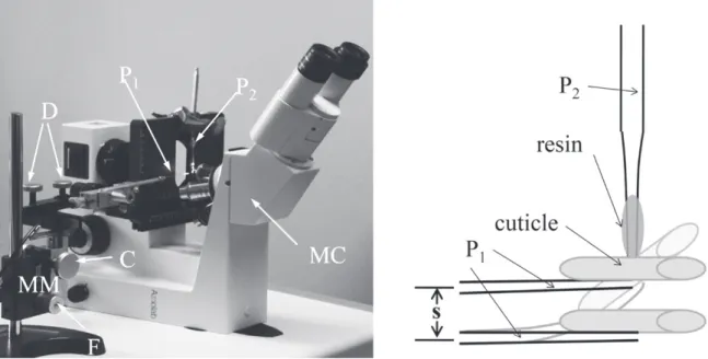
To measure the adhesive properties of the cuticles, these were mounted to fine, pulled-out glass pins (P1) being withdrawn from a similar glass pin P2 covered with resin (Figure 1). The adhesive properties were measured as forces necessary to provoke adhesive failure. These forces are called adhesive forces in the following.
PROduCTION OF GLASS PINS
The pins P1 and P2 were produced from glass bar fragments (diameter 1 mm, length 20 cm). With the Puller P-97 from Sutter Instruments Co the midpoint of the bars was heated up to 560°C.
With an acceleration of 110 m/s2, the pins were stretched; the resulting diameters of the pins’ tips ranged from 1 and 10 µm. For pin types P1, diameters of 3 to 4 µm were selected, types P2 of 10 µm.
FIxATION OF ThE CUTICLE SAMPLE ON P1
For analyzing the adhesive properties of the cuticles of the trochanter of C. sericeiventris and the mandible of T. angustula, the specimen were captured, knocked out with CO2 and dissected immediately.
All samples were fixed to the tip of P1 with warmed dental wax (65°C). P1 was mounted on the
micromanipulator (MM in Figure 1) and resin was installed on P2 immediately.
INSTALLATION OF RESIN ON P2
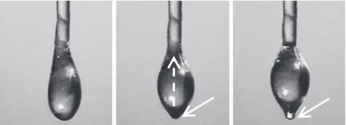
The dichloromethane cleaned P2 was dipped 2 - 3 mm into a mass of 2 g resin collected from a deposit of a colony of T. angustula. When the tip of P2 was covered completely by a drop of resin (length: 20-50 µm, width: 10-20 µm, see Figure 2), it was clipped perpendicular to P1 on the object table of the microscope MC (Figure 1). The behavior of the attached resin is described in Figure 2.
Figure 2 - Behavior of resin after the attachment on P2. Left - Resin from the bees hive immediately after the attachment on P2.
Middle - Once being attached to the glass pin it crawls upwards towards the base of P2 (dashed arrow). For a short moment just the tip
of the pin is covered with resin (white arrow). This is the moment to measure the adhesive properties on the cuticle samples. Right -About 60 seconds later the cover on the tip of P2 is vanishing (white arrow) and the resin has to be reinstalled to continue measurement.
MEASUREMENT
All treatments and measurements with resin were conducted at 19°C.
The adhesion forces were measured at the moment, when P2 was covered with only a thin, but still complete layer of resin (Figure 2, middle). P1 was slowly moved upwards to P2 until the cuticle sample touched the resin on P2. The two glass pins were kept in this position for 5 seconds so that resin and cuticle contacted each other. Then P1 was slowly withdrawn from P2 with a constant speed of 62.5 µm/s (corresponding to one turn of the micromanipulator’s fine adjustment), until
resin and cuticle disconnected. The distance s
(Figure 1) between position of contact and position of separation describes the deformation of P1 and depends on the stiffness of P1 as well as the adhesion forces between resin and cuticle.
CALIBRATION OF P1
CALCuLATION OF ThE ADHESIVE PROPERTIES
The adhesion forces FA is given by the product of mean distance s between position of contact and position of separation and the stiffness c of P1.
STATISTICAL EvALuATION
To proof the results statistically, the non-parametric Mann-Whitney Test has been used. Probability values lesser than 1% were considered as highly significant.
RESULTS
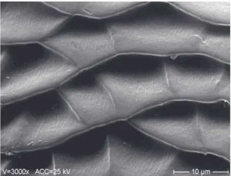
The cutting edge of the mandible of workers of
T. angustula is composed of a large blade and a pointed tooth. The surface of these structures are smooth (Figure 3) without microstructures. On several mandibles we found distinct traces of use like scratches or broken edges. On the other hand, the trochanter of the ant C. sericeiventris is scaled (Figure 4).
Figure 3 -Left – Smooth surface of the ventral side of the tip of the mandible of Tetragonisca angustula, right – mandible´s tip with distinct traces of use.
Calibration trials of all P1 show linear relation (R2 ≥ 0.998) between deformation in µm of P1 and weight in µg indicated by the balance (Figure 5), so that adhesion forces had been interpolated by linear regression.
During measurement of the adhesion forces, hydrophobic interaction has been observed between resin and the tested surfaces. The resin attached on P2 attracts the tested cuticles and the cover slip on
P1 with a jerk as soon as it reaches a critical distance somewhere between 1.5 µm and 1 µm.
Figure 6 shows that adhesion of resin on the structured cuticle of the trochanter of
C. sericeiventris is six times smaller than on the smooth cuticle of the tip of the mandible of T. angustula. These differences are highly significant using the Mann-Whitney-Test for independent samples.
Figure 5 - Calibration trial of glass pins P1 used to measure adhesion failure between resin and the surface of the mandible of Tetragonisca angustula and the trochanter of Camponotus sericeiventris.
Figure 6 - Adhesion of resin on the surface of the trochanter of Camponotus sericeiventris
and the mandible of Tetragonisca angustula in µg with electron-microscopical photos
DISCUSSION
The results of the adhesion force measurement refute our hypothesis that resin sticks lesser to the bees’ mandible than to other insects’ surfaces – even if the contact area between resinous and insect’s surface in experiment is smaller than in reality.
Adhesion on rough surfaces like the scaled trochanter of C. sericeiventris is lower than on smooth ones like the mandible of T. angstula
(Kendall 2001). Major contact areas covering hairs and various scales on the ant’s trochanter surface, will reduce adhesion further. As such adhesion reducing structures are lacking on the bee´s mandible, resin should stick more on the bee´s mandible than on the trochanter of the ant when contact area is increased.
however, these findings do not explain why resin, once attached to the mandible of T. angustula, is removed easily and without leaving residues behind. To manage that, the bee should
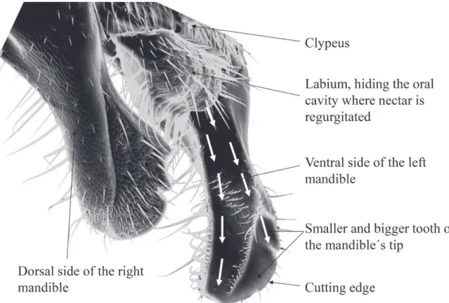
possess mechanisms to reduce adhesion of resin on the mandible. T. angustula might change the adhesive properties temporarily by lubricating their mandibles with adhesion reducing substances. Lubricating substances seem to be common within insects. The body surface of the mirid bug Pameridea roridulae (Reuter 1907) is covered by a lipid layer avoiding the bug getting stuck in the resinous surface of its host plant (Voigt and Gorb 2008). In stingless bees such lubricating substances might be secretions of the mandibular, salivary or other cephalic glands as proposed by Santos et al. (2009). As production of secretions is a complex and energy consuming processes, worker bees might even regurgitate nectar from the nectar crop. The liquid could run down the ventral side of the mandible via a small groove as illustrated in Figure 7. Once distributed, it reduces the adhesion of resin on the complete mandible.
This groove on the base of the ventral side of the mandible, also observed in other stingless bee species (Figure 8, Stort et al. 1986) and the honey bee Apis mellifera, is discussed by Goodman (2003) as a
feature to channel liquids to the tips of the mandibles. The groove might be an adaptation of bees to alter adhesive properties of the mandibles in order to form, cut and knead resins without sticking to them.
Figure 8 - Electron-microscopical photos showing the similarity of the mandible's ventral side of some Stingless Bees that collect resin. Left – right mandible of T. angustula, right – left mandible of Frieseomelitta varia (Lepeletier, 1836).
Further investigations have to test, if adhesion of resin is reduced on mandibles greased with nectar or honey. To prove our hypothesis, chemical research activities have to attest the existence of residues of the adhesion reducing substances like honey, nectar or sugar in resins treated by Stingless Bees.
ACKNOWLEDGMENTS
Thanks to Bernd Hoffmann and Wolfgang Rubner from the Institute of Bio- and Nanosystems, Institute 4 (Biological Layers), Research Center Jülich, Germany, for their help in developing a transportable apparatus to measure adhesion forces. We are grateful to Karin Ulmen from the Museum König in Bonn, Germany, to grant access to the SEM. Some parts of this research were supported by the DFG and the DAAD.
RESUMO
Muitas espécies das abelhas-sem-ferrão (Hymenoptera: Meliponini) como Tetragonisca angustula coletam resina para a defesa dos seus ninhos contra invasores como formigas ou abelhas cleptobióticas. Pequenas porções de resina são aderidas nos corpos e extremidades dos
invasores causando a sua imobilização. Observa-se que a resina é removida com facilidade das mandíbulas das abelhas, mas cola fortemente na cutícula dos invasores. Foi testada a hipótese de que a resina adere menos nas mandíbulas das abelhas-sem-ferrão do que na superfície dos invasores devido a estruturas especiais de superfície ou a propriedades adesivas dessas estruturas. As superfícies da mandíbula de T. angustula e do trocanter de Camponotus sericeiventris foram estudadas por microscopia eletrônica de varredura. Para medir suas propriedades de adesão, superfícies selecionadas foram montadas em uma alfinete de vidro fino e retiradas de uma ponta de vidro coberta com resina. A deformação do alfinete de vidro indica forças de adesão operando entre a resina e a superfície selecionada. O valor absoluto da força é calculado através da rigidez do alfinete. Foi observado que a resina adere mais na mandíbula da abelha com superfície lisa do que no trocanter estruturado da formiga. Uma nova hipótese a ser testada diz que as abelhas lubrificam as mandíbulas com néctar ou mel para reduzir a adesão da resina temporariamente.
REFERENCES
ARMBRUSTER WS. 1984. The role of resin in angiosperm pollination: Ecological and chemical considerations. Am J Bot 71: 1149-1160.
GASTAUER MM, CAMPOS LAO AND WITTMANN D. 2011.
Handling sticky resin by Stingless Bees. Rev Bras Ent 55: 234-240.
GOOdMAN J. 2003. Form and function in the Honey Bee. Cardiff: International Bee Research Association, 220 p. KENDALL K. 2001. Molecular Adhesion and Its Applications:
The Sticky Universe. New York, Boston, Dordrecht, London, Moscow: Kluwer Academic, 440 p.
MARSAIOLI AJ, PORTO ALM, GONçALvES RAC, OLIVEIRA
CMA, MANFIO GP AND BITTRICH V. 1999. The Ecosystem of Microorganisms, Bees, and Clusia Floral Resin and Oils, from the Chemistry Point of view. 2nd IUPAC
Conference on Biodiversity.
NOGuEIRA-NETO P. 1997. Vida e criação de abelhas indígenas sem ferrão. São Paulo: Editora Tecnapis, 445 p.
RADTKE R. 1994. Die kleptoparasitische neotropische Biene
Lestrimelitta limao: Verhalten und chemische Kommu-nikation bei Raubzügen auf Nester anderer Stachellosen Bienen. Ph.D. thesis, University of Tübingen, 172 p. ROuBIK DW. 1989. Ecology and natural history of tropical
bees. New York: Cambridge University Press, 514 p. SANTOS CGA, MEGIOLARO FL, SERRãO JE AND BLOChTEIN B.
2009. Morphology of the Head Salivary and Intramandibular Glands of the Stingless Bee Plebeia emerina (Hymenoptera: Meliponini) Workers Associated with Propolis. Ann Ent Soc Am 102: 137-143.
STORT AC, BuENO dE MORAES MM AND BARELLI N. 1986.
Scanning electron microscopy observations of the man-dibles of Scaptotrigona postica workers (Hymenoptera, Apoidea). J Apic Res 25: 65-70.
VOIGT D AND GORB S. 2008. An insect trap as habitat: cohesion-failure mechanism prevents adhesion of