ABSTRACT
Objective: To evaluate pulmonary function and functional capacity in children and adolescents with sickle cell disease. Methods: This was a cross-sectional study involving 70 children and adolescents (8-15 years of age) with sickle cell disease who underwent pulmonary function tests (spirometry) and functional capacity testing (six-minute walk test). The results of the pulmonary function tests were compared with variables related to the severity of sickle cell disease and history of asthma and of acute chest syndrome. Results: Of the 64 patients who underwent spirometry, 15 (23.4%) showed abnormal results: restrictive lung disease, in 8 (12.5%); and obstructive lung disease, in 7 (10.9%). Of the 69 patients who underwent the six-minute walk test, 18 (26.1%) showed abnormal results regarding the six-minute walk distance as a percentage of the predicted value for age, and there was a ≥ 3% decrease in SpO2 in 36 patients (52.2%). Abnormal pulmonary function was not signiicantly associated with any of the other variables studied, except for hypoxemia and restrictive lung disease. Conclusions: In this sample of children and adolescents with sickle cell disease, there was a signiicant prevalence of abnormal pulmonary function. The high prevalence of respiratory disorders suggests the need for a closer look at the lung function of this population, in childhood and thereafter.
Keywords: Anemia, sickle cell; Respiratory function tests; Exercise test.
Pulmonary function in children and
adolescents with sickle cell disease: have
we paid proper attention to this problem?
Ana Karine Vieira1, Cristina Gonçalves Alvim2,
Maria Cristina Marquez Carneiro3, Cássio da Cunha Ibiapina4
Correspondence to:
Cássio da Cunha Ibiapina. Avenida Professor Alfredo Balena, 110, CEP 30160-042, Belo Horizonte, MG, Brasil. Tel.: 55 31 3409-9772 or 55 31 9976-7871. Fax: 55 31 3409-9772. E-mail: cassioibiapina@terra.com.br Financial support: None.
INTRODUCTION
Sickle cell disease (SCD) is the most common monogenic disease in Brazil. The number of individuals with SCD in Brazil is estimated to range from 25,000 to 30,000.(1)
The incidence of SCD in the state of Minas Gerais, Brazil, is approximately 1:1,400 live births, according to the Minas Gerais State Neonatal Screening Program.(2) The
manifestations of this disease result from a predominance of sickle-shaped red blood cells, which leads to chronic hemolytic disease and vaso-occlusive phenomena.(2) SCD
leads to multisystem impairment, with lung involvement being a major cause of morbidity and mortality.(3)
Some studies have been published on assessment of pulmonary function in adults with SCD, showing that the major abnormality is restrictive lung disease (RLD). (4,5) Since the 1970s, studies have been published
on pulmonary function in the pediatric age group with SCD,(6) with conlicting results being reported; however,
most studies show that obstructive lung disease (OLD) is the most common abnormality.(7.8) RLD is also common
in some pediatric studies, highlighting the importance of pulmonary function testing in such children.(9,10)
Another important respiratory disease affecting children with SCD is asthma. Asthma is known to be a comorbidity impacting the course of SCD, leading to increased morbidity and mortality.(11,12) Several studies
have reported an association between asthma and an increased number of vaso-occlusive crises or acute
chest syndrome (ACS) episodes.(11-14) An association
between asthma and pulmonary hypertension in children was described by Hagar et al., raising the suspicion of a shared mechanism, possibly associated with chronic hemolysis.(15)
The mortality associated with lung disease is a serious problem in the SCD population.(16,17) It has been
demon-strated that abnormalities in pulmonary function tests are early objective signs of the development of chronic lung disease in SCD.(16) It has recently been published that
decreased FEV1 is associated with increased mortality in adults with SCD.(18) Nevertheless, few authors in Brazil
have attempted to investigate pulmonary function in patients with SCD.(19-21) We hypothesize that the onset of
pulmonary function abnormalities in SCD occurs as early as childhood. To address this challenge, the objective of the present study was to assess abnormalities in pulmonary function and functional capacity in children and adolescents with SCD, by means of spirometry and the six-minute walk test (6MWT), and to compare these abnormalities with clinical and laboratory variables in these patients.
METHODS
This was a descriptive analytical cross-sectional study. We included children and adolescents aged 8 to 15 years who were enrolled in the Minas Gerais State Neonatal
1. Fundação Hemominas, Belo Horizonte (MG) Brasil.
2. Departamento de Pediatria, Faculdade de Medicina, Universidade Federal de Minas Gerais, Belo Horizonte (MG) Brasil.
3. Grupo de Pneumologia Pediátrica, Faculdade de Medicina, Universidade Federal de Minas Gerais, Belo Horizonte (MG) Brasil.
4. Departamento de Pediatria, Faculdade de Medicina, Universidade Federal de Minas Gerais, Belo Horizonte (MG) Brasil.
Submitted: 9 March 2016. Accepted: 31 October 2016.
Screening Program, in the city of Belo Horizonte, Brazil,
had a conirmed diagnosis of SCD at age 1 year, and
were followed in the program between February of 2013 and February of 2014. We included patients with SS
phenotype or Sβ0-thalassemia—because they are known to have a more severe clinical course—who resided in the city of Belo Horizonte. We excluded patients who were unable to perform the respiratory maneuvers or the 6MWT because of cognitive or physical disability. We also excluded patients who had a severe comorbidity,
such as chronic inlammatory diseases, hematological
disorders, or neurological disorders.
Data were collected by interview, physical exami-nation, and medical chart review. The interview was conducted using a semi-structured data collection protocol. The physical examination consisted of measurement of vital signs and measurement of weight and height. The medical chart review consisted of identifying clinical and laboratory data relevant to the study. Baseline hemoglobin values and leukocyte and reticulocyte counts were obtained from the arithmetic mean of three complete blood counts before hydroxyurea therapy or blood transfusions.
Medical charts were reviewed for ACS, using the
following deinition: development of a new pulmo
-nary iniltrate involving at least one lung segment,
accompanied by at least another symptom, such as fever, chest pain, tachypnea, wheezing, cough, or hypoxemia.(22) The presence of a history of asthma
was based on a physician diagnosis recorded in the chart and on clinical and functional criteria established by the Global Initiative for Asthma.(23)
Spirometry and the 6MWT were performed in the outpatient clinic of a SCD referral center by the same
qualiied pulmonary function technician. The results were
interpreted by pediatric pulmonologists and reviewed by a pulmonologist specializing in pulmonary function.
Spirometry was performed with a Koko PFT spirometer (PDS Instrumentation, Inc., Louisville, CO, USA), with the patient seated and wearing a nose clip. All subjects underwent bronchodilator testing (400 µg of albuterol
aerosol), and a positive result was deined as a ≥ 12%
increase in FEV1 or a > 200 mL increase in absolute volume.(24) At least three curves were obtained. FVC,
FEV1, and FEF25‑75% values were derived from these curves and corrected for body temperature, pressure saturated. Tests were interpreted in accordance with the Brazilian Thoracic Association (2002) Guidelines for Pulmonary Function Testing.(25) Test results were
expressed as absolute values and as a percentage of predicted according to Mallozzi.(26) The spirometry indings were classiied as normal, RLD, OLD, or nonspeciic lung disease.(24,25) PEF was measured with
a Mini-Wright meter (Clement Clarke International, Essex, UK). The best of three consecutive readings was selected for analysis, using reference values provided by Polgar and Promadhat.(27) There was an interval
of at least two weeks between hospital admission or blood transfusion and the test.
The 6MWT was performed in accordance with the American Thoracic Society guidelines.(28) A wrist oximeter (Wrist 3100; Nonin Medical, Plymouth, MN,
USA) and a stopwatch were used. The patient was instructed to walk as fast as possible for six minutes. The walks were supervised by the physician responsible for the study. SpO2 was measured before, during, and immediately after the walk, with care being taken to provide a minimum one-minute interval for the oximeter curve to stabilize. The distance walked in six minutes—six-minute walk distance (6MWD)—was measured in meters. Two tests were performed within
30 minutes, and when there were conlicting results
between the tests, a third test was performed. A normal
result was deined as a 6MWD > 80% of predicted for age, a moderate result was deined as a 6MWD between 60% and 80% of predicted, and a severe result was deined as a 6MWD < 60% of predicted. (29) A signiicant decrease in SpO
2 (desaturation) was deined as that ≥ 3% relative to baseline.(30)
Percent predicted 6MWD was calculated following the equation proposed by Priesnitz et al.(31) Because
of the lack of studies in Brazil investigating 6MWD reference values in adolescents, the equation proposed by Priesnitz et al.(31) was also used for the patients
aged 13 to 15 years.
The study population was characterized with descriptive statistics. Variables were compared with the chi-square test and Fisher’s exact test. The Student’s t-test was used to compare independent groups. Continuous variables were tested for normality with the Shapiro-Wilk test and/or the Kolmogorov-Smirnov test. The three spirometry-based groups were compared with ANOVA and the Kruskal-Wallis test. The level
of statistical signiicance was set at p < 0.05. The
sample size calculation was based on the prevalence
of abnormal spirometry (37%) reported in a study
conducted in Brazil.(19) Therefore, for the sample size
of our study (70 cases), the margin of estimation
error is 6.3% within a 95% CI. A multivariate logistic
regression analysis was performed to determine the factors associated with the outcome measure “oxygen desaturation”. The baseline variables were used to build
the inal model. Given that the initial model included non‑signiicant variables, it had to be reduced to test the signiicance of the remaining variables, that is, we
sequentially removed the variable with the highest p
value until we reached a inal model with statistically signiicant variables.
informed consent using forms written in language appropriate for their age.
RESULTS
We aimed to recruit 100 patients with SCD by contact-ing their families over the phone or via a primary health care clinic and inviting these families to participate. Of the targeted 100 patients, 2 had died and 1 had a disabling comorbidity. In addition, 9 families were not located and 18 declined to participate. The sample
therefore consisted of 70 patients, of whom 31 (44.3%) were male and 39 (55.7%) were female. The mean
age was 11 ± 2.3 years (range, 8-15 years). Sixty-six individuals had the SS homozygous phenotype, whereas
4 had Sβ0‑thalassemia. Concomitant α‑thalassemia trait was found in 29.1% of the participants.
Thirty‑nine patients (55.7%) were being treated with hydroxyurea, and 10 (14.3%) were on a chronic transfusion regimen. Comorbid asthma was identiied in 23 patients (33.3%). In the univariate analysis,
a diagnosis of asthma was not associated with the number of vaso-occlusive crises or ACS episodes.
Forty‑four patients (63.1%) had at least one ACS episode, and, of those, 10 (23.1%) experienced two
or more episodes.
The descriptive statistics for the spirometric and 6MWT variables are presented in Table 1. Spirometry was completed successfully in 64 of the 70 patients included. Two patients were unable to perform the maneuvers, and 4 did not undergo spirometry because of problems with scheduling. However, these 4 were not excluded, because they underwent the 6MWT. Of
the patients who underwent spirometry, 15 (23.4%)
had abnormal pulmonary function tests. No patient was
classiied as having mixed obstructive‑restrictive lung disease or nonspeciic lung disease. The spirometry
results are presented in Table 2.
The results of the univariate analysis comparing the spirometry reports with intervening variables are
presented in Table 2. The patients reported as having OLD were older than were those reported as having normal spirometry results or RLD. The group of patients
classiied as having RLD showed lower baseline pulse oxygen saturation (89.8% ± 5.4%) than did the group
of patients with OLD and the group of patients with
normal spirometric results (92.3% ± 4.6% and 94.5% ± 4.4%, respectively; p = 0.02).
The 6MWT was administered to 69 patients, and 1 patient, who had undergone spirometry, declined to undergo the test. The results are presented in Table 2. The baseline SpO2 values reveal that hypoxemia was common in this population: 37 (52.9%) had values < 95%, with a mean of 93.6%. Eighteen patients (26.1%) had a 6MWD < 80% of predicted for their age, and only 1 patient had a 6MWD < 60%. There was a ≥ 3%
decrease in SpO2 in 36 patients (52.2%). Univariate analysis showed that a history of ACS was the only
variable of interest that had a statistically signiicant
association with oxygen desaturation, there being an inverse association between the number of ACS episodes
and the level of oxygen desaturation (< 3% vs. ≥ 3%),
as shown in Table 3. The median (interquartile range) for ACS was 1.0 (0.0-3.0) for the group of patients with
desaturation < 3% and 1.0 (0.0‑2.0) for the group of patients with desaturation ≥ 3%.
In the univariate analysis, none of the variables of
interest were found to have a statistically signiicant
association with 6MWD as a percentage of predicted (Table 3). In contrast, age and hypertransfusion were
found to have signiicant associations with absolute
values of 6MWD (Table 3).
The multivariate analysis of possible predictors of oxygen desaturation is presented in Table 4. An interesting observation is that, as in the univariate analysis, a history of ACS was found to be a protective factor against oxygen desaturation during the 6MWT
(p < 0.05). According to the measures of accuracy
of the logistic regression model, the sensitivity and
speciicity were 48.4% and 59.0%, respectively.
Table 1. Patient descriptive statistics for the spirometric and six-minute walk test variables.
Variable Minimum Maximum Median Mean ± SD
PEF, % of predicted 50.0 106.3 81.0 79.4 ±13.9
FVC, % of predicted 63.0 115.0 85.0 85.0 ± 10.6
FEV1, % of predicted 57.0 117.0 78.5 78.7 ± 10.7
FEV1/FVC ratio 84.0 112.0 0.86 0.86 ± 0.05
FEF25-75% 43.0 125.0 72.0 76.0 ± 17.6
Baseline SaO2, % 78.0 99.0 94.0 93.6 ± 4.8
Final SaO2, % 72.0 99.0 91.0 89.3 ± 7.6
Desaturation, % −3.0 −26.0 −2.0 −4.6 ± 6.3
6MWD, m 380 640 520.0 527.3 ± 51.4
6MWD, % of predicted 56.4 114.8 85.1 84.9 ± 8.3
Baseline HR, bpm 60.0 120 87.0 87.9 ± 13.7
Final HR, bpm 91.0 175.0 131.0 132.4 ± 18.8
Baseline RR, breaths/min 12.0 28.0 16.0 18.0 ± 3.4
Final RR, breaths/min 20.0 36.0 28.0 27.7 ± 3.6
DISCUSSION
The present study showed that pulmonary func-tion, as measured by spirometry, was abnormal in approximately one fourth of the patients with SCD, the most common abnormality being RLD. A high rate of desaturation during the 6MWT was found, which was not associated with 6MWD or with spirometry-assessed pulmonary function.
A history of asthma was present in one third of the patients, which is double the prevalence observed in the population of children and adolescents in the city of
Belo Horizonte (17.8%), according to the International
Study of Asthma and Allergies in Childhood.(32) A similar
prevalence was found in a retrospective cohort study conducted by Williams et al.,(33) who demonstrated that 35.9% of the patients had a diagnosis of asthma and
that a decline in FEV1 as a percentage of predicted is associated with progression to pulmonary dysfunction.
These indings underscore the need for the recognition
of asthma, as well as the importance of longitudinal follow-up of pulmonary function, in this population.
Lung disease patterns in SCD are heterogeneous and
can change over time. In the present study, 11% of the patients were found to have OLD; of those, 57% had
a clinical diagnosis of asthma. Similarly, in a cohort of patients studied by Boyd et al.(12) 13% were found to have OLD, and of those 13%, 77% had a diagnosis of asthma; in addition, an association was found between
OLD and increased rates of hospitalization for pain or
ACS. Another important study showed that 63% of the
patients with asthma had normal spirometry results
and that only 40% of the patients classiied as having
OLD had a history of asthma.(34) These indings indicate
that there may be several inlammatory mechanisms
involved in the genesis of airway obstruction in SCD.
The prevalence of OLD (12.5%) found in the present
study was also similar to those found in other studies, such as the ones conducted by Boyd et al. and by Tassel
et al., both of which reported a prevalence of 13%.(12,35)
One of the limitations of the present study is the lack
of whole‑body plethysmographic conirmation of RLD.
Longitudinal studies have shown that there is a decline in lung volume and pulmonary function over the years
in children with SCD; however, the pathophysiology
of respiratory disorders in childhood has not been fully elucidated.(9,35) This decline was reported in a
cohort study by Lunt et al.,(36) who demonstrated that
a history of ACS episodes was the only independent factor associated with reduced lung volumes. Some biological markers, such as leukocytosis, are known to be associated with SCD severity, but their relationship with pulmonary function has only recently been the subject of investigation.(37) A study by Tassel et al.(35)
showed that the decline in pulmonary function in childhood was directly associated with two markers of severity of the underlying disease: leukocytosis and hemolysis. In the present study, an association was noted between baseline hypoxemia and RLD, indicating that this group of patients may tend to experience greater disease severity, given that baseline hypoxemia is associated with the degree of anemia and of hemolysis.(30)
The 6MWT has grown in importance in the follow-up of SCD because studies in adults show a relationship between 6MWD and tricuspid regurgitant jet velocity, which is used to estimate pulmonary artery systolic
pressure on echocardiography. This inding suggests Table 2. Comparative statistics across three spirometry‑based patient groups (N = 64) for the variables of interest.a
Variables Groups p*
Normal RLD OLD
(n = 49) (n = 8) (n = 7)
Age, years 11.0 ± 2.3 11.9 ± 1.7 13.3 ± 1.5 0.045
Gender, % Male Female
44.9 55.1
25.0 75.0
71.4 28.6
0.241**
Baseline hemoglobin, g/dL 8.1 ± 1.2 7.4 ± 0.5 8.0 ± 0.5 0.151
Fetal hemoglobin, % 11.2 ± 8.4 11.7 ± 9.8 12.1 ± 6.7 0.875
Baseline leukocytes/µL 14.542 ± 2.988 14.222 ± 2.680 14.269 ± 4.931 0.979
Reticulocytes, % 14.6 ± 5.6 13.6 ± 3.9 14.9 ± 6.6 0.604
SaO2, % 94.5 ± 4.6 89.8 ± 5.4 92.3 ± 4.4 0.020
ACS episodes 1.7 ± 2.1 1.4 ± 1.5 0.4 ± 0.5 0.245
Asthma, % 28.6 37.5 57.1 0.274**
Hydroxyurea therapy, % 55.1 28.6 57.1 0.457**
Hypertransfusion, % 16.3 0.0 16.7 0.667**
α-thalassemia trait, % 30.8 0.0 28.6 0.638** Desaturation, %
< 3
≥ 3 45.854.2
50.0 50.0
28.6 71.4
0.693**
6MWD, % of predicted 84.1 ± 7.4 84.5 ± 11.1 86 ± 9.3 0.879**
RLD: restrictive lung disease; OLD: obstructive lung disease; ACS: acute chest syndrome; and 6MWD: six‑minute
walk distance. aValues expressed as mean ± SD, except where otherwise indicated. *Kruskal-Wallis test, except
that the 6MWT can be used as a noninvasive measure of severity of pulmonary hypertension and functional capacity in this population.(38) Studies in the pediatric
population remain scarce. An important study of
children and adolescents carried out by Minniti et al. observed that elevated tricuspid regurgitant jet velocity was associated with a decline in SpO2 during the 6MWT but not with a shorter 6MWD, as occurs
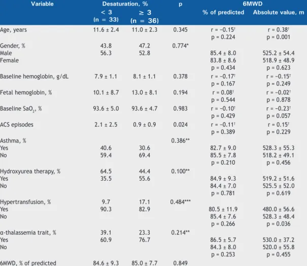
Table 3. Comparative statistics (N = 69 patients) between the outcome measures (oxygen desaturation and six‑minute
walk distance) and the variables of interest.*
Variable Desaturation, % p 6MWD
< 3 (n = 33)
≥ 3 (n = 36)
% of predicted Absolute value, m
Age, years 11.6 ± 2.4 11.0 ± 2.3 0.345 r = −0.15†
p = 0.224
r = 0.38†
p = 0.001 Gender, %
Male Female
43.8 56.3
47.2 52.8
0.774*
85.4 ± 8.0 83.8 ± 8.6 p = 0.434
525.2 ± 54.4 518.9 ± 48.9 p = 0.623
Baseline hemoglobin, g/dL 7.9 ± 1.1 8.1 ± 1.1 0.378 r = −0.17†
p = 0.167
r = −0.15†
p = 0.249
Fetal hemoglobin, % 10.1 ± 8.7 13.0 ± 8.1 0.194 r = 0.08†
p = 0.544
r = −0.02†
p = 0.878
Baseline SaO2, % 93.6 ± 5.0 93.6 ± 4.7 0.983 r = −0.10†
p = 0.429
r = −0.23†
p = 0.057
ACS episodes 2.1 ± 2.5 0.9 ± 0.9 0.024 r = −0.11†
p = 0.389
r = 0.15†
p = 0.229 Asthma, %
Yes No
40.6 59.4
30.6 69.4
0.386**
82.7 ± 9.0 85.5 ± 7.8 p = 0.210
528.3 ± 55.3 518.2 ± 49.1 p = 0.456 Hydroxyurea therapy, %
Yes No
64.5 35.5
44.4 55.6
0.100**
84.9 ± 9.3 84.4 ± 7.0 p = 0.781
519.2 ± 51.6 525.5 ± 52.0 p = 0.619 Hypertransfusion, %
Yes No
9.7 90.3
17.1 82.9
0.484***
80.5 ± 11.9 85.4 ± 7.6
p = 0.266
480.0 ± 56.6 528.3 ± 48.4 p = 0.036
α-thalassemia trait, %
Yes No
39.1 60.9
23.3 76.7
0.214**
86.5 ± 5.7 84.3 ± 8.0 p = 0.253
530.0 ± 37.2 520.0 ± 55.8 p = 0.455
6MWD, % of predicted 84.6 ± 9.3 85.0 ± 7.7 0.849
ACS: acute chest syndrome; and 6MWD: six‑minute walk distance. *Student’s t‑test for independent samples,
except where otherwise indicated. **Chi-square test. ***Fisher’s exact test. †Pearson’s correlation coeficient for
continuous variables (r value) and its respective p value.
Table 4. Multivariate logistic regression analysis for identiication of factors associated with six‑minute walk test
desaturation (≥ 3%).
Variable (Wald) chi-square p
Initial model
Intercept −1.116 0.157 0.692
Gender 0.213 0.134 0.715
Baseline hemoglobin 0.183 0.355 0.551
SaO2 0.170 0.058 0.810
Asthma −0.158 0.065 0.799
Hydroxyurea therapy 0.085 0.016 0.901
Acute chest syndrome −0.309 2.024 0.155
6MWD, % of predicted 0.003 < 0.001 0.996
Final model
Intercept 0.570 2.642 0.104
Acute chest syndrome −0.393 4.207 0.040
in adults.(39) In addition, a study conducted by Waltz
et al. reported a decline in SpO2 during the 6MWT in
patients with HbSS genotype (in 34%) and in patients with HbSC (in 18%).(40)
The 6MWD as a percentage of predicted for age was
below normal in 26.1% of the patients; however, severe
impairment was found in only 1 patient. A similar result was reported by Dedeken et al.,(29) who found that 30% of a sample of 46 children and adolescents had
abnormal 6MWT. The role of the 6MWT in children and
adolescents with SCD has yet to be fully determined; however, it seems that > 3% desaturation during the
6MWT may serve as an early marker of development of pulmonary hypertension in this population.(30)
In the present study, 52% of the patients had a signiicant decrease in post‑6MWT pulse SpO2, a prevalence that is higher than that reported in other studies, such as the ones by Waltz et al.(40) (34%)
and by Campbell et al.(30) (8%), the latter of which
involved patients with other phenotypes (SC, SD, and
others). This difference needs to be conirmed in other studies in Brazil, because the present study is the irst
of its kind in the country and shows the importance of functional assessment in this population.
In the logistic regression analysis, a history of ACS was surprisingly found to be a protective factor against oxygen desaturation during the 6MWT. This is likely
a relection of further treatment intensiication in the
patients who experienced ACS, including hydroxyurea
therapy and/or chronic transfusion. This inding was
also reported in a study that sought to understand the differences between adults and children with pulmonary hypertension.(15) That study demonstrated
that a history of ACS was a protective factor against pulmonary hypertension in the adult population.(15) In the present study, the low sensitivity and speciicity
of the multivariate analysis model do not allow the
elucidation of this complex pathophysiological process, and it is possible that clinical variables other than the ones we measured are associated.
As far as limitations of this study are concerned, we acknowledge that it was conducted in a single blood bank and was cross-sectional and that important tests, such as echocardiography and whole-body plethysmography were not performed, because they were not part of the research protocol. Therefore, further longitudinal studies with a large number of centers are warranted to verify the results obtained here and to determine which clinical and laboratory variables in SCD are of importance in clinical practice, because, in the literature, there is a lack of data on
the factors actually inluencing the development of
lung function impairment.
Finally, the present study showed a relevant prevalence
of abnormal pulmonary function and of signiicantly
decreased SpO2 during the 6MWT in a sample of patients aged 8 to 15 years, indicating the early onset of chronic
lung disease in SCD. The common inding of obstructive
and of restrictive pulmonary function abnormalities in this population underscores the need for a closer look at lung function, with the aid of objective measures, such as spirometry, in childhood and thereafter, in order to screen for chronic lung disease.
In summary, the data presented here, indicating that the prevalence of pulmonary function abnormalities in patients with SCD may be higher in Brazil, emphasize how important it is that studies of lung function in this population be conducted in the country. This subject must be researched extensively and thoroughly, including incorporating other methods of assessment, such as whole-body plethysmography and
echocar-diography, and is a large ield of research yet to be
explored in Brazil.
REFERENCES
1. Cançado RD, Jesus JA. Sickle cell disease in Brazil [Article in Portuguese]. Rev Bras Hematol Hemoter. 2007;29(3):204-6.
2. Fernandes AP, Januário JN, Cangussu CB, Macedo DL, Viana MN. Mortality of children with sickle cell disease: a population study. J Pediatr (Rio J). 2010;86(4):279-84. http://dx.doi.org/10.2223/ jped.2005
3. Darbari DS, Kple-Faget P, Kwagyan J, Rana S, Gordeuk VR, Castro O. Circumstances of death in adult sickle cell disease patients. Am J Hematol. 2006;81(11):858-63. http://dx.doi.org/10.1002/ajh.20685
4. Klings ES, Wyszynski DF, Nolan VG, Steinberg MH. Abnormal pulmonary function in adults with sickle cell anemia. Am J Resp Crit Care Med. 2006;173(11):1264-9. http://dx.doi.org/10.1164/ rccm.200601-125OC
5. Young RC Jr, Rachal RE, Reindorf CA, Armstrong EM, Polk OD Jr, Hackney RL Jr, et al. Lung function in sickle cell hemoglobinopathy patients compared with healthy subjects. J Nat Med Assoc. 1988;80(5):509-14.
6. Wall MA, Platt OS, Strieder DJ. Lung function in children with sickle cell anemia. Am Rev Respir Dis. 1979;120(1):210-4.
7. Koumbourlis AC, Hurlet-Jensen A, Bye MR. Lung function in infants with sickle cell disease. Pediatr Pulmonol. 1997;24(4):277-81. http://dx.doi.org/10.1002/(SICI)1099-0496(199710)24:4<277::AID-PPUL6>3.0.CO;2-H
8. Santoli F, Zerah F, Vasile N, Bachir D, Galacteros F, Atlan G. Pulmonary function in sickle cell disease with or without acute chest
syndrome. Eur Resp J. 1998;12(5):1124-9. http://dx.doi.org/10.1183/ 09031936.98.12051124
9. Boyd JH, DeBaun MR, Morgan WJ, Mao J, Strunk RC. Lower airway obstruction is associated with increased morbidity in children with sickle cell disease. Pediatr Pulmonol. 2009;44(3):290-6. http://dx.doi. org/10.1002/ppul.20998
10. MacLean JE, Atenafu E, Kirby-Allen M, MacLusky IB, Stephens D, Grasemann H, et al. Longitudinal decline in lung volume in a population of children with sickle cell disease. Am J Resp Crit Care Med. 2008;178(10):1055-9. http://dx.doi.org/10.1164/rccm.200708-1219OC
11. Boyd JH, Macklin EA, Strunk RC, DeBaun MR. Asthma is associated with acute chest syndrome and pain in children with sickle cell anemia. Blood. 2006;108(9):2923-7. http://dx.doi.org/10.1182/ blood-2006-01-011072
12. Boyd JH, Macklin EA, Strunk RC, DeBaun MR. Asthma is associated with increased mortality in individuals with sickle cell anemia. Haematologica. 2007;92(8):1115-8. http://dx.doi.org/10.3324/ haematol.11213
13. Bernaudin F, Strunk RC, Kamdem A, Arnaud C, An P, Torres M, et al. Asthma is associated with acute chest syndrome, but not with an increased rate of hospitalization for pain among children in France with sickle cell anemia: a retrospective cohort study. Haematologica. 2008;93(12):1917-8. http://dx.doi.org/10.3324/haematol.13090
Painful episodes in children with sickle cell disease and asthma are temporally associated with respiratory symptoms. J Pediatr Hematol Oncol. 2006;28(8):481-5. http://dx.doi.org/10.1097/01. mph.0000212968.98501.2b
15. Hagar RW, Michlitsch JG, Gardner J, Vichinsky, EP, Morris CR. Clinical differences between children and adults with pulmonary hypertension and sickle cell disease. Br J Haematol. 2008;140(1):104-12.
16. Hamideh D, Alvarez O. Sickle cell disease related mortality in the United States (1999-2009). Pediatr Blood Cancer. 2013;60(9):1482-6. http://dx.doi.org/10.1002/pbc.24557
17. Powars D, Weidman JA, Odom-Maryon T, Niland JC, Johnson C. Sickle cell chronic lung disease: prior morbidity and the risk of pulmonary failure. Medicine (Baltimore). 1988;67(1):66-76. http:// dx.doi.org/10.1097/00005792-198801000-00005
18. Low forced expiratory volume is associated with earlier death in sickle cell anemia. Blood. 2015;126(13):1544-50. http://dx.doi. org/10.1182/blood-2015-05-644435
19. Souza LC, Viegas CA. Quality of sleep and pulmonary function in clinically stable adolescents with sickle cell anemia. J Bras Pneumol. 2007;33(3):275-81. http://dx.doi.org/10.1590/S1806-37132007000300008
20. Fonseca CS, Araújo-Melo CA, Carvalho RM, Barreto-Neto J, Araújo JG, Cipolotti R. Lung function in patients with sickle cell anemia. Rev Paul Pediatr. 2011;29(1):85-90. http://dx.doi.org/10.1590/S0103-05822011000100014
21. Ohara DG, Ruas G, Walsh IA, Castro SS, Jamami M. Lung function and six-minute walk test performance in individuals with sickle cell disease. Braz J Phys Ther. 2014;18(1):79-87. http://dx.doi. org/10.1590/S1413-35552012005000139
22. Castro O, Brambilla DJ, Thorington B, Reindorf CA, Scott RB, Gillette P, et al. The acute chest syndrome in sickle cell disease: incidence and risk factors. The Cooperative Study of Sickle Cell Disease. Blood. 1994;84(2):643-9.
23. Global Initiative for Asthma [homepage on the Internet]. Bethesda: Global Initiative for Asthma. [cited 2016 Feb 01]. Global Strategy for Asthma Management and Prevention 2012. [Adobe Acrobat document, 128p.]. Available from: http://www.ginasthma.org/local/ uploads/iles/GINA_Report_March13.pdf
24. Lung function testing: selection of reference values and interpretative strategies. American Thoracic Society. Am Rev Respir Dis. 1991;144(5):1202-18. http://dx.doi.org/10.1164/ajrccm/144.5.1202
25. Sociedade Brasileira de Pneumologia e Tisiologia. Diretrizes para testes de função pulmonar. J Pneumol. 2002;28(Suppl 3):S1-S238.
26. Mallozi MC. Valores de referência para espirometria em crianças e adolescentes, calculados a partir de uma amostra da cidade de São Paulo [thesis]. São Paulo: Escola Paulista de Medicina/Universidade Federal de São Paulo; 1995.
27. Polgar G, Promadhat V. Pulmonary function testing in children: techniques and standards. Philadelphia: WB Saunders; 1971.
28. ATS Committee on Proiciency Standards for Clinical Pulmonary Function Laboratories. ATS statement: guidelines for the six-minute walk test. Am J Resp Crit Care Med. 2002;166(1):111-7. http:// dx.doi.org/10.1164/ajrccm.166.1.at1102
29. Dedeken L, Chapusette R, Lê PQ, Heijmans C, Devalck C, Huybrechts S, et al. Reduction of the six-minute walk distance in children with sickle cell disease is correlated with silent infarct: results from a cross-sectional evaluation in a single center in Belgium. PloS One. 2014; 9(9):e108922. http://dx.doi.org/10.1371/journal.pone.0108922
30. Campbell A, Minniti CP, Nouraie M, Arteta M, Rana S, Onyekwere O, et al. Prospective evaluation of haemoglobin oxygen saturation at rest and after exercise in paediatric sickle cell disease patients. Br J Haematol. 2009;147(3):352-9. http://dx.doi.org/10.1111/j.1365-2141.2009.07854.x
31. Priesnitz CV, Rodrigues GH, Stumpf Cda S, Viapiana G, Cabral CP, Stein RT, et al. Reference values for the 6-min walk test in healthy children aged 6-12 years. Pediatric Pulmonol. 2009; 44(12):1174-9. http://dx.doi.org/10.1002/ppul.21062
32. Solé D, Wandalsen GF, Camelo-Nunes IC, Naspitz CK. Prevalence of symptoms of asthma, rhinitis, and atopic eczema among Brazilian children and adolescents identiied by the International Study of Asthma and Allergies in Childhood (ISAAC) - Phase 3. J Pediatr (Rio J). 2006;82(5): 341-6. http://dx.doi.org/10.1590/S0021-75572006000600006
33. Williams SN, Nussbaum E, Yoonessi L, Morphew T, Randhawa I. Progression and prognostic indicators of bronchial disease in children with sickle cell disease. Lung. 2014;192(3):385-93. http://dx.doi. org/10.1007/s00408-014-9572-y
34. Koumbourlis AC, Lee DJ, Lee A. Longitudinal changes in lung function and somatic growth in children with sickle cell disease. Pediatric Pulmonol. 2007;42(6):483-8. http://dx.doi.org/10.1002/ ppul.20601
35. Tassel C, Arnaud C, Kulpa M, Fleurence E, Kandem A, Madhi F, et al. Leukocytosis is a risk factor for lung function deterioration in children with sickle cell disease. Respir Med. 2011;105(5):788-95. http:// dx.doi.org/10.1016/j.rmed.2010.12.024
36. Lunt A, McGhee E, Sylvester K, Rafferty G, Dick M, Rees D, et al. Longitudinal assessment of lung function in children with sickle cell disease. Pediatr Pulmonol. 2016;51(7):717-23. http://dx.doi. org/10.1002/ppul.23367
37. Okpala I. Leukocyte adhesion and the pathophysiology of sickle cell disease. Curr Opin Hematol. 2006;13(1):40-4. http://dx.doi. org/10.1097/01.moh.0000190108.62414.06
38. Anthi A, Machado RF, Jison ML, Taveira-Dasilva AM, Rubin LJ, Hunter L, et al. Hemodynamic and functional assessment of patients with sickle cell disease and pulmonary hypertension. Am J Resp Crit Care Med. 2007;175(12):1272-9. http://dx.doi.org/10.1164/ rccm.200610-1498OC
39. Minniti CP, Sable C, Campbell A, Rana S, Ensing G, Dham N, et al. Elevated tricuspid regurgitant jet velocity in children and adolescents with sickle cell disease: association with hemolysis and hemoglobin oxygen desaturation. Haematologica. 2009;94(3):340-7. http://dx.doi. org/10.3324/haematol.13812