* Study carried out at the Adolfo Lutz Institute, Santos Regional Laboratory, Santos, Brazil.
1. Assistant Technician in Scientific and Technological Research. Adolfo Lutz Institute, Santos Regional Laboratory, Santos, Brazil. 2. Scientific Researcher. Adolfo Lutz Institute, Santos Regional Laboratory, Santos, Brazil.
3. Support Technician in Scientific and Technological Research. Adolfo Lutz Institute, Santos Regional Laboratory, Santos, Brazil. 4. Pharmacist. Adolfo Lutz Institute, Santos Regional Laboratory, Santos, Brazil.
Correspondence to: Andréa Gobetti Vieira Coelho. Rua Oswaldo Cruz, 67/94, Boqueirão, CEP 11045-101, Santos, SP, Brazil. Tel 55 13 3232-5112. Fax 55 13 3322-3151. E-mail: [email protected]
Submitted: 25 January 2007. Accepted, after review: 16 April 2007.
Detection of cord factor for the presumptive identification
of
Mycobacterium tuberculosis
complex*
Andrea Gobetti Vieira Coelho1, Liliana Aparecida Zamarioli2,
Clemira Martins Pereira Vidal Reis3, Bruno Francisco de Lima Duca4
Abstract
Objective: Virulent strains of the Mycobacterium tuberculosis complex, under certain appropriate conditions, grow as characteristic ropes, bundles or serpentine cords known as cord factor or growth in cords. The objective of the present study was to evaluate cord factor detection as a method of achieving presumptive identification of the M. tuberculosis complex, comparing it to conventional typing tests. Methods: A total of 743 strains were analyzed from January of 2002 to December of 2005 in the Mycobacteria Sector of the Adolfo Lutz Institute, located in the city of Santos, Brazil. Samples were obtained from clinical specimens collected from patients with respiratory symptoms treated at basic health clinics in the greater metropolitan area of Santos. Ziehl-Neelsen-stained smears were prepared, 301 (40.5%) in MB/BacT broth and 442 (59.5%) on solid media, either Lowenstein-Jensen or Ogawa-Kudoh. Results: The sensitivity, specificity, positive predictive value and negative predictive value obtained during the performance comparison of the two methods (cord factor detection and conventional typing) using both isolation media were, respectively, 98.5, 88, 97 and 93%. The method was more sensitive on solid medium (100%), and the difference in sensitivity between the two media types was only 2.7%. Conclusions: Taking into consideration the results obtained, we conclude that, in laboratories with a high incidence of M. tuberculosis complex isolation and limited economic resources, cord factor detection is a fast and valid criterion for identifying these mycobacteria using liquid or solid medium. It also enables subsequent conclusive identification tests, as well as additional sensitivity tests when necessary.
53% of the new cases are concentrated in 10 of those cities.(8)
The greater metropolitan area of Santos, a coastal city located in the state of São Paulo, is the third most populated region in the state, with approxi-mately 1,500,000 people distributed in 9 cities. This region comprises 8 of the 73 cities considered priorities for the Programa Nacional de Controle da Tuberculose (PNCT, National Tuberculosis Control Program). In 2002, 2003 and 2004, the incidence of TB in the greater metropolitan area of Santos was 95.2, 99.2 and 94.9 per 100,000 inhabitants,
respectively.(8,9) In view of the current
epidemio-logical situation of this disease, the rapid diagnosis of mycobacterioses is a constant challenge for the PNCT.
As a consequence of the glycolipid trehalose dimycolate, present in the bacterial cell wall, and under appropriate conditions, we observe, in the virulent strains of the bacillus of TB, the growth
of the M. tuberculosis complex into microscopic
serpentine-like cords, denominated cord factor, or growth in cords, in which the acid-fast bacilli
(AFB) are in parallel arrays.(10) Studies have evaluated
the use of cord factor detection in liquid medium as a reliable presumptive result in the rapid early
identification of the M. tuberculosis complex in
laboratories that use automated methodology to
isolate mycobacteria.(11)
The method has also been used as a triage in the
presumptive identification of the M. tuberculosis
complex, together with the evaluation of the colony morphology in solid medium, as a practical and low cost pre-test to determine whether additional
iden-tification or sensitivity tests are needed.(11,12)
In view of this, and as a Regional Laboratory, we carried out this study with the objective of evaluating the presence of the cord factor as
Introduction
Tuberculosis (TB) is one of the most ancient diseases, with wide geographic distribution, consti-tuting a serious public health problem worldwide
and in Brazil.(1) It occurs in developed countries, as
well as in countries with emerging economies that present deep developing contrasts, and is associated with high poverty indices, making it a public health
priority in developing countries.(2)
The etiologic agent of TB is Mycobacterium
tuberculosis, and its most common clinical pres-entation is pulmonary. The most common form of transmission is the person-to-person contact from an infection source (infected individual) with
pulmonary lesions.(3)
The World Health Organization (WHO) estimates that TB accounts for 2.7 million deaths annu-ally, 95% of which occur in developing countries. Projections made in 1995 indicate that, by the year 2005, 11.9 million new cases of the disease will occur annually. Brazil is ranked 16th among the countries that account for 80% of the total
cases of TB worldwide,(4,5) and presents the greatest
number of cases in Latin America.(6) In addition, it
is among the 22 countries that the WHO considers
priorities,(7) with an incidence rate of 60 per 100,000
inhabitants and a mortality rate of 7.8 per 100,000 inhabitants.(5)
The state of São Paulo reports approximately 21,000 cases annually, which is, in absolute numbers, the highest of all states in Brazil. In 2005, the inci-dence of TB in the state of São Paulo was 43.9 cases per 100,000 inhabitants. Although it was not the highest in Brazil, it was close to the national average which was, in 2004, 44.1 per 100,000 inhabitants. The state of São Paulo comprises 645 cities, and
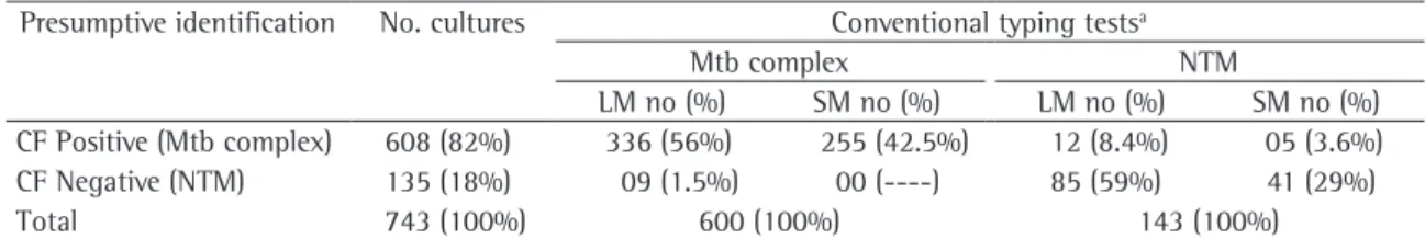
Table 1 - Comparison of presumptive results of cord factor detection and conventional typing tests of 743 strains analyzed in the 2002-2005 period.
Presumptive identification No. cultures Conventional typing testsa
Mtb complex NTM
LM no (%) SM no (%) LM no (%) SM no (%)
CF Positive (Mtb complex) 608 (82%) 336 (56%) 255 (42.5%) 12 (8.4%) 05 (3.6%)
CF Negative (NTM) 135 (18%) 09 (1.5%) 00 (----) 85 (59%) 41 (29%)
Total 743 (100%) 600 (100%) 143 (100%)
aResults obtained from Gen-Probe tests (DNA probes), morphological analysis of growth and other traditional biochemical methods;
was submitted to the Gen-Probe test (DNA probes), morphological analysis of growth and other
tradi-tional biochemical methods.(14,15)
The study was approved by the Ethics in Human Research Committee of the Adolfo Lutz Institute.
Results
The prevalence of mycobacteria species of the M. tuberculosis complex in the period of the study (2002-2005) was 81%; therefore, presumptive iden-tification of this species is an important diagnostic resource.
Based on the results obtained from the 743 strains analyzed, we determined the sensitivity and specifi-city, as well as positive and negative predictive values, for the presence of the cord factor in the
presumptive identification of the M. tuberculosis
complex in liquid and on solid medium, in rela-tion to convenrela-tional typing tests. To that end, we divided this analysis into three phases: evaluation of cord factor detection in liquid medium; evalua-tion of cord factor detecevalua-tion on solid medium; and, finally, evaluation of the performance of the method in relation to the total strains analyzed (Table 1).
In the presumptive identification of the M. tuberculosis complex, cord factor detection presented sensitivity, specificity, positive predictive value and negative predictive value of 97.3, 87.6, 96.5 and 90.4%, respectively, in liquid medium, compared with 100, 89, 98 and 100%, respectively, on solid medium.
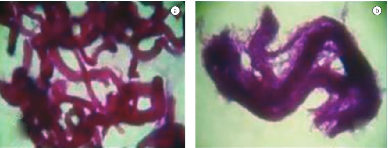
In Figures 1 and 2, we can observe microscopic images of M. kansasii and M. tuberculosis, respec-tively, isolated using both types of media.
presumptive identification of the M. tuberculosis
complex, using mycobacteria strains isolated in a liquid medium (MB/BacT broth) and on a solid medium (Lowenstein-Jensen or Ogawa-Kudoh). We intend to demonstrate that this rapid, easy, sensi-tive low-cost method can be safely performed in our laboratory and in local laboratories that use the isolation of mycobacteria on solid media.
Methods
This study was conducted based on the analysis of a collection consisting of 743 mycobacteria strains (2002-2005), 301 (40.5%) in liquid medium and 442 (59.5%) on solid medium, isolated from clinical samples collected from patients experiencing respi-ratory symptoms or clinically suspected of having pulmonary TB or mycobacteriosis and treated at the basic health clinics in the greater metropolitan area of Santos, using techniques recommended by the
National Ministry of Health.(13)
Ziehl-Neelsen-stained smears were prepared according to the Guidebook for Tuberculosis Bacteriology(13):
• smeared strain in liquid medium: performed
directly on slide, from the sediment obtained from 5 mL centrifuged in liquid medium.
• smeared strain on solid medium: performed
directly on slide with sterile distilled water, from an isolated strain.
Slides were analyzed, and the presence of AFB and cord factor formation was noted.
The identification of the strain as belonging to the M. tuberculosis complex was confirmed by the analysis of the records of conventional typing test results previously performed, in which the strain
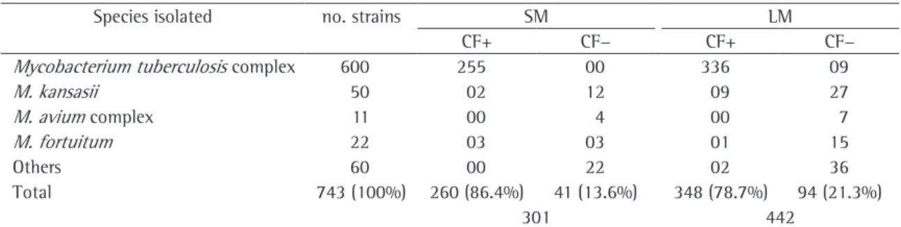
Table 2 - Species isolated from 743 strains analyzed according to the result of presumptive identification of the cord factor in the period 2002-2005.
Species isolated no. strains SM LM
CF+ CF− CF+ CF−
Mycobacterium tuberculosis complex 600 255 00 336 09
M. kansasii 50 02 12 09 27
M. avium complex 11 00 4 00 7
M. fortuitum 22 03 03 01 15
Others 60 00 22 02 36
Total 743 (100%) 260 (86.4%) 41 (13.6%) 348 (78.7%) 94 (21.3%)
301 442
and classified according to the presence or absence of the cord factor, can be seen in Table 2.
Discussion
The sensitivity, specificity, positive predic-tive value and negapredic-tive predicpredic-tive value found in our analysis are comparable to those reported
in the literature.(16) Other authors,(17) examining
cord factor detection as presumptive result of the M. tuberculosis complex isolated in liquid medium, report 90% sensitivity, similar to that obtained in our study.
In the comparison of the performance of the method in both isolation media and in the final identification with conventional typing tests, the sensitivity, specificity, positive predictive value and negative predictive value obtained were, respec-tively, 98.5, 88, 97 and 93%.
Of the 743 strains studied, 608 (81.8%) were
identified as belonging to the M. tuberculosis
complex by identifying the cord factor. Of those, 591 (97.2%) were confirmed by conventional typing tests and 17 (2.8%) were identified as nontubercu-lous mycobacteria (NTM).
The species identified using conventional methods, isolated in liquid and on solid medium,
b a
a b
Figure 1 -Mycobacterium kansasii, Ziehl-Neelsen staining, optical microscopy (1600×): a) in liquid isolation medium, absence of cord factor; and b) on solid isolation medium, absence of cord factor.
It is possible to observe the cord factor in NTM, since they produce ‘pseudo cords’, that is, incomplete growth in cords, and the interpretation depends on the experience of the laboratory techni-cian (Figure 1). Of the total strains analyzed, 17 NTM (12%) were described as cord factor positive, and 53% of those were identified as M. kansasii. These values are considered high when compared with
those of other authors.(11,16) However, we
empha-size that such studies do not reveal the isolation of M. kansasii. In addition, our values are lower than
the 16.9% presented by one well-known author.(12)
Analyzing the performance of the method using both isolation media, only 1.5% of the strains of the M. tuberculosis complex were identified as cord factor negative, 100% of them in liquid medium. Such differences seem insignificant and are lower than the approximately 10% presented by other authors.(12,17)
The sensitivity of the method was greater on solid medium. We found only a 2.7% difference in sensitivity when analyzing the method using both isolation media, a quite relevant number, since most public health laboratories in Brazil use solid medium in the isolation of mycobacteria, confirming the viability of this method.
Statistical calculations to determine concord-ance values among methods revealed the following: 96% overall concordance, 69% expected concord-ance and 87% adjusted concordconcord-ance (kappa).
Based on the positive and negative predictive values obtained in our study, we can conclude that identifying growth in cords is a real and rapid
crite-rion for the identification of the M. tuberculosis
complex isolated in liquid or on solid medium, enabling us to refer to conclusive identification tests as well as additional sensitivity tests that are deemed necessary, in laboratories with a high prevalence of M. tuberculosis and in which other techniques for early identification are unavailable.
References
1. Rosemberg J. Tuberculose. Panorama global. Óbices para seu controle. 2nd ed. Fortaleza: Secretaria de Estado da Saúde do Ceará; 1999.
2. Hijjar MA, Oliveira MJPR, Teixeira GM. A tuberculose no Brasil e no mundo. Bol Pneumol Sanit. 2001; 9(2):9-15. 3. Murray CJL, Stylbo K, Rouillon A. Tuberculosis. In: Jamison
DT. Disease control proiorities in developing countries. Oxford: Oxford Medical Publication. Oxford University press; 1993. p. 233-59.
4. Sociedade Brasileira de Pneumologia e Tisiologia. II
Consenso Brasileiro de Tuberculose. Diretrizes Brasileiras para Tuberculose 2004. J Bras Pneumol. 2004;30(1):S1-S55. 5. World Health Organization. Global tuberculosis control
surveillance, planning, financing: WHO report 2003. Geneva; WHO/TB; 2003. p. 1-316.
6. Santos Filho ET. Política de TB no Brasil: Uma Perspectiva da Sociedade Civil – Tempos de Mudanças para o Controle da Tuberculose no Brasil. Rio de Janeiro: Open Society Institute; 2006. p. 85.
7. Natal S. Emergência da resistência às drogas. Bol Pneumol Sanit. 2002. 10(2):57-70.
8. Tuberculose no Estado de São Paulo: Indicadores de Morbimortalidade e Indicadores de Desempenho. Bol Epidmiol Paulista 2006; 3(Supl 4):S1-S3.
9. Sistema estadual de Análise de Dados – SEADE [homepage on the Internet]. São Paulo: Fundação SEADE [cited 2005 Apr 05]. Available from: http://www.seade.gov.br
10. Levinson W, Jawetz E. Microbiologia médica e imunologia. 7th ed. Rio de Janeiro: Guanabara Koogan; 2005. p. 631. 11. Badak FZ, Goksel S, Sertoz R, Guzelant A, Kizirgil A,
Bilgic A. Cord formation in MB/BacT medium is a reliable criterion for presumptive identification of Mycobacterium tuberculosis complex in laboratories with high prevalence of M. tuberculosis. J Clin Microbiol. 1999;37(12):4189-91. 12. Monteiro PHT, Martins MC, Ueki SYM, Giampaglia CMS,
Telles MAS. Cord formation and colony morphology for the presumptive identification of Mycobacterium tuberculosis complex. Braz J Microbiol. 2003;34(2):171–4.
13. Fundação Nacional de Saúde. Centro de Referência Professor Helio Fraga. Manual de Bacteriologia da Tuberculose. 2nd ed. Rio de Janeiro: Ministério da Saúde; 1994.
14. Collins CH, Grange JM, Yates MD. Tuberculosis bacteriology: organization and practice. 2nd ed. London: Butterworth-Heinemann; 1997. p. 139.
15. Kent PT, Kubica GP. Public health mycobacteriology - a guide for level III laboratory. Atlanta: Centers for Disease
Control, US Department of Health and Public Services, 1985. p. 207.
16. McCarter YS, Ratkiewicz IN, Robinson A. Cord formation in BACTEC medium is a reliable, rapid method for presumptive identification of Mycobacterium tuberculosis complex. J Clin Microbiol. 1998; 36(9):2769-71.