vol. 45, n. 1, jan./mar., 2009
Ar
*Correspondence: M. B. R. Pierre. Universidade Federal do Rio de Janeiro, Faculdade de Farmácia, Av. Carlos Chagas Filho, 373 - 21941.590 - Rio de Janeiro - RJ, Brazil. E-mail: [email protected]
Influence of ceramide 2 on
in vitro
skin permeation and retention
of 5-ALA and its ester derivatives, for Photodynamic Therapy
Maria Bernadete Riemma Pierre*
1, Renata Fonseca Vianna Lopez
2,
Maria Vitória Lopes Badra Bentley
21Faculdade de Farmácia, Departamento de Medicamentos, Universidade Federal do Rio de Janeiro, 2Faculdade de Ciências Farmacêuticas de Ribeirão Preto, Universidade de São Paulo
Photodynamic therapy (PDT) based on topical 5-aminolevulinic acid (5-ALA), an endogenous precursor of protoporphyrin, is an interesting approach for the treatment of skin cancer. However, 5-ALA is a hydrophilic molecule and such a characteristic limits its appropriate cutaneous penetration and retention.
In this way, more lipophilic molecules, such as esteriied 5-ALA derivatives, have been under investigation
in order to improve the skin penetration of this molecule. Drug formulation can also alter 5-ALA skin
penetration. Therefore, the aim of this work was to study the inluence of ceramide 2 - the main lipid
of the SC- on the cutaneous delivery of 5-ALA and its ester derivatives in vitro, using Franz diffusion cell. The skin permeation of all studied drugs was decreased in the presence of ceramide, representing a desirable characteristic in order to avoid the risk of systemic side effects. Nevertheless, the SC and [epidermis + dermis] retention after 16 h has also been decreased in the presence of ceramide, as compared to control. In conclusion, ceramide was not a good adjuvant, meaning that research of other vehicles could be useful to improve cutaneous delivery of 5-ALA.
Uniterms: 5-aminolevulinic acid. Ceramide 2. In vitro/Skin permeation. In vitro/Skin retention. Photodynamic therapy.
A Terapia Fotodinâmica (TFD) tópica com um precursor das poririnas endógenas, o ácido 5-aminolevulínico
(5-ALA), constitui uma nova modalidade para o tratamento do câncer de pele. Entretanto, o 5-ALA é
uma molécula hidrofílica, o que limita sua penetração e retenção cutânea apropriadas. Moléculas mais lipofílicas, tais como derivados esteriicados do 5-ALA, estão sob intensa investigação para melhorar a penetração cutânea desta molécula. A formulação que contém o fármaco também pode alterar a penetração cutânea do 5-ALA. Desta forma, o objetivo deste trabalho foi estudar a inluência da ceramida 2 – o principal lipídeo do EC- sobre a penetração cutânea de 5-ALA e seus derivados esteriicados usando células de difusão de Franz. A permeação de todas as drogas estudadas através da pele foi diminuída na presença de ceramida, o que é desejável, evitando riscos de efeitos colaterais sistêmicos. Entretanto, a retenção no EC e [epiderme + derme] também foi diminuída na presença da ceramida, após 16 horas, comparado ao controle. Concluindo, a ceramida não foi um bom adjuvante, sendo necessária a pesquisa de outros veículos para melhorar a liberação cutânea do 5-ALA.
Unitermos: Ácido 5-aminolevulínico. Ceramida 2. Permeação cutânea/in vitro. Retenção cutânea/in vitro. Terapia fotodinâmica.
INTRODUCTION
Exogenous 5-aminolevulinic acid (5-ALA) is
Despite the promising results obtained with 5-ALA, a significant limitation for its topical application is the poor ability of this molecule to diffuse through biological membranes like the stratum corneum (SC), due to its higher hydrophilicity. Then, a high dose of 5-ALA must be admi-nistered, in order to increase PpIX in the tissue at a level that is useful for PDT treatment. Hence, improving the uptake of 5-ALA would be expected, to increase the eficiency of PDT (Lopez et al., 2004).
There are several methods to improve 5-ALA skin penetration: (i) changing the drug formulation by adding a penetration enhancer (Pierre et al., 2006) or by packaging it into a drug delivery system, e.g., liposomes (Pierre et al., 2001); (ii) applying an electrical current (Lopez et al., 2001); (iii) modulating the biosynthetic pathway by the addition of iron chelators (Curnow et al., 1998) and (iv) increasing the lipophilicity of 5-ALA by its own esteriication, for example (De Rosa et al., 2003). In topical applications, esters can im-prove penetration depth as a result of their enhanced lipophi-licity and also yield a more homogeneous tissue distribution (Van Den Akker et al., 2000) leading to shorter application times, lower drug doses, lower cost, reduced side-effects (e.g. pain), and improved drug stability (Kloeck et al., 1998; Gaullier et al., 1997).
It is known that the main skin barrier resides on the stra-tum corneum (SC) due to the combination of unique properties of its components: corneocytes and lipidic barrier (Scheuplein et al., 1969; Kessner et al., 2008). The lipids, 2% to 10% of the SC, form a hydrophobic layer between cells, avoiding the diffusion of water and hydrophilic substances across SC. Therefore, intercellular lipids have an important role in the maintenance of SC barrier function (Feingold, 2007). Efforts to modulate this barrier, in order to increase the penetration of drugs to or through the skin have been focused.
SC lipids are composed mainly by free fatty acids, cho-lesterol, ceramides and cholesteryl sulfates, which represent approximately 25 %, 25 %, 40 % and 10 % (w/w) of the SC lipids, respectively (Yardley, Summerly, 1981; Long et al., 1985). In this way, ceramides (CER) constitute the major group of lipids in the mammalian SC.
The CER consist of a long-chain or sphingoid base linked to a fatty acid via an amide bond (Figure 1a). CER are formed as key intermediates in the biosynthesis of all com-plex sphingolipids, in which the terminal primary hydroxyl group is linked to carbohydrate, phosphate, etc. Unlike the sphingoid precursors, they are not soluble in water.
The CER of the SC can be subdivided in three main subgroups, based on the nature of their head group archi-tectures: sphingosine (S), phytosphingosine (P) or 6-hydro-xysphingosine (HS) (Figure 1b). Through an amide bond, long-chain non-hydroxy (N) or α-hydroxy (A) fatty acids
with varying acyl chain lengths are chemically linked to the sphingosine bases (Wertz, 1992).
The CER consist of, at least, nine different species with a wide range of long to very long fatty acid chains (Jeong, Oh, 2007). The human skin CERs are classiied by Arabic numbers, changing from 1 to 9 according to their position on thin layer chromatography plate (Robson et al., 1994). On the other hand, the Roman numbers indicate the commercially available cera-mide types, so that CER III corresponds to CER 2 while CER IV to CER 5 of the human skin (Motta et al., 1994).
The CER 2 represents the most important fraction between all other ceramides that are present in the SC (18% w/w) (Wertz et al., 1986). According to Wertz and Downing (1983), they protect the epidermal barrier from water loss.
The proposal of this work was to study the in vitro inluence of ceramide 2 on the skin penetration of the hydro -philic 5-ALA and its lipo-philic esters derivatives. Since ceramides constitute an abundant component in the SC, their exogenous addition could lead to a “reservoir” effect, incre-asing the retention of lipophilic drugs and facilitating their penetration into the skin. In this case, they could improve the effect of topical delivery of 5-ALA or its derivatives in 5-ALA based-PDT treatment of skin cancers.
MATERIALS AND METHODS
Chemicals
5-ALA hydrochloride was purchased from Sigma Chemical Co. (St. Louis, MO, USA). Other 5-ALA esters FIGURE 1 -(a) General structure of the ceramides. (b)
Representative molecular structure of SC ceramides according to WERTZ (1992). NS: long-chain non-hydroxy fatty acid sphingosine, AS: α-hydroxy fatty acid sphingosine, NP: long-chain non-hydroxy fatty acid phytosphingosine and AP:
(Table 1) were synthesized as previously described (Kloeck et al., 1996), with purity over 95%. Ceramide 2 (CER III) that corresponds to synthetic non-hydroxylated fatty acid N-acyl sphingosine, prepared from the action of phospholipase C, a bovine brain sphingomielin, was purchased from Sigma Aldrich Chemie GmbH (Schnelldorf, Germany). All other chemicals were of analytical grade.
Skin
Permeation experiments were performed in full thi-ckness pig ear skins. Tissue was obtained less than 2 h after animal slaughter (Frigoríico Pontal Ltda, Brazil) and used at once or stored frozen for a maximum of 30 days before use.
Fluorimetric assay of 5-ALA and its esters
The amounts of 5-ALA were determined after conversion of 5-ALA, H-ALA and O-ALA onto its luorescent derivatives, by reaction with acetylacetone and formaldehyde (OISHI et al., 1996); the derivative was estimated spectroluorometri -cally using a spectrofluorometer (FLUOROLOG 3-Spex, Ivon Jobin), with excitation at 378 nm and emission at 464 nm (bandwidth 1.5/2.5 nm), by reference to standard curves.
Determination of partition coefficients for 5-ALA and its ester derivatives
Partition coefficient octanol/water
The measurements of the 1-octanol/water partition coeficients were carried out using the shake-lask method according to the OECD guideline 107 (OECD, 1995). The 5-ALA and its ester derivatives were dissolved in water previously saturated with octanol at a concentration of 60 µg/mL, and mixed with the same volume of octanol previously saturated with water. All samples were shaken for 30 minutes, centrifuged to separate the two phases, and then the amount of 5-ALA and its esters in the aqueous phase was quantiied. The amount of drugs in octanol phase was calculated by subtraction of the measured concentration (C) from the initial concentration. The partition coeficients (Ko/w) were calculated using the following equation:
C octanol Ko/w = ––––––– C water
Skin/water partition coefficient
This methodology was based on the method propo-sed previously by Scheuplein et al. (1969). Full-thickness skin was excised from the dorsal surface of 4-6 weeks old hairless mice HRS/J strain (Jackson Laboratories, Bar Harbour, ME). Skin samples (≈ 150 mg) were cut and transferred to extraction tubes containing 3.0 mL of 5-ALA aqueous solution or its esteriied esters, at a concentration of 60 µg/mL. The skin and drug aqueous solutions were stirred for 6 minutes, iltered by ilter paper and then by nitrate cellulose membrane of 0.45 µm porosity. The KS/W was determined in the aqueous phase by spectroluori -metric assay of 5-ALA and its derivatives, according to the following equation: KS/W= Cskin/Cwater, where Cskin was
calculated by the difference between the measured con-centration and the initial concon-centration in the water.
In vitro skin permeation studies
Mixtures containing CER III at 2.0% (w/w) and 5-ALA, H-ALA or O-ALA at 1.0 % (w/w) were obtai-ned after their dissolution in chloroform: methanol (2:1) solvent mixture, followed by drying under nitrogen low, forming a thin ilm. Next, this ilm was dispersed in pro -pylene glycol (PG). Control solutions (without CER) were represented by 5-ALA or its derivatives at a 1% concen-tration, dispersed in PG.
Full thickness porcine ear dorsal skins were ex-cised and mounted in a modified Franz diffusion cell (Microette-Hanson Research, Chatsworth, CA, USA) with the dermal side facing downwards into receptor medium: 7 mL of isotonic phosphate buffer, pH 5.0 for 5-ALA and pH 7.2 for its ester derivatives (De Rosa et al., 2000). The donor compartment was filled with 120 µL of the mixtures containing 5-ALA, H-ALA or O-ALA at 1% concentration (w/w), and CER III (2% w/w) in PG. The total available diffusion area of the cell was 1.7 cm2. The system was maintained at 37°C,
giving a temperature of 32 ºC measured at the SC, and the receptor medium was stirred at 300 rpm. At regular intervals for up to 16 hours, 1 mL of the receptor phase was removed for determination of the total drug content permeated through the skin by luorescence derivatiza -tion, followed by spectroluorimetric assay and replaced by an equal volume of fresh receptor solution.
The amount of drug permeated was calculated ac-cording to the equation:
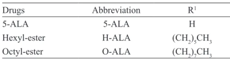
TABLE I - 5-ALA and alkyl esters: general structure = HCl·NH2−CH2−CO−CH2−CH2CO−OR1
Drugs Abbreviation R1
5-ALA 5-ALA H
Hexyl-ester H-ALA (CH2)5CH3
Q real, t = (C measured, t x Vr + (Va x ∑ n-1 C a),
where Q= accumulated permeated amount; Q real= real value at the time t; C measured, t= concentration measured from the sample at time t; V r= volume of the diffusion cell; Va = volume of the removed sample and Ca = concentration of the removed sample.
Finally, the amount of drugs permeated through the skin was divided by the skin area, and these values were plotted as a function of the time (µg/cm2).
In vitro skin retention studies
At the end of the above described experiment (after 16 h), the amount of drug present into the skin was also evaluated. For that, the skin was removed from the diffu-sion cell and pinned to a piece of ParailmTM with the SC
face up. That part of the skin, which had been exposed to the formulation (1.7cm2), was then tape-stripped 10
times using Scotch Book Tape n° 845 (3M, St Paul, MN). The tape-strips were subsequently immersed in 10 mL of methanol in a vial, and shaken for 1 min to extract the permeant, before an aliquot of the resulting solution was subjected to derivatization and luorimetric analyses, to evaluate the compound in the SC. The remaining skin [epidermis without SC + dermis], was cut into small pie-ces and homogenized with 5 mL of methanol for 1 min, sonicated for 30 min and iltered. An aliquot of the iltered homogenate was then analyzed by luorimetric assay, to determine the compound quantity in the skin without the SC (“viable epidermis”).
RESULTS AND DISCUSSION
Drugs that are highly hydrophilic or lipophilic cannot lead to an optimum passive transport through the skin and others biological barriers. Then, the 5-ALA es-teriication aims to increase their lipophilic properties, in order to verify if this modiication in the 5-ALA molecule could really improve skin penetration of the drug. Then, partition coeficient studies were realized to evaluate (i) the lipophilicity grade, represented by KO/W and (ii) the afinity to the full thickness skin (KS/W) of 5-ALA and its more lipophilic derivatives. Table II shows partition coe-ficients studies of 5-ALA, H-ALA and O-ALA.
As can be seen at Table II, the Ko/w of 5-ALA is low, indicating that this molecule is a highly hydrophilic com-pound. Also, its Ks/w was low, meaning 5-ALA limited afinity to the skin. On the contrary, the esteriication of the drug with hexyl and octyl alcohol increased signiican -tly its lipophilicity. Table II shows that this lipophilicity
increases with the alkyl chain length of the alcohol used in the esteriication. In this way, H-ALA is about 3 times more lipophilic than 5- ALA, and O-ALA is about 6 times more lipophilic than the mother molecule. The esteriica -tion improves as well the afinity of the drug for the skin, as showed by KS/W results.
Recently, stratum corneum/water partition coefi -cients of 5-ALA and several alkyl esters were determined by De Rosa et al. (2003) and also showed this effect, that is, an increase in the K with the alkyl chain length of the ALA ester.
However, the KSC/W of ALA esters (De Rosa et al., 2003) is much higher than the KS/W (Table II). These results indicated that the afinity of ALA esters for the SC is signi -icantly higher than their afinity for the viable epidermis, pointing out retention of these esters in the most external barrier of the skin, i.e, the SC. They also indicate that the esters can penetrate the skin thanks to improvement of drug skin afinity. It is well known (Aulton, 2005) that to have an eficient topical effect, a drug has to release itself from the formulation and enter, by partition and diffusion, into the skin. Therefore, the more drug enters the skin, the more drug can diffuse to deeper layers of the tissue. In this way, the afinity of ALA esters for the SC can lead to an improved amount of the drug into skin. It can improve the 5-ALA bioavailability in the tissue and, possibly, might even lead to a more homogeneous distribution of the re-sulting photosensitizer.
The SC lipidic lamellae consists mainly of cerami-des, cholesterol, free fatty acids and cholesteryl sulphate, however it is devoid of phospholipids, which are bilayer forming components in all other cellular and intracellular membranes (Wertz et al, 1986). It was suggested that lipids such as ceramides and cholesterol participate of the bilayer formation in the SC, providing selectivity in the transcu-taneous permeation of lipophilic and hydrophilic subs-tances (Gray, Yardley, 1975). In this way, the similarity TABLE II - Octanol/water (KO/W) and skin/water (KS/W) partition
coeficients (K) of 5-ALA and its n-alkyl esters
Partition
coeficients 5-ALA H- ALA O-ALA
K O/Wa 0.055
(± 0.007)
0.1720 (± 0.035)
0.3205 (± 0.010)
K S/Wb 0.032
(± 0.007)
0.2740 (± 0.135)
0.8265 (± 0.020)
K sc/wc 0.042 8.169 10.522
a, bStatistic test ONE WAY ANOVA (Tukey’s multiple
of composition of the drug delivery system with the main SC lipid, the CER III, could lead to a higher interaction between formulation and skin, and consequently, a higher drug accumulation/retention in the tissue. Thus, in order to verify the inluence of CER III on cutaneous delivery of 5-ALA and its esters, the in vitro skin permeation and retention studies were carried out.
According to Robert (1997), the skin permeability to polar solutes can be favored by hydrocarbon based vehicles, as CER. In this way, we have expected an im-proved permeation for 5-ALA dispersed in CER formu-lation. However, it was not observed in the experiment of in vitro skin permeation of our work. Figure 2shows the cumulative amount of 5-ALA, H-ALA and O-ALA present in the receptor solution after permeation through full-thickness porcine ear skin, after 16 h. It is possible to notice that the presence of CER did not increase the permeation of 5-ALA and its esters. In the case of 5-ALA, it has even decreased signiicantly the amount of drug in the receptor solution.
A reasonable explanation for the results obtained is that the propylene glycol, also present in the formulations, had changed solubility pattern of the skin (William, Barry, 2004), increasing the partition coeficient of the drug (to -gether with CER) to this layer and, therefore, decreasing its permeation through the skin. These results, however, can be advantageous, once systemic risk effects could be avoided by this low permeation and for a topical treatment, the drug should be present into the skin and not in the circulation (represented by the receptor solution in the in vitro experiments).
To verify the inluence of CER in the skin retention of 5-ALA and its esters, their presence in the SC and “viable epidermis” was also evaluated. As can be seen in Figure 3, drugs skin retention was not improved by CER presence in the formulation. Moreover, for H-ALA and O-ALA CER presence has even decreased drug amount in the SC (a) and viable epidermis (b), respectively. Therefore, CER III was not eficient to improve neither 5-ALA/esters derivatives permeation nor retention, when dispersed in a propylene glycol solution.
It has been postulated (Coderch et al., 2003; Bou-wstra, Ponec, 2006) that formulations containing lipids identical to those in skin, in particular, some ceramide supplementation could have two divergent actions: (i) improve intercellular lamellae disorder or (ii) increase the skin lamellar phase, which could dificult the penetration of some drugs. These at irst sight contradictory effects are dependent of the formulation that contains the ceramide. It seems that dispersion of CER in propylene glycol leads to increase of the skin lamellar phase (Jager et al., 2003).
Probably, dispersion changes skin pattern, making dificult in this case, the drugs diffusion.
According to Figure 3 (a, b), the H-ALA retention is higher than 5-ALA and O-ALA. The same result in SC was observed by De Rosa et al. (2003) for these drugs dis-persed in an O/W emulsion, that is, the H-ALA retention in SC was also higher than 5-ALA and O-ALA.
These authors veriied also a small amount of O-ALA through the skin, and it is probably related to the small release of this drug from the formulation (O/A emulsion). They have postulated that due to the fact that O-ALA is highly lipophilic, its release from this vehicle is also dificult, because its interaction with the oil phase of the emulsion is also high. The same behavior can be observed in Figure 2 (indicating a low skin permeation of O-ALA) and Figure 3, where the skin retention of O-ALA (SC as well as in epidermis plus dermis) is low, probably due to its high afinity to the vehicle, in this case, CER III in propylene glycol.
The 5-ALA low retention is much probably related to its hydrophilicity, which becomes difficult the drug skin penetration. In conclusion, CER III dispersed in a propylene glycol vehicle did not improve the skin retention and permeation of 5-ALA and its ester derivatives . The composition of the formulation where CER III is dispersed inluences directly its performance. The development of appropriate vehicles for 5-ALA and its ester derivatives FIGURE 2 - In vitropermeation of 5-ALA, H- ALA and O-ALA
(μg/cm2) through porcine skin, from PG solution and mixture
with ceramide (2.0% w/w) in PG, after 16 h of permeation experiments. Percentage of 5-ALA and its ester derivatives in the formulation: 1.0% (w/w). Values are expressed as S. M. E
(n=4) for each group. Values are signiicantly different only
is required to improve their skin penetration/retention equilibrium.
ACKNOWLEDGEMENTS
Fundação de Amparo à Pesquisa do Estado de São Paulo - FAPESP, Brazil, supplied inancial support.
REFERENCES
AULTON, M. Delineamento de Formas farmacêuticas. 2. ed. Rio de Janeiro, Artmed Editora Ltda, 2005. 677p.
BOUWSTRA J.A; PONEC M. The skin barrier in healthy and diseased state. Biochim Biophys Acta. Amsterdam, v.1758, n.12, p.2080-2095, 2006.
CODERCH, L., LOPEZ, O., DE LA MAZA, A., PARRA, J.
L. Ceramides and skin function. Am. Clin. Dermatol., Auckland, v.4, n.2, p.107-129, 2003.
CURNOW, A., McILROY, B. W., POSTLE-HACON, M. J., PORTER, J. B., Mac ROBERT, A. J. and. BOWN, S. G. Enhancement of 5- aminolaevulinic acid induced photodynamic therapy using hydroxypyridinone iron chelating agents. Br. J. Cancer, London, v.78, n.10, p.1278-1282, 1998.
DE ROSA, F. S., MARCHETTI, J. M., THOMAZINI, J.
A., TEDESCO, A. C. BENTLEY, M. V. L. B. A vehicle for photodynamic therapy of skin cancer: influence of dimethylsulphoxide on 5-aminolevulinic acid in vitro cutaneous permeation and in vivo protoporphyrin IX
accumulation determined by confocal microscopy.J.
Control Rel., Amsterdam, v.65, n.3, p.359-366, 2000.
DE ROSA, F.S., TEDESCO, A. C., LOPEZ, R. F. V., PIERRE,
M. B. R., LANGE, N., MARCHETTI, J. M. , ROTTA, J. C. G, BENTLEY, M. V. L. B. In vitro skin permeation and retention of 5-aminolevulinic acid ester derivatives for photodynamic therapy. J. Control. Rel., Amsterdam, v.89, n.2, p.261-269, 2003.
FEINGOLD, K. R. The role of epidermal lipids in cutaneous permeability barrier homeostasis. Thematic review series: Skin Lipids. J. Lipid. Res., Bethesda, v.48, n.12, p.2531-2546, 2007.
GAULLIER, J. M., BERG, K., PENG, Q., ANHOLT, H., SELBO, P. K., MA, L. W. AND MOAN, J. Use of 5-aminolevulinic acid esters to improve photodynamic therapy on cells in culture. Cancer Res., Baltimore, v.57, n.8, p.1481-1486, 1997.
GRAY, G. M. AND YARDLEY, H. J. Different population of pig epidermal cells: isolation and lipid composition. J. Lipids Res., Bethesda, v.16, n.6, p.441-447, 1975.
JAGER, M. W., GOORIS, G. S.; DOLBNYA, I. P.; BRAS, W.;
PONEC, M.; BOUWSTRA, J.A. The phase behaviour of
skin lipid mixtures based on synthetic ceramides. Chem. Phys.Lipids, Amsterdam, v.124, n.2, p.123-134, 2003.
JEONG, T.H; OH, S. G. Influence of the Ceramide (III) and Cholesterol on the Structure of a Non-hydrous
Phospholipid-based Lamellar Liquid Crystal Structural and
Thermal Transition Behaviors. Bull. Korean Chem. Soc., Seoul, v.28,n.6, p.1021-1030, 1026, 2007.
KESSNER D, RUETTINGER A, KISELEV MA, WARTEWIG S, NEUBERT RH. Properties of Ceramides and Their Impact on the Stratum Corneum Structure: Part 2: Stratum Corneum Lipid Model Systems. Skin Pharmacol. Physiol.,
Basel, v.21, n.2, p.58-74, 2008.
KLOECK, J; AKKERMANS, W.; BEIJERSBERGEN VAN HENEGOUWEN, G. M. J. Derivatives of 5-aminolevulinic acid for protodynamic therapy: enzimatic conversion into protoporphyrin. Photochem. Photobiol. Oxford, v.67, n.1, p.150-154, 1998.
LONg, S.A., WERTZ, P.W., STRAuSS, J. S., DOWNINg, D.
T. Human stratum corneum polar lipids and desquamation.
Arch. Dermatol. Res., Berlin, v.227, n.4, p.284-287, 1985.
LOPEZ, R. F. V.; BENTLEY, M. V.; DELgADO-CHARRO,
M.B ; GUY, R.H . Iontophoretic delivery of 5-aminolevulinic acid (ALA): effect of pH. Pharm. Res, New York, v.18, n.3, p.311-315, 2001.
LOPEZ, R. F, LANgE, N., guY, R., BENTLEY, M. V.
Photodynamic therapy of skin cancer: controlled drug delivery of 5-ALA and its esters. Adv. Drug. Deliv. Rev.,
Amsterdam, v.56, n.1, p.77-94, 2004.
MOTTA, S., MONTI, M., SESANA, S., MELLESI, L., GHIDONI, R., CAPUTO, R. Abnormality of water barrier function in psoriasis. Rule of ceramide fractions.Arch. Dermatol., Chicago, v.130, n.4, p.452-456, 1994.
MURMUR, E. S., SCHUMULTS, C. D., GOLDBERG, D. J. A review of laser and photodynamic therapy for the treatment of nonmelanoma skin cancer. Dermatol. Surg., New York, v.30, suppl.2, p.264-271, 2004.
OECD, 1995. OECD guideline for testing of chemicals N°
107. Partition coeficient (n-octanol/water): shake-lask
method. OECD. Available at: http://www.oecd.org/ dataoecd/17/35/1948169.pdf. Access on: 20th. Feb. 2009.
OISHI, H., NOMIYAMA, H., NOMIYAMA, K., TOMUKINI, K. Fluorometric HPLC determination of 5- Aminolevulinic acid (ALA) in the plasma and urine of lead workers: biological indicators of lead exposure. J. Anal. Toxicol., Niles, v.20, n.2, p.106-110, 1996.
PIERRE, M. B. R., TEDESCO, A. C., MARCHETTI J. M., BENTLEY M. V. L. B. Stratum corneum lipid liposomes for the topical delivery of 5-aminolevulinic acid in photodynamic therapy of skin cancer: preparation and in vitro permeation study. B. M. C. Dermatology, v.1, n.5, p.1471-1476, 2001. Available at: http://www.biomedcentral. com/1471-5945/1/5. Access on: 19th. Aug. 2008.
PIERRE, M. B. R.; RICCI, E. JR, TEDESCO, A. C. and BENTLEY, M. V. Oleic Acid as Optimizer of the Skin Delivery of 5-Aminolevulinic Acid in Photodynamic Therapy. Pharmaceutical Research, New York, v.23, n.2, p.360-366, 2006.
ROBERT, M.S. Target drug delivery to the skin and deeper tissues: role of physiology, solute structure and disease,
Clin. Exp. Pharmacol. Physiol., Oxford, v.24, n.11, p.874-879, 1997.
ROBSON, K. J., STEWART, M. E., MICHELSEN, S., LAZO,
N. D., DOWNING, D. T. J. 6-Hydroxy-4-sphingenine in human epidermal ceramides. Lipid Res., Bethesda, v.35, n.11, p.2060-2068, 1994.
SCHEUPLEIN, R. J., BLANK, I. H., BRAUNER, G. J., MCFARLANE, D. J. Percutaneous absorption of steroids. J. Invest. Dermatol., New York, v.52, n.1, p.63-70, 1969.
VAN DEN AKKER, J. T. H. M.; IANI, V.; STAR, W. M.; STERENBORG, H. J. C. M. AND MOAN, J. Topical Application of 5-Aminolevulinic Acid Hexyl Ester and 5-Aminolevulinic Acid to Normal Nude Mouse Skin: Differences in Protoporphyrin IX Fluorescence Kinetics and the Role of the Stratum Corneum. Photochem. Photobiol., Oxford, v.72, n.5, p.681-689, 2000.
WERTZ P. W. AND DOWNINg, D. T. Ceramides of pig epidermis: structure determination. J. Lipid Res., Bethesda, v.24, n.6, p.759-765, 1983.
WERTZ, P. W., ABRAHAM, W., LANDMAN, L., DOWNINg,
D. T. 1986. Preparation of liposomes from stratum corneum lipids. J. Invest. Dermatol., New York, v.87, n.5, p.582-584, 1986.
WILLIAMS, A. C., BARRY, B. W. Penetration enhancers. Adv. Drug Deliv. Rev., Amsterdam, v.56, n.5, p.603-618, 2004.
YARDLEY, H. J., SUMMERLY, R. Lipid composition and metabolism in normal and diseased epidermis. Pharmacol. Ther., Oxford, v.13, n.2, p.357-383, 1981.
ZEITOuNI, N. C., OSEROFF, A. R., SHIEH, S. Photodynamic therapy for nonmelanoma skin cancers. Current review and update. Mol. Immunol., Oxford, v.39 , n.17-18, p.1133-6, 2003.
![FIGURE 3 - In vitro retained amount of 5-ALA and its esters into: (a) SC and (b) [epidermis without SC + dermis] in porcine ear skin, after 16 h of permeation study from PG solution and mixture with ceramide (2.0% w/w) in PG](https://thumb-eu.123doks.com/thumbv2/123dok_br/15412317.586695/6.892.71.422.117.583/figure-retained-epidermis-porcine-permeation-solution-mixture-ceramide.webp)
