Arq Neuropsiquiatr 2001;59(2-B):421-423
NASO-ETHMOID SCHWANNOMA WITH
INTRACRANIAL EXTENSION
Case report
Mario G. Siqueira
1, Erik Jennings
1, Osmar J.S. Moraes
1, Marco Tulio S. Santos
1,
Nelci Zanon
1, Belmiro J. Mattos
2, Luiz Belmonte Netto
3ABSTRACT - Intranasal schwannomas are rare lesions, specially when they present with an intracranial extension. The fifth case in the medical literature of a naso-ethmoid schwannoma with extension into the anterior cranial fossa is presented. The magnetic resonance findings and the details of the combined intracranial / transfacial operative approach used are described. The possible origin and the clinical characteristics of this rare lesion are reviewed.
KEY WORDS: schwannoma, intranasal schwannoma, paranasal sinus tumor.
Schwannoma naso-etmoidal com extensão intracraniana: relato de caso
RESUMO - Schwannomas intranasais são lesões raras, principalmente quando apresentam um extensão intracraniana. Estamos apresentando o quinto caso da literatura médica de um schwannoma naso-etmoidal com extensão para o interior da fossa craniana anterior. São descritos os achados da ressonância magnética e os detalhes da via de acesso cirúrgico combinada intracraniana/transfacial. A possível origem e as características clínicas dessa lesaõ rara são revistas.
PALAVRAS-CHAVE: schwannoma, schwannoma intranasal, tumor de seio paranasal.
Serviços de Neurocirurgia1, Cirurgia de Cabeça e Pescoço2 e Patologia3, Hospital Santa Marcelina, São Paulo, SP, Brasil.
Received 28 November 2000, received in final form 14 February 2001. Accepted 19 February 2001.
Dr. Mario G. Siqueira - Rua Virgilio de Carvalho Pinto, 381/apt 42 - 05415-030 São Paulo SP - Brasil. E-mail: mgsiqueira@uol.com.br
Any nerve with a schwann cell sheath may give
origin to a Schwannoma and so, this neoplasm may
develop in almost any part of the body
1, 2.
Although up to 45% of all schwannomas occur
in the head and neck region
3, the involvement of
the nasal cavity and paranasal sinuses is rare, with
approximately 40 cases reported
3-18. From those
ca-ses, only four have been associated with intracranial
extension into the anterior cranial fossa
4-8,18.
CASE REPORT
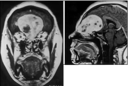
A 40-year-old white woman presented to the Depart-ment of Neurosurgery of the Hospital Santa Marcelina in march, 1995 with a 3-year history of frontal headaches, gradual loss of the olfaction, altogether with a bulging deformity in the frontal area. Eight months prior to ad-mission the patient became anosmic and developed a pa-inful swelling of the frontal area, over the mentioned de-formity. At examination she had no neurological abnor-malities besides a bilateral anosmia. Magnetic resonance imaging revealed a mass lesion that filled the superior part of the naso-ethmoid complex and extended superiorly into the anterior cranial fossa (Fig 1). The tumor was sucessfully
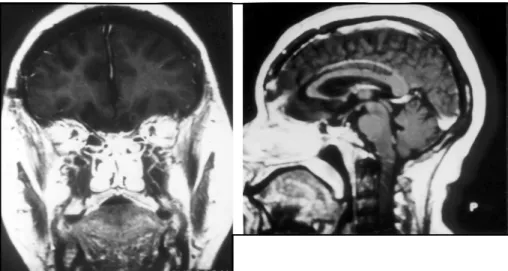
excised through a combined intracranial and transfacial procedure. A bifrontal craniotomy was performed and after extradural elevation of the right frontal lobe a white and smooth tumor was displayed. After the tumor was debulked its intracranial portion was completely removed extradurally to the level of the cribiform plate. The projec-tion of the tumor into the ethmoid sinus and nasal cavity was then totally removed through a lateral rhinotomy. Microscopic examination revealed a tumor that consisted of regions of dense spindle cells, arranged in short bundles and forming interlacing fascicles, and of regions of a loose, myxoid matrix. The histological diagnosis, confirmed by immunohistochemical studies, was of a benign schwan-noma (Fig 2). Shortly after the surgery the patient returned to her previous work and more than five years after the surgical treatment she is doing quite well, with a persis-tent bilateral anosmia. Recent image studies demonstrated the absence of any residual or recurrent tumor (Fig 3).
DISCUSSION
422 Arq Neuropsiquiatr 2001;59(2-B)
Fig 2. Photomicrograph showing resected tumor consisting of compact interlacing fas-cicles of spindle-shaped cells (Antoni A pat-tern). H&E, original magnification X 160.
Fig 1. Pre-operative magnetic reso-nance images. T1-weighted coronal (A) and sagittal (B) images obtained after IV administration of gadolinium shows a large mass lesion occupying the superior part of the naso-ethmoid complex and extending into the an-terior cranial fossa.
the cavities and foramina in which they arise. The
tumor in the present case, as well as the other four
reported in the literature, presumably arose from one
of the intranasal nerves and grew until it eroded the
floor of the frontal fossa to reach the intracranial
compartment. The precise origin of the intranasal
schwannomas is obscure, as there are many nerve
branches in the region. They may arise from any of
the following nerves
18: (a) general sensory branches
of the ophthalmic and maxillary divisions of the
trige-minal nerve; (b) autonomic fibers (parasympathetic)
from the sphenopalatine ganglion and (c) autonomic
fibers (sympathetic) derived from the carotid plexus.
As the olfactory nerves are covered by glial cells they
cannot give rise to nerve sheath tumors
1.
All the cases of nasal schwannomas with
intracra-nial extension, including the one we are reporting,
presented with a long history of nasal symptoms.
The tumors were large at the diagnosis and all of
them were completely resected by an intracranial
approach or by a combined intracranial / transfacial
approach.
The CT or MR images are non specific. The
featu-res pfeatu-resented are those of a benign, slow growing
tumor of greater signal intensity than polyps or
mucoceles
8.The differential diagnosis must include
papilloma, sarcoma, carcinoma, and lymphoma
19.
occu-Arq Neuropsiquiatr 2001;59(2-B) 423
rence, it should be part of the differential diagnosis
of intranasal lesions elected to be submited to
bi-opsy and CT or MR imaging should be taken prior to
the biopsy procedure.
REFERENCES
1. Batsakis JG. Tumors of the peripheral nervous system, ed 2. Baltimore: Williams & Wilkins, 1979:313-333.
2. Rosai J, Ackerman LV. Ackerman’s Surgical Pathology, ed 8. St Louis: Mosby, 1996:Vol 2.
3. Hawkins DB, Luxford WM. Schwannoma of the head and neck in chil-dren. Laringoscope 1980;12:1921-1926.
4. Bavetta S, McFall MR, Afshar F, et al. Schwannoma of the anterior cra-nial fossa and paranasal sinuses. Br J Neurosurg 1993;7:697-700. 5. Calcaterra TC, Rich JR, Ward PW. Neurilemmoma of the sphenoid
si-nus. Arch Otolaryngol 1974;100:383-385.
6. Dutt PK. A case of nasal neurilemmoma. J Laryngol Otol 1969;83:1209-1213.
7. Enion DS, Jenkins A, Miles JB, Diengdoh JV. Intracranial extension of a naso-ethmoid schwannoma. J Laryngol Otol 1991;105:578-581. 8. Gatscher S, Love S, Coakham HB. Giant nasal schwannoma with
in-tracranial extension. J Neurosurg 1998;89:161.
Fig 3. Post-operative magnetig reso-nance images. T1-weighted coronal (A) and sagittal (B) images obtained after IV administration of gadolinium demonstrated the absence of any re-sidual or recurrent tumor. Some de-gree of residual atrophy of the fron-tal lobe is shown.
9. Gignoux M, Labayle J. Les tumeurs nerveuses de fossa nasales en tumeurs de l’ethmoid. Paris: Masson 1949:44-54.
10. Harkins W. Neurinoma of the ethmoid sinuses. Ann Otol Rhinol Laryngol 1949;58:498- 506.
11. Iwamura S, Suriura S, Nomura V. Schwannoma of the nasal cavity. Arch Otolaryngol 1972;96:176-177.
12. Kaufman SM, Conrad LP. Schwannoma presenting as a nasal polyp. Laryngoscope 1976;86:595-596.
13. Khalfifa M, Bassyouni A. Nasal schwannoma. J Laryngol Otol 1981;95:503-507.
14. Perzin KH, Panyu H, Wechter S. Nonepithelial tumors of the nasal cavity, paranasal and nasopharynx: A clinicopathologic study. XII: Schwann cell tumors (neurilemmoma, neurofibroma, malignant schwannoma). Cancer 1982;50:2193-2202.
15. Robitaille Y, Seemayer TA, Deiry AR. Peripheral nerve tumors involv-ing paranasal sinuses: a case report and review of the literature. Can-cer 1975;35:1254-1258.
16. Shugar MA, Montgomery WW, Reardon EJ. Management of paranasal schwannomas. Ann Otol Rhinol Laryngol 1982;91:65-69.
17. Verma PL, Marwaha AR. Intranasal schwannoma. J Laryngol Otol 1970;84:1069-1071.
18. Zovickian J, Barba D, Alksne JF. Intranasal schwannoma with exten-sion into the intracranial compartment: Case report. Neurosurgery 1986;19:813-815.