Audiological characterization of children under oncologic
treatment
Caracterização audiológica de crianças em tratamento oncológico
Érica Alessandra Caldas1, Luciane Maria Oliveira Brito2, Patrícia Andréia Caldas3, Savya Cybelle Milhomem
Rocha4, Edson Diniz Ferreira Filho4, Maria Bethânia da Costa Chein2
ABSTRACT
Purpose: To investigate the effects of cancer treatment on the auditory system of children. Methods: The study population comprised 12 children, aged 2 to 12 years, who had been diagnosed with cancer and recommended radiotherapy with/without chemotherapy. Distortion pro-duct otoacoustic emissions (DP-OAE) and transient evoked otoacoustic emissions OAE (TE-OAE) were measured before treatment and after six months of treatment. Results: Out of the 24 ears, only two ears (8.3%) failed the TE-OAE and DP-OAE tests, and no significant change was observed after cancer treatment. Conclusion: Cancer treatment over a period of six months did not cause hearing impairment in this group of patients.
Keywords: Child; Toxicity; Neoplasms; Hearing; Hearing loss
RESUMO
Objetivo: Investigar os efeitos do tratamento oncológico no sistema auditivo de crianças. Métodos: A amostra foi constituída por 12 crian-ças de 2 a 12 anos de idade, com diagnóstico de câncer e indicação para radioterapia associada ou não à quimioterapia. Foram realizadas pesquisas das emissões otoacústicas produto de distorção (EOA-PD) e emissões otoacústicas transientes (EOA-TE), antes e após seis meses de tratamento. Resultados: Das 24 orelhas, apenas duas (8,3%) falharam para as EOA-TE e EOA-PD e não foi observada alteração significativa após o tratamento oncológico. Conclusão: O tratamento oncológico, durante um período de seis meses, não ocasionou alterações auditivas nesta amostra.
Descritores: Criança; Toxicidade; Neoplasias; Audição; Perda auditiva
This work was carried out at the Post-Graduate Program in Maternal and Child Health, Universidade Federal do Maranhão – UFMA – São Luís (MA), Brazil. (1) Post-Graduate Program (Master’s Degree) in Maternal and Child Health, Universidade Federal do Maranhão – UFMA – São Luís (MA), Brazil.
(2) Post-Graduation Department in Maternal and Child Health, Universidade Federal do Maranhão – UFMA – São Luís (MA), Brazil.
(3) Post-Graduate Program (Master’s Degree) in Speech Pathology, School of Philosophy and Sciences, Universidade Estadual Paulista “Júlio de Mesquita Filho” – UNESP – Marília (SP), Brazil.
(4) Universidade CEUMA – UNICEUMA – São Luís (MA), Brazil.
Funding: Fundação de Amparo à Pesquisa ao Desenvolvimento Científico do Maranhão (FAPEMA). Conflict of interests: No
Authors’ contribution: EAC was responsible for the study design, data collection, data analysis and drafting of the article; LMOB was responsible for the study design; PAC and SCMR were responsible for revision of article; EDFF was responsible for the statistical treatment of data; MBCC was responsible for the con-ception and design of the study, data analysis, supervising the study and drafting of the article.
Correspondence address: Érica Alessandra Caldas. Centro de Pesquisa do CBBS, Cidade Universitária, Av. dos Portugueses, 1966, Bacanga, São Luís (MA), Brazil, CEP: 65080-805. E-mail: [email protected]
INTRODUCTION
Hearing loss can produce several psychosocial alterations, since auditory deterioration and problems associated with speech comprehension negatively affect the sociability of an individual. The consequences can be even more serious during the course of speech acquisition in children, since speech and language acquisition depends on the hearing ability. Thus, early diagnosis and especially prevention of hearing loss in children is essential, as it can cause a delay in speech acquisition, reducing the child’s sociability in school and among family. This can result in social and emotional disorders that can persist well into adulthood(1).
It is known that cancer therapy can lead to hearing dysfunc-tion. It is estimated that there will be approximately 11840 new cases of cancer in children and adolescents up to the age of 19 in Brazil in 2014, of which 2790 will be in north-eastern Brazil(2).
In some developing countries, childhood cancer comprises 3%–10% of all cancers and death by cancer is considered to be the second leading cause of childhood mortality (4%–5%). In developed countries, on the other hand, childhood cancer represents approximately 1% of all cancers, and its mortality rate is much lower than that in developing countries (approxi-mately 1%), infectious diseases being the main cause of death(2).
There are different therapies for cancer and each one causes different functional changes in the individual, some being re-versible and some irrere-versible. These changes can occur early or late, depending on the therapy and the age of the child(3).
One such change is ototoxicity, which is defined as the reaction that produces structural lesions in the inner ear, le-ading to irreversible sensorineural, descending, and bilateral auditory alterations(4). These alterations initially involve the
basal portion of the cochlea, affecting high frequencies, and may progress towards the cochlear apex, affecting middle and low frequencies(5).
Approximately 200 drugs are considered ototoxic, including polyfunctional alkylating agents, antimetabolites, antitumor antibiotics and mitotic inhibitors(6).
The degeneration of the organ of Corti caused by the use of ototoxic drugs can be avoided through adequate auditory monitoring(7), and the assessment of otoacoustic emissions
(OAE) is one of the indicated tests(1,8).
The OAE test has a higher sensitivity and specificity than other methods for the assessment of auditory function. The recording of emissions, with the aim of monitoring cochlear function in ototoxic drug users, displays altered responses before any changes in auditory threshold are recorded(8).
According to the criteria proposed by the American Speech Language Hearing Association (ASHA)(6), audiologic
evalua-tion must be performed prior to the start of drug therapy or, at most , 24 hours after the administration of the first dose of chemotherapy and within the first 72 hours in case of antibiotic therapy. In the case of a decrease or absence of response in
a previously present frequency, a complete audiological re--evaluation and reassessment of therapy protocol is suggested.
Thus, the objective of this study was to assess the effects of cancer therapy on the auditory system of children by the measurement of otoacoustic emissions.
METHODS
The study was started after obtaining approval from the Ethics and Research Committee of the University Hospital of the Universidade Federal do Maranhão (UFMA),
accord-ing to consubstantiated opinion nº 123.444 and followaccord-ing the Resolution of the National Council of Health/Ministry of Health no. 466/2012.
The study population comprised 12 children of both gen-ders, aged 2 to 12 years, who had been diagnosed with cancer and recommended chemotherapy, with or without radiotherapy. None of the children exhibited any otologic symptoms.
After consent to include the children in the study was obtained from their guardians, all children were subjected to two evaluations for the detection of auditory disorders: the first before starting the therapy (T0) and the second after six months of therapy (T1).
We used a structured questionnaire developed for this study, to collect demographic data, and information such as variables of the pathology underlying the oncological therapy, and comorbidities that could impair hearing. These data were provided by the children’s guardians or obtained by the analysis of hospital records, to ensure absence of history of audiologi-cal and otologiaudiologi-cal disorders and risk factors for hearing loss.
In all children, the external acoustic meatus was examined with an otoscope (Pocket Jr WelchAllyn®), to identify any type of alteration, and if needed, to refer to an otolaryngologist, for later assessment of OAE.
Due to weakness in the children, the OAE were measured while they lay in bed. Care was taken to eliminate as much of background noise as possible, so as to ensure accurate evalu-ation of the OAE responses.
The equipment used was the OtoRead® Otoacoustic Emissions instrument to assess Transient-Evoked Otoacoustic Emissions (TEOAE) and Distortion Product Otoacoustic Emissions (DPOAE), duly calibrated according to standards 8253.1, Resolution 364.9 ISO3741 and using frequencies of 1.5, 2, 2.5, 3, 3.5 and 4 kHz for TEOAE and 2, 3, 4 and 5 kHz for DPOAE.
The possible results of this examination were classified as: “pass”, which indicates the absence of alterations identified by this method; and “failed”, which demonstrates the absence of OAEs in at least three frequency bands(9).
The presence of OAE was defined as a signal-to-noise ratio
≥ 5 dB(10).
RESULTS
We studied 12 children, the majority of whom were boys (58.3%), had brown skin (83.3%), and were residents of the city of São Luís (MA) (66.7%).
The most frequent oncologic diagnosis was acute lym-phocytic leukemia (ALL) (58.3%). The other types of cancer– pilocytic astrocytoma, acute myeloid leukemia (AML), lym-phoma, retinoblastoma and Wilms’ tumor–were present with approximately equal frequencies (approximately 8.3 % each).
The child with Wilms’ tumor was provided abdominal radiotherapy (total dose of 540 cGy, divided into 6 doses of
90 cGy each) associated with chemotherapy; the remaining 11 (91.6 %) children received only chemotherapy.
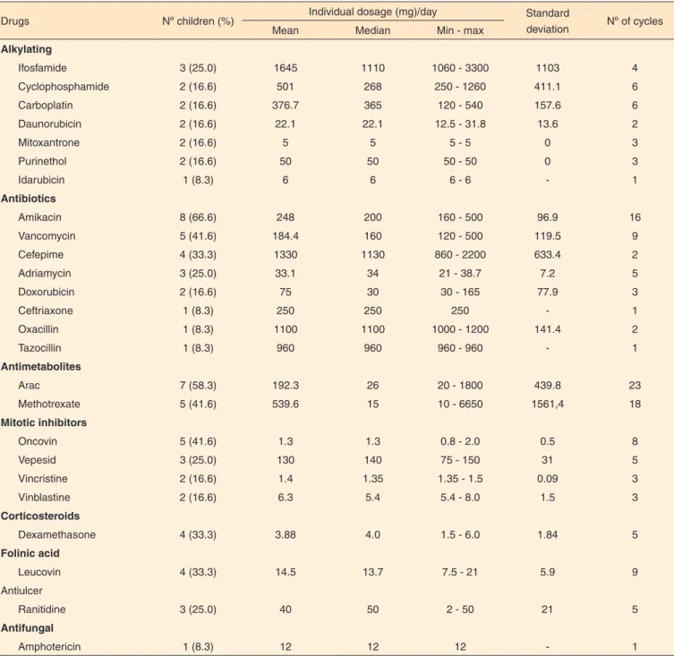
Amikacin was the most frequently used anti-cancer drug (66.6 %) (Table 1).
Of a total of 24 ears (12 children) examined at T0, none failed the OAE test. At T1, one child failed the DPOAE and TEOAE tests in both ears. This boy, aged 2 years and 10 mon-ths, had AML and was subjected to 4 cycles of chemotherapy, consisting of amikacin (180 mg/m2/day), amphotericin B (12
mg/m2/day), ARAC (26 mg/m2/day), idarubicin (6 mg/m2/day),
mitoxantrone (5 mg/day), tazocin (960 mg/day), vancomycin (120 mg/m2/day) and vepesid (960 mg/m2/day).
Table 1. Frequency of use, drug, dose and nº cycles (IMOAB, 2013)
Drugs Nº children (%) Individual dosage (mg)/day Standard
deviation Nº of cycles
Mean Median Min - max
Alkylating
Ifosfamide 3 (25.0) 1645 1110 1060 - 3300 1103 4
Cyclophosphamide 2 (16.6) 501 268 250 - 1260 411.1 6
Carboplatin 2 (16.6) 376.7 365 120 - 540 157.6 6
Daunorubicin 2 (16.6) 22.1 22.1 12.5 - 31.8 13.6 2
Mitoxantrone 2 (16.6) 5 5 5 - 5 0 3
Purinethol 2 (16.6) 50 50 50 - 50 0 3
Idarubicin 1 (8.3) 6 6 6 - 6 - 1
Antibiotics
Amikacin 8 (66.6) 248 200 160 - 500 96.9 16
Vancomycin 5 (41.6) 184.4 160 120 - 500 119.5 9
Cefepime 4 (33.3) 1330 1130 860 - 2200 633.4 2
Adriamycin 3 (25.0) 33.1 34 21 - 38.7 7.2 5
Doxorubicin 2 (16.6) 75 30 30 - 165 77.9 3
Ceftriaxone 1 (8.3) 250 250 250 - 1
Oxacillin 1 (8.3) 1100 1100 1000 - 1200 141.4 2
Tazocillin 1 (8.3) 960 960 960 - 960 - 1
Antimetabolites
Arac 7 (58.3) 192.3 26 20 - 1800 439.8 23
Methotrexate 5 (41.6) 539.6 15 10 - 6650 1561,4 18
Mitotic inhibitors
Oncovin 5 (41.6) 1.3 1.3 0.8 - 2.0 0.5 8
Vepesid 3 (25.0) 130 140 75 - 150 31 5
Vincristine 2 (16.6) 1.4 1.35 1.35 - 1.5 0.09 3
Vinblastine 2 (16.6) 6.3 5.4 5.4 - 8.0 1.5 3
Corticosteroids
Dexamethasone 4 (33.3) 3.88 4.0 1.5 - 6.0 1.84 5
Folinic acid
Leucovin 4 (33.3) 14.5 13.7 7.5 - 21 5.9 9
Antiulcer
Ranitidine 3 (25.0) 40 50 2 - 50 21 5
Antifungal
Amphotericin 1 (8.3) 12 12 12 - 1
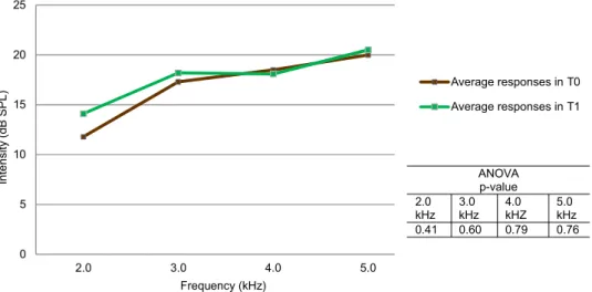
Figures 1 and 2 present the results of the comparison betwe-en the mean frequbetwe-encies obtained at T0 and T1 in the DPOAE and TEOAE tests. There was no significant difference in any of the frequencies tested.
DISCUSSION
We agree with published literature(4) that emphasizes the
importance of setting up audiology services in oncological therapy centers, because the practice of auditory monitoring is not prevalent in Brazil, which leads to a smaller sample size and hinders data collection.
In the present study, only two ears (8.3%), of a single child, failed the TEOAE and DPOAE tests after oncological therapy. This child was treated with amikacin and vancomycin at mean doses of 180 mg/m2/day and 120 mg/m2/day, respectively. The
ototoxic effect of vancomycin has already been proven(11,12). It
is especially notable when vancomycin is used in association
with other potentially ototoxic drugs(13), which was not the
case with this child.
The same methodology was followed in another study conducted on 250 newborns(14). In addition, that study also
employed audiometry with visual reinforcement, tympanome-try, and brainstem auditory evoked potential test, conducted at 3 time points (at the time of discharge from hospital, after three months of discharge, and after six months of discharge). These newborns were treated with a combination of amikacin, cefotaxime, furosemide, ceftazidime, and vancomycin, and hearing loss was found in 11.6% of them.
Cisplatin and other platinum derivatives are the most com-mon ototoxic agents described in literature(7,8,15-18). Currently,
an increase in the number of studies on ototoxic medications has led to awareness regarding their adverse effects, and hence the use of these drugs has become less common(1).
In our sample, only two children were administered car-boplatin in 4 cycles between T0 and T1 (doses of 120, 240,
0 5 10 15 20
1.5 2.0 2.5 3.0 3.5 4.0
Intensity
(dB SPL)
Frequency (kHz)
Average responses in T0
Average responses in T1
ANOVA p-value 1.5
kHz 2.0 kHz
2.5 kHz
3.0 kHz
3.5 kHz
4.0 kHz 0.38 0.70 0.38 0.49 0.17 0.67
Note: T0 = Assessment before initiating therapy; T1 = Assessment after six months of therapy; TEOAE = Transient-Evoked Otoacoustic Emission; dB SPL = decibel of sound pressure level
Figure 2. Mean responses found at T0 and T1 for TEOAE, by frequency 0
5 10 15 20 25
2.0 3.0 4.0 5.0
Intensity
(dB SPL)
Frequency (kHz)
Average responses in T0
Average responses in T1
ANOVA p-value 2.0
kHz 3.0 kHz
4.0 kHZ
5.0 kHz 0.41 0.60 0.79 0.76
Note: T0 = Assessment before initiating therapy; T1 = Assessment after six months of therapy; DPOAE = Distortion Product Otoacoustic Emissions; dB SPL = decibel of sound pressure level
360 and 400 mg/m2/cycle, respectively for the 4 cycles), with
a mean interval of 20 days between cycles. These children had been diagnosed with retinoblastoma and pilocytic astrocytoma and passed the TEOAE and DPOAE tests at the end of the fourth cycle.
The same was observed(16) in a previous study on 18
chil-dren (age range: 9 months to 9 years) who were administered carboplatin in 4 to 6 cycles (560 mg/m2/cycle). In this study,
all the children passed the TEOAE test at frequencies of 1, 1.5, 2, 3, and 4 kHz, i.e. they did not exhibit ototoxicity.
Researchers examined 25 children with retinoblastoma who had been administered carboplatin (cumulative dose of 2240 mg/m2), by tympanometry, OAE test, audiometry, and
BAEP test. They concluded that carboplatin does not present any danger of ototoxicity, unless when used in combination with other drugs(15).
Nevertheless, in a retrospective cohort study(17) on 60
children treated for retinoblastoma, from 1996 to 2005, who used two chemotherapy schemes (which included carboplatin), the authors concluded that carboplatin is associated with a significant risk of development of hearing loss. Furthermore, the study reports that the age at the onset of therapy plays a significant role in the development of hearing loss: younger age is associated with a higher susceptibility to hearing loss. This supports our results in which the child who failed the OAE tests was 2 years and 10 months old.
In another study, 67 patients (8 to 23 years old) receiving therapy with carboplatin and cisplatin, were examined by pure tone audiometry; 38% of them exhibited hearing loss(18).
When comparing our study and the studies cited above, it should be noted that studies with a larger sample size had a higher percentage of children with hearing loss.
However, in a study(7) investigating the alteration of the
auditory thresholds in patients with childhood cancer who had been treated with antineoplastic drugs (cisplatin and vincristine + actinomycin D + other drugs), eight out of ten individuals were found to have hearing loss. Five of them exhibited significant changes at 6 kHz. This indicates the importance of high frequen-cies (6 and 8 kHz), which were not tested in our study due to the limitation of the equipment used. There is a consensus among researchers(5,7,19) that the auditory alterations caused by the use
of ototoxic drugs initially involve high frequencies; hence, it is important to use equipment that can attain these frequencies.
When comparing the mean signal-to-noise ratio by frequen-cies between responses at T0 and T1 in this study, there was a deterioration at 1.5 and 4 kHz in the TEOAE test. However, there was a slight improvement at 2 and 3 kHz in the DPOAE test and at 2, 3, and 3.5 kHz in the TEOAE test.
The same was observed in another study(19), which reported
constant improvement in DPOAE responses, rather than dete-rioration, and attributed this improvement to cochlear lesion or dysfunction in the area close to the response, which favors the capture conditions of the “response sound” through these
injured regions, causing them to be captured with greater ease by the device probe, or, even, by an irritation of the CCE before the lesion, as occurs in the cells of the vestibular system.
This was also reported by other researchers(20), who found
an increase in the signal/noise ratio at 1 and 2 kHz, after a dose of 120 mg/m2 of cisplatin; however, there was a decrease after
a dose of 240 mg/m2. The authors explained this variation by
chemical changes caused by cisplatin in cells; the increase at low dose may indicate lesions and subsequent cell death.
Hence, we stress the importance of auditory assessment, during not only oncological therapy, but also post- therapy. Hearing loss by ototoxicity may appear up to six months after exposure to the drug and, if during this period, the hearing thresholds are not stabilized, monitoring should be continued(21).
Likewise, patients considered to be at high risk of ototoxicity are those who most need monitoring. These include patients with renal function defects, those receiving high doses of ototoxic drugs or prolonged treatment, very young or very old individuals, those with symptoms of hearing loss, newborn infants, or those suffering from malnutrition or having genetic predisposition to hearing loss(21).
Monitoring allows prevention of the onset of hearing loss and modification of anti-cancer therapy to minimize its effects on the quality of life of the patients. It also facilitates patient education through guidelines and required referrals(1).
Limitations of the study
The Instituto Maranhense de Oncologia Aldenora Belo
(IMOAB) treats children with cancer from different locations. Majority of the patients have already received some kind of therapy, thus making it difficult to obtain a large sample size for conducting any study.
In the present study, tests were not conducted at 6 and 8 kHz, because of the limitation of the equipment used. The lack of an audiology section and specialized equipment prevented acquisition of more accurate data.
A study with a longer observation period will make more detailed monitoring possible, along with detection of higher auditory variations. In addition, it will be possible to standar-dize chemotherapy for the children participating in the study to a greater extent.
CONCLUSION
There were no significant changes in global responses, as well as responses characterized by frequency, in the DPOAE and TEOAE tests, after 6 months of cancer therapy.
ACKNOWLEDGEMENTS
REFERENCES
1. Jacob LCB, Aguiar FP, Tomiasi AA, Tschoeke SN, Bitencourt RF. Monitoramento auditivo na ototoxicidade. Rev Bras Otorrinolaringol. 2006;72(6):836-4. http://dx.doi.org/10.1590/ S0034-72992006000600017
2. Instituto Nacional de Câncer. Quimioterapia. 2012 [citado 28 jan 2014]. Disponível em: http://www.inca.gov.br/conteudo_view. asp?ID=101
3. Liberman PHP. Avaliação auditiva em pacientes tratados de câncer na infância [dissertação]. São Paulo: Fundação Antônio Prudente; 2005. 4. Silva AM, Latorre MRDO, Cristofani LM, Odone Filho VO. A
prevalência de perdas auditivas em crianças e adolescentes com câncer. Rev Bras Otorrinolaringol. 2007;73(5):608-14. http://dx.doi. org/10.1590/S0034-72992007000500005
5. Liberman PHP, Goffi-Gomez MVS, Schultz C, Lopes LF. Quais as frequências audiométricas acometidas são responsáveis pela queixa auditiva nas disacusias por ototoxicidade após o tratamento oncológico? Arq Int Otorrinolaringol. 2012;16(1):26-31. http:// dx.doi.org/10.7162/S1809-48722012000100003
6. American Speech-Language-Hearing Association (ASHA). Audiologic management of individuals receiving cochleotoxic drug terapy. RockAmerican Speech-Language-Hearing Association; 1994. 7. Almeida EOC, Umeoka WG, Viera RC, Moraes IF. Estudo de alta
frequência em pacientes curados de câncer tratados com cisplatina. Rev Bras Otorrinolaringol. 2008;74(3):382-90. http://dx.doi. org/10.1590/S0034-72992008000300012
8. Yilmaz S, Oktem F, Karaman E. Detection of cisplatin-induced ototoxicity with transient evoked otoascustic emission test before pure tone audiometer. Eur Arch Otorhinolaryngol. 2010;267(7):1041-4. http://dx.doi.org/10.1007/s00405-009-1165-7
9. Cavalcante JMS, Isaac ML. Análise das emissões otoacústicas transientes em recém-nascidos a termo e pré-termo. Brazilian J Otorrinolaryngol. 2013;79(5):582-8. http://dx.doi.org/10.5935/1808-8694.20130104
10. Sousa LCA, Piza MRT, Alvarenga KF, Cóser PL. Eletrofisiologia da audição e emissões otoacústicas: princípios e aplicações clínicas. São Paulo: Novo Conceito; 2008. Capítulo 10, Emissões otoacústicas (EOA). p.110-27.
11. Salt AN. Pharmacokinetics of drug entry into cochlear fluids. Volta Rev. 2005;105(3):277-98.
12. Forouzesh A, Moise PA, Sakoulas G. Vancomycin ototoxicity: a reevaluation in an era of increasing doses. Antimicrob Agents Chemother. 2009;53(2):483-6. http://dx.doi.org/10.1128/ AAC.01088-08
13. Shields RK, Martello JL, Potoski BA. Is vancomycin ototoxicity a significant risk? Antimicrob Agents Chemother. 2009;53(10):4572-3. http://dx.doi.org/10.1128/AAC.00537-09
14. Câmara MFS, Azevedo MF, Lima JWO, Sartorato EL. Efeito de fármacos ototoxicos na audição de recém nascidos de alto-risco. Rev Soc Bras Fonoaudiol. 2010;15(3):376-82. http://dx.doi.org/10.1590/ S1516-80342010000300011
15. Smits C, Swen SJ, Goverts T, Moll AC, Imhof SM, Schouten-van-Meeteren AY. Assessment of hearing in very young children receiving carboplatin for retinoblastoma. Eur J Cancer. 2006;42(4):492-500. http://dx.doi.org/10.1016/j.ejca.2005.11.004 16. Amorin AM, Azevedo MF, Carvalho CAF, Macedo CRPD. Emissões
otoacústicas evocadas por estímulo transiente em crianças portadoras de retinoblastoma submetidas a tratamento quimioterápico com carboplatina. Int Arch Otorhinolaryngol. 2007;11(4):375-9. 17. Qaddoumi I, Bass JK, Wu J, Billups CA, Wozniak AW, Merchant
TE et al. Carboplatin-associated ototoxicity in children with retinoblastoma. J Clinical Oncology. 2012;30(10):1034-41. http:// dx.doi.org/10.1200/JCO.2011.36.9744
18. Knight KRG, Kraemer DF, Neuwelt EA. Ototoxicity in children receiving platinum chemotherapy: underestimating a commonly occurring toxicity that may influence academic and social development. J Clin Oncol. 2005;23(34):8588-96. http://dx.doi. org/10.1200/JCO.2004.00.5355
19. Valejjo JC, Silva MN, Oliveira JAA, Carneiro JJ, Rocha LSO, Figueiredo JFC et al. Detecção precoce de ototoxicidade usando emissões otoacústicas produtivas de distorção. Rev Bras Otorrinolaringol. 2001;67(6):845-51. http://dx.doi.org/10.1590/ S0034-72992001000600014
20. Garcia AP, Iório MCM, Petrilli AS. Monitoramento da audição de pacientes expostos à cisplatina. Rev Bras Otorrinolaringol. 2 0 0 3 ; 6 9 ( 2 ) : 2 1 5 2 1 . h t t p : / / d x . d o i . o rg / 1 0 . 1 5 9 0 / S 0 0 3 4 -72992003000200011