INTRODUCTION
Quadricuspid aortic valve (QAV) is a rare congenital heart disease with an incidence of 0.00028-0.00033% in autopsy series[1], 0.0059-0.0065% for patients undergoing transthoracic
echocardiographic examinations[2] and 0.05-1% for those receiving
aortic valve replacements for aortic regurgitation (AR)[3,4]. With
the advent of echocardiography and other imaging diagnostic techniques, QAVs are increasingly reported[5].
Debates remain in the management strategies of the patients with a QAV in terms of surgical indication, surgical procedure of choice and antibiotic prophylaxis against infective endocarditis.
Quadricuspid Aortic Valve: A Comprehensive
Review
Shi-Min Yuan
1, MMed, PhD
Abstract
Quadricuspid aortic valve (QAV) is a rare congenital heart disease. The functional status of QAV is predominantly a pure aortic regurgitation. Clinical manifestations of patients with a QAV depend on the functional status of the QAV and the associated disorders. Significant valvular regurgitation and (or) stenosis is often present with subsequent operation performed at the fifth to sixth decade of life. The functional status of QAV is predominantly regurgitant; whereas pure stenotic QAV can be as few as in only 0.7% of the patients. QAV is usually an isolated anomaly, but other congenital heart defects can be present in
18-32% of the patients. About one-fifth of them require a surgical operation. Tricuspidalization is a preferred technique for QAV repair. As not all the patients with a QAV necessarily warrant a surgical operation, decision-making in patient selection and surgical procedure of choice are crucial. Antibiotic prophylaxis against infective endocarditis is necessary in the QAV patients with unequal-sized cusps.
Keywords: Aortic Valve Insufficiency. Cardiac Surgical Procedures. Heart Valve Diseases.
DOI: 10.5935/1678-9741.20160090
1The First Hospital of Putian, Teaching Hospital, Fujian Medical University, Putian, Fujian Province, China.
This study was carried out at The First Hospital of Putian, Teaching Hospital, Fujian Medical University, Putian, Fujian Province, People’s Republic of China.
No inancial support. No conlict of interest.
Correspondence Address: Shi-Min Yuan
Longdejing Street, 389 – Chengxiang District – Putian – Fujian Province, People’s Republic of China
E-mail: [email protected]
Article received on June 9th, 2016 Article accepted on July 25th, 2016
Abbreviations, acronyms & symbols
AR QAV
=Aortic regurgitation =Quadricuspid aortic valve
The aim of the present study is to describe the clinical features and treatment strategies of QAV.
MECHANISMS
The mechanisms of QAV development remain unclear. It was believed to be anomalous septation of the conotruncus and abnormal septation of one of the endocardial cushions as a result of an inlammatory episode[6]. Aberrant cusp formation may
represent abnormal fusion of the aorticopulmonary septum or abnormal mesenchymal proliferation in the truncus arteriosus[7].
CLASSIFICATIONS
There are two classiication schemes. The Hurwitz & Roberts[8]
classiication, based on the relative size of the supranumerary cusp, divides QAVs into 7 types from A to G, to which Vali et al.[9] supplemented with a type H (Figure 1). Types A, B and C
by focusing on the position of the supernumerary cusp: type I, supernumerary cusp between the left and right coronary cusps; type II, supernumerary cusp between the right and non-coronary cusps; type III, supernumerary cusp between the left and non-coronary cusps; and type IV, unidentiied supernumerary cusp as of two equal-sized smaller cusps (Figure 2). Types I and II of the simpliied classiication are the same as types A and B of Hurwitz & Roberts[8]. Nakamura et al.[11] reviewed 42 patients with a QAV,
and disclosed that the four types accounted for 23.8%, 30.9%, 7.1% and 4.9%, respectively. They also found the location of the supernumerary cusp did not inluence the clinical outcomes[11].
Pirundini et al.[4] found type II QAV account for 39%.
AORTIC VALVE FUNCTION
The functional status of QAV is predominantly a pure AR[4,12],
i.e., AR in QAV is more common than aortic stenosis[4], even
though its primary incompetency may develop into subsequent stenosis at a later stage[1]. Tutarel & Westhof-Bleck[13] reported
that the functional status of QAV was regurgitant in 74.7%, combined stenosis and regurgitation in 8.4%, stenotic in 0.7%, and normally functioning in 16.2%. Yotsumoto et al.[14] reported
that, among 616 patients for an aortic valve operation, 9 (1.46%) patients had a QAV, all of whom had signiicant AR except one with combined aortic stenosis and mild AR. They also found 55.6% (5/9) of the AR patients had a cusp fenestration. Janssens et al.[15] reported that AR was present in 56% (39/70) of the patients
with a QAV. Tsang et al.[2] described that 23% of the patients with
a QAV had progression of AR during a mean follow-up of 5.5±3.7 years, and an association between morphological characteristics of QAV and severity of AR was found. Unequal shear stress may lead to lealet ibrosis and incomplete coaptation[16]. Restriction
and thickening of the aortic cusp, apparent restriction and cusp prolapse were also considered the most probable mechanisms of AR[17]. Thickened cusps with poor coaptation[1], very thin and
symmetrical cusps[18], ibrous thickening, myxoid degeneration
and severe calciication of the valve have been observed[14].
Progressive cusp ibrosis with subsequent failure of cusp coaptation over time has been suggested as the key mechanism in AR[2]. Unequal distribution of stress and incomplete coaptation
of the cusps lead to the progression of AR[15].
ASSOCIATED DISORDERS
QAV is usually an isolated anomaly, but other congenital heart defects can be present in 18-32% of the patients[2,19], including
coronary artery and coronary ostium anomalies, atrial septal defect[20], ventricular septal defect[21], patent ductus arteriosus[22],
tetralogy of Fallot[23], sinus of Valsalva istula[24], subaortic
ibromuscular stenosis[25], mitral valve regurgitation[26,27], mitral
valve prolapse[28], hypertrophic non-obstructive cardiomyopathy
(with echocardiographic evidence of massive left ventricular hypertrophy and asymmetric septal hypertrophy)[15], and
transposition of the great arteries[29], etc. Moreover, QAV was
once found in a patient with Ehlers-Danlos syndrome[19].
CORONARY ANOMALIES
Coronary artery and coronary ostium anomalies are the most frequent associated disorders[15]. Saccular aneurysm of the
non-coronary sinus and a single coronary ostium[30], abnormal
take-of of the right coronary artery with a small supernumerary coronary artery near the left ostium[18] and displaced right
coronary oriice[31] have been reported to be associated with QAV.
Malformation and displacement of coronary ostia is found in 10% of patients with a QAV[32,33]. However, Tsang et al.[2] reported a
lower prevalence of the malformation with an incidence of only 2%, while the left coronary ostium occluded by a small accessory aortic valve cusp was found[2].
AORTIC DILATION
Attaran et al.[7] stated that in the patients studied the QAV
was rarely associated with ascending aortic aneurysm and they once asserted that only 2 such cases reported in the literature. Nevertheless, Godefroid et al.[18] and Bauer et al.[34] reported
Fig. 1 - Hurwitz & Roberts[8] classification of quadricuspid aortic valve with Vali et al.[9] supplement.
Fig. 2 - Nakamura et al.[11] simplified classification of quadricuspid aortic valve.
earlier three cases of aortic root dilation altogether. Moreover, a recent report on dysfunctional QAV surgery suggested 42% (13/31) patients had an ascending aortic diameter of ≥ 4 cm, and 7 (53.8%) patients of whom were performed concomitant repair of ascending aorta. Tsang et al.[2] observed that aortic dilation
was present in 29% (14/48) patients, including aortic root dilation in 36% (5/14), tubular ascending aorta dilation in 36% (5/14), and both aortic root and tubular ascending aorta dilation in 29% (4/14). Of these aortic dilation cases, 79% (11/14) were mild and 21% (3/14) were moderate. The mechanism of aortic root dilation in QAV was considered a result of elastic disruption of the aortic ring[18].
INFECTIVE ENDOCARDITIS
Infective endocarditis was found in 1.4% of the cases[6]. A
small supernumerary cusp can be a predictive risk factor of infective endocarditis[35]. In patients with four equally sized cusps
the risk of infective endocarditis is lower because of the lack of asymmetry or low disturbance. In valves with unequal cusps, uneven distribution of stress and incomplete juxtaposition during diastole may lead to progressive aortic insuiciency and gradual deterioration over the years, and thus increasing the risk for endocarditis[5]. However, a 75-year-old man with a type A QAV
with four equal-sized cusps was once reported to be afected by infective endocarditis[36]. The identiication of a QAV with AR is
important as for the high risk of endocarditis[37]. Takeda et al.[35]
reported a case of type F QAV with AR and infective endocarditis that warranted a valve replacement with a Medtronic Freestyle bioprosthesis. Pirundini et al.[4] reportedthat one of their three
patients with a QAV had recurrent endocarditis and severe AR and underwent aortic valve replacement with a bioprosthesis. Debates remained concerning the prophylaxis of infective endocarditis in patients with a QAV. Some authors advised unconditional antibiotic prophylaxis[38], others recommended
prophylaxis only in patients with AR with a small supernumerary cusp other than in those with trivial or mild AR with equal-sized cusps[15,35]. However, the American College of Cardiology/
American Heart Association (ACC/AHA) 2008 update on guidelines for infective endocarditis does not recommend prophylactic antibiotic treatment for the patients without the evidence of active infection[39].
CLINICAL FEATURES
The function of the QAV is usually kept normal when the patient is at the age of <18 years, and it is worsening at >40 years[6]. Signiicant valvular disorder is often present with
subsequent operation performed at the ifth to sixth decade of life[10]. The patients’ age at diagnosis was reported to be 49
(range, 6-78) years[15]. Most of the authors described a slight male
predominance, but Janssens et al.[15] presented a larger
male-to-female ratio (62% vs. 38%).
Clinical manifestations of the patients with a QAV depend on the functional status of the QAV and the associated disorders. Patient can be asymptomatic until the sixth decade of life[6].
Palpitations[18], chest pain[40], shortness of breath, fatigue and
pedal edema[41], and syncope[15] can be present. Congestive heart
failure can be the presenting symptom[1]. Salum et al.[42] reported
a 56-year-old female patient with a QAV presenting with severe heart failure, heart enlargement and progressive AR. In extreme cases, sudden cardiac death may occur[15,43]. A decrescendo
diastolic murmur at the left sternal boarder can be audible[44].
In patients with severe AR with left heart failure, S3 or S4 may
be auscultated[44]. Electrocardiogram may show incomplete or
complete right bundle branch block and signs of left ventricular hypertrophy[18].
DIAGNOSIS
Echocardiography was the leading mode of detection of QAVs. In majority of the patients, the diagnosis of QAV was made by echocardiography (51%), followed by surgery (22.6%), autopsy (15.6%), and aortography (6.5%)[18]. In a literature
review including 70 cases of QAV[15], the diagnosis was made
by transthoracic or transesophageal echocardiography (26/70, 31.7%), necropsy (25/70, 35.7%), surgery (15/70, 21.4%), and angiography (4/70, 5.7%). The screening value of transthoracic echocardiography and diagnostic accuracy of transesophageal echocardiography for QAVs were praised[6]. Two-dimensional
transthoracic echocardiography became available in the 1970s, and it was not used for the diagnosis of QAV until 1984[45,46]. It
could delineate aortic valve morphology (number of cusps, degree of thickening and vegetations) and function (coaptation, regurgitation, or stenosis), aortic root size and left ventricular size, etc.[5,47]. Nowadays, transesophageal echocardiography has
become a preferred diagnostic tool of QAVs, for not only displaying the morphology of the QAV, but also disclosing the displaced coronary ostium[12]. Transesophageal echocardiography usually
shows a QAV with four cusps, coaptation defect and AR[48]. On
the short axis view of the aortic valve in diastole, the commissural lines formed by the adjacent cusps shows an “X” coniguration other than the “Y” coniguration of the normal tricuspid aortic valve[12]. Color Doppler may conirm AR with central jet due to
incomplete coaptation of the cusps[5,6].
Cardiac computed tomography may accurately show the status of QAV, such as the failed coaptation of the lealets and signiicant AR[41]. Additionally, it may also clearly demonstrate
the location of coronary ostia, dimensions of the aorta and the conditions of the coronary arteries[49]. Cardiac magnetic
resonance imaging may also deine the morphology of QAV, AR volume and calciication of the lealets as well[48,50].
SURGICAL INDICATIONS
The surgical indications for QAV are severe AR[2], severe aortic
stenosis[51], or dysfunctional QAV associated with other lesions,
such as occlusion of the left coronary ostium[2]. In patients
with a QAV with AR, 66.7% (26/39) required an aortic valve replacement[15]. George et al.[52] summarized previously published
15 cases of QAV and noted that only 3 (20%) required a surgical operation, in whom the surgical indications were aortic stenosis and severe AR in one, and AR associated with severe mitral valve prolapse in two patients. Tutarel[53] performed simultaneously
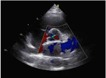
calciication of the ascending aorta. We recently reported a patient with QAV (type D in Hurwitz & Roberts’[8] classiication
and type III in Nakamura et al.’[11] classiication) with mild AR as
identiied by transthoracic echocardiography (Figure 3), who was initially referred to us due to mild exertional dyspnea, and she was advised a regular follow-up[54]. However, she soon went
to a provincial hospital, where she received an aortic valve replacement with a St. Jude Medical mechanical prosthesis. As such, an excessive treatment was seen in both surgical indication and surgical procedure of choice.
et al.[58] included abnormal commissure detachment, thickened
tissue excision, lealet approximation and subcommissural annuloplasty in their surgical technique of aortic valve repair. Song et al.[55] presented their tricuspidization of QAV for eight
consecutive patients with an at least moderate AR. Their surgical key points are pericardial lealet reconstruction, sinotubular junction reduction and commissure coaptation suture. The latter two teams[55,58] emphasized the importance of subcommissural
annuloplasty and sinotubular ixation in the maintenance of the coaptation of the neocusps.
Luciani et al.[59] reported their bicuspidization technique for a
68-year-old male patient with a type G QAV by joining two small non-coronary cusps to the left coronary cusp while preserving the right coronary cusp. The patient was asymptomatic at 18-month follow-up.
Additionally, Ross procedure (subcoronary technique) was reported as an alternative of treatment of QAV for decreasing the risk of aortic root dilation[60]. Manouguian’s operation was once
performed in a QAV patient with narrow annulus associated with aortic steno-insuiciency and mitral insuiciency[27].
POSTOPERATIVE COMPLICATIONS
Postoperative complications are seldom. Tsang et al.[2]
reported three postoperative complications, including progressive AR, transient ischemic attack and cardiac arrest in one patient each. Pirundini et al.[4] reported that a patient had
postoperative complete heart block, which was believed to be a result of conduction system impairment by manipulation of the supernumerary cusp of QAV that was most commonly located between the right and non-coronary coronary cusps. The overall survival rates of QAV patients were 89.9% and 84.9% at 5- and 10-year follow-up, respectively[2].
PROGNOSIS
The non-tricuspid aortic valves are less amenable to repair and durability of repair was almost uncertain as there are limited cases and scanty of reports concerning the long-term outcomes[17]. In the early years, Yotsumoto et al.[14] reported one
patient with a QAV failed for aortic valve repair and was converted to valve replacement. Song et al.[55] reported eight patients
with QAV with signiicant AR in each patient. Tricuspidization with new aortic valve lealet created with bovine pericardium resulted in signiicantly improved hemodynamics in all patients and showed satisfactory early and mid-term results with no reoperative requirements. Idrees et al.[33] reported the long-term
outcomes of QAV patients undergoing aortic valve repair and aortic valve replacement. Three (42.9%, 3/7) patients with aortic valve repair developed regurgitation and (or) stenosis of the aortic valve and one of the three required reoperation for aortic valve replacement at 13 years after the initial operation. In comparison, 2 (8.7%, 2/23) patients developed aortic stenosis after aortic valve replacement, but without the need of re-replacement of the aortic valve. One patient of the aortic valve replacement group developed infective endocarditis and warranted re-replacement of the aortic valve. Figure 4 shows the management protocols and late results of QAV patients undergoing surgical operations. Fig. 3 - Echocardiography of a type D/type III quadricuspid aortic
valve with mild aortic regurgitation.
SURGICAL TECHNIQUES
Aortic valve replacement is not an optimal solution for these young patients, because they are exposed to valve-related risks, such as thromboembolism, prosthetic valve degeneration, infective endocarditis and bleeding, and therefore aortic valve repair could be a promising option[17]. The target of aortic valve
repair is to restore an accurate coaptation and low transvalvular gradient with no turbulent low and therefore to achieve a favorable long-term durability[55]. In addition, transcatheter aortic
valve replacement is not recommended for those patients with severe AR[44]. Anyway, aortic valve repair started late and the
choice of the procedure is usually determined on the disease severity, condition of QAV, and surgeon’s preference[33].
The most common repair technique is the aortic valve tricuspidalization. Iglesias et al.[25] reported a case of QAV, in whom
tricuspidalization by conjoining the rudimentary and right aortic valve lealets and resection of subaortic stenosis were performed. Langer et al.[56] described their QAV repair technique of neocusp
creation by rudimentary commissure detachment, adjacent cusp approximation and neocusp augmentation. Schmidt et al.[17]
used pericardial patch augmentation and triangular resection of cusp tissue in their aortic repair technique. Kawase et al.[57]
Fig. 4 - Management and prognosis of patients with a quadricuspid aortic valve[33].
AR=aortic regurgitation; AS=aortic stenosis; AV=aortic valve; AVR=aortic valve replacement; IE=infective endocarditis; QAV=quadricuspid aortic valve
CONCLUSION
QAV is a rare congenital heart disease. Most of the patients with a QAV develop aortic valve incompetency at the ifth to sixth decade of life. About one-ifth of them require a surgical operation. Although tricuspidalization is a preferred repair technique for QAV with signiicant AR, the associated aortopathy could be a predictive risk factor of late failure of aortic repair. As not all the patients with a QAV necessarily warrant a surgical operation, decision-making in patient selection and surgical procedure of choice are crucial. The aortic valve repair of panegyric was started later and the procedural choice was determined by the feasibility concerning the QAV condition and
surgeon’s preference. Antibiotic prophylaxis against infective endocarditis is necessary in the QAV patients with unequal-sized cusps.
Author’s roles & responsibilities
REFERENCES
1. Cheema MA. Quadricuspid aortic valve as cause of congestive cardiac failure: case history. Pak Heart J. 1990;23(1):14-5.
2. Tsang MY, Abudiab MM, Ammash NM, Naqvi TZ, Edwards WD, Nkomo VT, et al. Quadricuspid aortic valve: characteristics, associated structural cardiovascular abnormalities, and clinical outcomes. Circulation. 2016;133(3):312-9.
3. Olson LJ, Subramanian R, Edwards WD. Surgical pathology of pure aortic insuiciency: a study of 225 cases. Mayo Clin Proc. 1984;59(12):835-41. 4. Pirundini PA, Balaguer JM, Lilly KJ, Gorsuch WB, Taft MB, Cohn LH, et
al. Replacement of the quadricuspid aortic valve: strategy to avoid complete heart block. Ann Thorac Surg. 2006;81(6):2306-8.
5. Malviya A, Jha PK, Ashwin, Mishra J, Srivastava P, Mishra A. Quadricuspid aortic valve: a case report and literature review. Egypt Heart J. 2015. (in press) 6. Savino K, Quintavalle E, Ambrosio G. Quadricuspid aortic valve: a case report and review of the literature. J Cardiovasc Echography. 2015;25:72-6. http://www.jcecho.org/text.asp?2015/25/3/72/166077.
7. Attaran RR, Habibzadeh MR, Baweja G, Slepian MJ. Quadricuspid aortic valve with ascending aortic aneurysm: report of a case and discussion of embryological mechanisms. Cardiovasc Pathol. 2009;18(1):49-52. 8. Hurwitz LE, Roberts WC. Quadricuspid semilunar valve. Am J Cardiol.
1973;31(5):623-6.
9. Vali Y, Rajendra R, Nishtar S. A previously undescribed type of quadricuspid aortic valve: type H. J Heart Valve Dis. 2010;19(6):792-3. 10. Jagannath AD, Johri AM, Liberthson R, Larobina M, Passeri J, Tighe D,
et al. Quadricuspid aortic valve: a report of 12 cases and a review of the literature. Echocardiography. 2011;28(9):1035-40.
11. Nakamura Y, Taniguchi I, Saiki M, Morimoto K, Yamaga T. Quadricuspid aortic valve associated with aortic stenosis and regurgitation. Jpn J Thorac Cardiovasc Surg. 2001;49(12):714-6.
12. Timperley J, Milner R, Marshall AJ, Gilbert TJ. Quadricuspid aortic valves. Clin Cardiol. 2002;25(12):548-52.
13. Tutarel O, Westhof-Bleck M. Functional status of the quadricuspid aortic valve/an uncommon coincidence of congenital quadricuspid aortic valve accompanied by hypertrophic obstructive cardiomyopathy. Anadolu Kardiyol Derg. 2008;8(1):86.
14. Yotsumoto G, Iguro Y, Kinjo T, Matsumoto H, Masuda H, Sakata R. Congenital quadricuspid aortic valve: report of nine surgical cases. Ann Thorac Cardiovasc Surg. 2003;9(2):134-7.
15. Janssens U, Klues HG, Hanrath P. Congenital quadricuspid aortic valve anomaly associated with hypertrophic non-obstructive cardiomyopathy: a case report and review of the literature. Heart. 1997;78(1):83-7. 16. Feldman BJ, Khandheria BK, Warnes CA, Seward JB, Taylor CL, Tajik
AJ. Incidence, description and functional assessment of isolated quadricuspid aortic valves. Am J Cardiol. 1990;65(13):937-8.
17. Schmidt KI, Jeserich M, Aicher D, Schäfers HJ. Tricuspidization of the quadricuspid aortic valve. Ann Thorac Surg. 2008;85(3):1087-9. 18. Godefroid O, Colles P, Vercauteren S, Louagie Y, Marchandise B.
Quadricuspid aortic valve: a rare etiology of aortic regurgitation. Eur J Echocardiogr. 2006;7(2):168-70.
19. Dotti MT, De Stefano N, Mondillo S, Agricola E, Federico A. Neurological involvement and quadricuspid aortic valve in a patient with Ehlers-Danlos syndrome. J Neurol. 1999;246(7):612-3.
20. Sousa L, Pinto F, Nogueira G, Kaku S, Antunes AM. Quadricuspid aortic valve and atrial septal defect. Rev Port Cardiol. 2001;20(3):329-30. 21. Demirkol S, Balta S, Arslan Z, Unlu M, Kucuk U, Iyisoy A. Association
of quadricuspid aortic valve and ventricular septal defect in a patient who had undergone atrial septal defect surgery. Kardiol Pol. 2013;71(5):546.
22. Seol SH, Kim U, Cho HJ, Kim DK, Kim DI, Kim DS. Quadricuspid aortic valve with patent ductus arteriosus. Tex Heart Inst J. 2010;37(6):726-7. 23. Suzuki Y, Daitoku K, Minakawa M, Fukui K, Fukuda I. Congenital
quadricuspid aortic valve with tetralogy of Fallot and pulmonary atresia. Jpn J Thorac Cardiovasc Surg. 2006;54(1):44-6.
24. Egred M, Patel JC, Metcalfe MJ. Sinus of Valsalva fistula with quadricuspid aortic valve, a irst reported association. Int J Cardiol. 2005;101(1):151-2.
25. Iglesias A, Oliver J, Muñoz JE, Nuñez L. Quadricuspid aortic valve associated with ibromuscular subaortic stenosis and aortic regurgitation treated by conservative surgery. Chest. 1981;80(3):327-8.
26. Irisawa T, Yoshiya K, Yokosawa T, Iwamatsu T, Arai K, Aoki T. A case of quadricuspid aortic valve associated with mitral regurgitation. Kyobu Geka. 1993;46(7):618-21.
27. Sakamoto Y, Saitoh F, Ohnishi K, Kurosawa H, Takakura H. A case of quadricuspid aortic valve associated with mitral insuiciency. Nihon Kyobu Geka Gakkai Zasshi. 1994;42(8):1235-7.
28. Konrad R, Costa MNS, Salamé CK. Valva aórtica quadricúspide: uma revisão completa. Rev Bras Ecocardiogr Imagem Cardiovasc. 2009;22(3):39-52.
29. Erdmenger J, Vázquez-Antona C, Becerra R, Romero A, Roldan J, Buendía A, et al. Quadricuspid aortic valve in a patient with d-transposition of the great arteries. Arch Cardiol Mex. 2005;75(4):460-2.
30. Finch A, Osman K, Kim K-S, Nanda NC, Willman B, Soto B, et al. Transesophageal echocardiographic indings of an infected quadricuspid aortic valve with an anomalous coronary artery. Echocardiography. 1994;11(4):369-75.
31. Lanzillo G, Breccia PA, Intonti F. Congenital quadricuspid aortic valve with displacement of the right coronary oriice. Scand J Thorac Cardiovasc Surg. 1981;15(2):149-51.
32. Tutarel O. The quadricuspid aortic valve: a comprehensive review. J Heart Valve Dis. 2004;13(4):534-7.
33. Idrees JJ, Roselli EE, Arafat A, Johnston DR, Svensson LG, Sabik JF 3rd, et al. Outcomes after repair or replacement of dysfunctional quadricuspid aortic valve. J Thorac Cardiovasc Surg. 2015;150(1):79-82.
34. Bauer F, Litzler PY, Tabley A, Cribier A, Bessou JP. Endocarditis complicating a congenital quadricuspid aortic valve. Eur J Echocardiogr. 2008;9(3):386-7. 35. Takeda N, Ohtaki E, Kasegawa H, Tobaru T, Sumiyoshi T. Infective
endocarditis associated with quadricuspid aortic valve. Jpn Heart J. 2003;44(3):441-5.
36. Matsukawa T, Yoshii S, Hashimoto R, Muto S, Suzuki S, Ueno A. Quadricuspid aortic valve perforation resulting from bacterial endocarditis: 2-D echo- and angiographic diagnosis and its surgical treatment. Jpn Circ J. 1988;52(5):437-40.
37. Bilge AK, Buğra Z, Tayyareci Y, Rüzgar O, Umman S, Meriç M. An uncommon coincidence of congenital quadricuspid aortic valve accompanied by hypertrophic obstructive cardiomyopathy. Anadolu Kardiyol Derg. 2007;7(4):E7-8.
38. Kawanishi Y, Tanaka H, Nakagiri K, Yamashita T, Okada K, Okita Y. Congenital quadricuspid aortic valve associated with severe regurgitation. Asian Cardiovasc Thorac Ann. 2008;16(5):e40-1.
39. Nishimura RA, Carabello BA, Faxon DP, Freed MD, Lytle BW, O’Gara PT, et al. ACC/AHA 2008 Guideline update on valvular heart disease: focused update on infective endocarditis: a report of the American College of Cardiology/American Heart Association Task Force on Practice Guidelines endorsed by the Society of Cardiovascular Anesthesiologists, Society for Cardiovascular Angiography and Interventions, and Society of Thoracic Surgeons. J Am Coll Cardiol. 2008;52(8):676-85.
40. Hayakawa M, Asai T, Kinoshita T, Suzuki T. Quadricuspid aortic valve: a report on a 10-year case series and literature review. Ann Thorac Cardiovasc Surg. 2014;20(Suppl):941-4.
41. Karlsberg DW, Elad Y, Kass RM, Karlsberg RP. Quadricuspid aortic valve deined by echocardiography and cardiac computed tomography. Clin Med Insights Cardiol. 2012;6:41-4.
43. Kurosawa H, Wagenaar SS, Becker AE. Sudden death in a youth. A case of quadricuspid aortic valve with isolation of origin of left coronary artery. Br Heart J. 1981;46(2):211-5.
44. Choji CL, Selvaraj N, Prather J. Female adolescent with quadricuspid aortic valve. J Am Osteopath Assoc. 2015;115(9):570-2.
45. Herman RL, Cohen IS, Glaser K, Newcomb EW 3rd. Diagnosis of incompetent quadricuspid aortic valve by two-dimensional echocardiography. Am J Cardiol. 1984;53(7):972.
46. Chandrasekaran K, Tajik AJ, Edwards WD, Seward JB. Two-dimensional echocardiographic diagnosis of quadricuspid aortic valve. Am J Cardiol. 1984;53(11):1732-3.
47. Küçükoglu MS, Erdogan I, Okçün B, Baran T, Mutlu H, Uner S. Quadricuspid aortic valve abnormality associated with aortic stenosis and aortic insuiciency. J Am Soc Echocardiogr. 2002;15(1):90-2.
48. Pouleur AC, le Polain de Waroux JB, Pasquet A, Watremez C, Vanoverschelde JL, El Khoury G, et al. Successful repair of a quadricuspid aortic valve illustrated by transoesophageal echocardiography, 64-slice multidetector computed tomography, and cardiac magnetic resonance. Eur Heart J. 2007;28(22):2769.
49. Chapman CB, Kohmoto T, Kelly AF, Thornton F, Keevil JG. Cardiac computed tomography and quadricuspid aortic valve: a case report. WMJ. 2010;109(4):219-21.
50. Kajinami K, Takekoshi N, Mabuchi H. Images in cardiology. Non-invasive detection of quadricuspid aortic valve. Heart. 1997;78(1):87. 51. Mecozzi G, Pratali S, Milano A, Nardi C, Bortolotti U. Severe quadricuspid
aortic valve stenosis after mediastinal irradiation. J Thorac Cardiovasc Surg. 2003;126(4):1198-9.
52. George BA, O’Hayre TA, Schussler JM. Association between congenitally quadricuspid aortic valve and mitral valve prolapse. Proc (Bayl Univ Med Cent). 2013;26(3):272-4.
53. Tutarel O. Quadricuspid aortic valves and anomalies of the coronary arteries. J Thorac Cardiovasc Surg. 2004;127(3):897.
54. Yuan SM, Yan SL. Quadricuspid aortic valve: a case report. Cor Vasa. 2016;58(5):579-80.
55. Song MG, Yang HS, Lee DH, Shin JK, Chee HK, Kim JS. Mid-term results in patients having tricuspidization of the quadricuspid aortic valve. J Cardiothorac Surg. 2014;9:29.
56. Langer F, Aicher D, Kissinger A, Wendler O, Lausberg H, Fries R, et al. Aortic valve repair using a diferentiated surgical strategy. Circulation. 2004;110(11 Suppl 1):II67-73.
57. Kawase I, Ozaki S, Yamashita H, Uchida S, Nozawa Y, Matsuyama T, et al. Original aortic valve plasty with autologous pericardium for quadricuspid valve. Ann Thorac Surg. 2011;91(5):1598-9.
58. Williams L, Peters P, Shah P. Tricuspidization of quadricuspid aortic valve. Ann Thorac Surg. 2013;95(4):1453-5.
59. Luciani GB, Morjan M, Faggian G, Mazzucco A. Repair of quadricuspid aortic valve by bicuspidization: a novel technique. Interact Cardiovasc Thorac Surg. 2010;11(3):348-50.
![Fig. 2 - Nakamura et al. [11] simplified classification of quadricuspid aortic valve](https://thumb-eu.123doks.com/thumbv2/123dok_br/15421163.590371/2.914.484.846.134.255/fig-nakamura-et-simplified-classification-quadricuspid-aortic-valve.webp)
![Fig. 4 - Management and prognosis of patients with a quadricuspid aortic valve [33] .](https://thumb-eu.123doks.com/thumbv2/123dok_br/15421163.590371/5.914.66.829.122.769/fig-management-prognosis-patients-quadricuspid-aortic-valve.webp)
