288
1. São José do Rio Preto Pediatric Cardiovascular Surgery Service – Hospital de Base – São José do Rio Preto Medical School, SP, Brazil.
Correspondence address: Ulisses Alexandre Croti
Hospital de Base – FAMERP – Avenida Brigadeiro Faria Lima, 5544. CEP 15090-000 – São José do Rio Preto – SP - Brasil
Fone (Fax): 17 – 3201-5025 / 3222-6450 E-mail: [email protected]
Ulisses Alexandre CROTI1, Domingo Marcolino BRAILE1, Sírio HASSEM SOBRINHO1, Carlos Henrique DE
MARCHI1
Rev Bras Cir Cardiovasc 2008; 23(2): 288-289
CLINICAL-SURGICAL CORRELATION
RBCCV 44205-989
Doença valvar mitral reumática submetida à plastia com anel de Gregori-Braile
Surgical repair of rheumatic mitral valve disease
with Gregori-Braile’s Ring
Article received on Abril 20th, 2008 Article accepted on May 21st, 2008 CLINICAL DATA
We report a case of a 13-year-old male child, weighing 40 kg, born in Porto Velho, RO. At the age of 8 years, he developed a clinical picture of inappetence and weight loss, when a cardiac murmur was observed. On examination, rheumatic disease with cardiac alteration was found. A therapy with penicillin benzathine, 1,200,000 U, every 21 days, plus furosemide and captopril was introduced, along with clinical follow-up. The child‘s mother denies an articular clinical picture, tonsillitis, or skin infections. However, when he arrived at the Hospital Service, he complained of fatigue to mild and severe exertion; otherwise, he was in good general health, red-faced, hydrated, eupneic, and acyanotic. Patient presented ictus cordis impulsive and shifted to the left, systolic fremitus at left sternal border, regular rhythm of two normal sounds, hyperphonetic second heart sound at pulmonary focus, systolic murmur 4+/6+ irradiating towards the axilla, and diastolic murmur 2+/6+, both at mitral
focus. Lungs were free. Abdomen with the liver at 2 cm from the right costal border was observed. The pulses were normal and peripheral saturation at 98%.
289 CROTI, UA ET AL - Surgical repair of rheumatic mitral valve disease
with Gregori-Braile’s Ring
Rev Bras Cir Cardiovasc 2008; 23(2): 288-289
ELECTROCARDIOGRAM
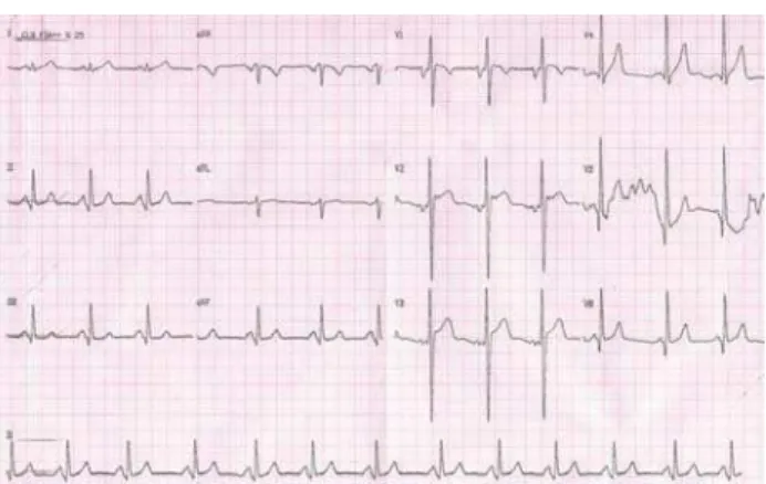
An electrocardiogram evidenced sinusal rhythm with a heartbeat of 75 bpm. The SAP angle was +60º and the SÂQRS was +90º with a PR interval of 0.16s, the QRS was 0.08s and the QTc was 0.40s. There was sign of left atrial overload. Left ventricular overload was also evidenced by QRS complexes with wide S waves in V1 and wide R waves in V5 and V6, besides a potential right ventricular overload through QRS axis deviation to the right (Figure 1).
RADIOGRAM
Radiogram showed visceral situs solitus, cardiothoracic index of 0.48, and moderate enlarged left atrium. Pulmonary vascular prominence with pulmonary vasculature inversion was also observed suggesting pulmonary venocapillary congestion.
ECHOCARDIOGRAM
Echocardiogram showed situs solitus with levocardia. Thickening of both aortic and mitral valves, severe mitral stenosis, moderate-to-severe mitral regurgitation, slight aortic and tricuspid valve insufficiency, and slight pericardial effusion were all observed. Electrocardiogram also showed a transvalvar mitral gradient of 15 mmHg, a mitral valve area of 1.2 cm2, right ventricular systolic pressure of 30 mmHg,
ejection fraction of 70.4% (Teich index), and left ventricular diastolic and systolic diameters were 50 mm and 30 mm, respectively; aorta and left atrium diameters were 27 mm and 30 mm, respectively. Block score was used to assess mitral cusp mobility, valvar and subvalvar calcification and thickening. The Block score was nine.
DIAGNOSIS
The adequate evaluation of every heart valve and the severity degree of their impairment should always be considered along with the clinical picture for referral to surgical treatment. In this young patient, echocardiogram provided all necessary data for treatment. Catheterism is useful when it is necessary to check pulmonary artery pressure or even after the age of 40 years in order to study the coronary arteries. Nearly 25% of the patients may have a significant coronary disease. On differential diagnosis, mitral valve stenosis, Lutembacher syndrome, Shone syndrome, and cor triatriatum should be taken into consideration.
OPERATION
An approach using a longitudinal median sternotomy was performed. Cardiopulmonary bypass (CPB) support was established with cannulation of the aorta, and both the superior and inferior vena cava. CBP was started with hypothermia at 28ºC. Intermittent and antegrade hypothermic blood cardioplegia at 4ºC was repeated at every
20 minutes. Left atrium was opened and mitral valve examined. A fusion of both anterolateral and posteromedial commissures were found (Figure 2). Bilateral commissurotomy and anterolateral papilotomy were started. Some tendinous cords were fenestrated in order to improve the posterior cusp mobility. The ring presented a posterior dilation to the right as usual besides malcoaptation between both anterior and posteriors cusps. At this moment, we have chosen to implant a rigid open ring (Gregori-Braile) considering the patient’s age, which poses growth potential and growing requirements. Seven 2-0 polyester threads were initially fixed in the mitral ring and afterwards in the external portion of the prosthesis [1,2]. Perfusion and myocardial ischemia times were 72 minutes and 57 minutes, respectively. The patient was discharged from the hospital at postoperative day 4 with the echocardiogram showing an excellent outcome with no presence of stenosis and minor mitral valve regurgitation.
REFERENCES
1. Gregory F Jr, Takeda R, Silva S, Façanha L, Meier MA. A new technique for repair of mitral insufficiency caused by ruptured chordae of the anterior leaflet. J Thorac Cardiovasc Surg. 1988;96(5):765-8.
2. Cordeiro CO, Gregori F Jr, Gregori TEF, Murakami AN, Abrão A. Resultados da operação reconstrutora da valva mitral em pacientes com idade inferior a 15 anos. Rev Bras Cir Cardiovasc. 2004;19(2):115-9.