ORIGINAL ARTICLE
Protein fraction of
Calotropis procera
latex protects
against 5-fluorouracil-induced oral mucositis associated
with downregulation of pivotal pro-inflammatory mediators
Ana Paula F. Freitas&Flavio S. Bitencourt&
Gerly Anne C. Brito&Nylane Maria N. de Alencar& Ronaldo A. Ribeiro&Roberto Cesar P. Lima-Júnior& Marcio V. Ramos&Mariana L. Vale
Received: 25 April 2012 / Accepted: 25 June 2012 #Springer-Verlag 2012
Abstract Oral mucositis is an important dose-limiting and costly side effect of cancer chemotherapy. Soluble proteins obtained of the latex ofCalotropis procerahave been exten-sively characterized as anti-inflammatory in different experi-mentally induced inflammatory conditions, including arthritis and sepsis. In this study, the phytomodulatory laticifer pro-teins (LP) were challenged to regress the inflammatory events associated with 5-fluorouracil-induced oral mucositis. We also evaluated the expression of pro-inflammatory cytokines and inducible enzymes, such as cyclooxygenase-2 (COX-2) and inducible nitric oxide synthase (iNOS). Oral mucositis was induced in hamsters by two injections of 5-fluorouracil (5-FU; 60 and 40 mg/kg, i.p., on experimental days 1 and 2,
respectively). LP (5 mg/kg, i.p.) was injected 24 h before and 24 h after mechanical trauma of the cheek pouches. A normal control group received only saline. On day 10, the animals were sacrificed, and the cheek pouches were excised for macroscopic and histopathological analysis, myeloperox-idase activity measurement, and immunohistochemical as-sessment of tumor necrosis factor-α (TNF-α), interleukin-1β (IL-1β), iNOS, and COX-2. LP significantly inhibited macroscopic histopathological scores and myeloperoxidase activity compared with the FU control group. 5-Fluorouracil also induced marked immunostaining of
TNF-α, IL-1β, iNOS, and COX-2 on inflamed conjunctive and epithelial tissue compared with the normal control group. Such damage was significantly inhibited (p<0.05) by LP treatment compared with the 5-FU group. These findings demonstrate an anti-inflammatory effect of LP on 5-FU-induced oral mucositis. The protective mechanism appears to involve inhibition of the expression of iNOS, COX-2, TNF-α, and IL-1β.
Keywords Oral mucositis . 5-Fluorouracil .Calotropis procera. Inflammation . Chemotherapy toxicity
Introduction
Mucositis is a clinical term that describes a syndrome char-acterized by ulceration of the mucosa of the entire digestive tract and associated symptoms (Sonis and Fey2002). This side effect is common in cancer patients treated with various chemotherapeutic agents, particularly anti-metabolites (e.g., methotrexate and fluorouracil), and other agents, such as cisplatin, doxorubicin, and ifosfamide (Balis et al. 1985;
A. P. F. Freitas
Department of Clinical Medicine, Faculty of Medicine, Federal University of Ceará,
60430-140 Fortaleza, CE, Brazil
F. S. Bitencourt
:
N. M. N. de Alencar:
R. A. Ribeiro:
R. C. P. Lima-Júnior
:
M. L. Vale (*)Department of Physiology and Pharmacology, Faculty of Medicine, Federal University of Ceará, Rua Cel. Nunes de Melo, 1127,
60431-270 Fortaleza, CE, Brazil e-mail: marianavale@yahoo.com
G. A. C. Brito
Department of Morphology, Faculty of Medicine, Federal University of Ceará,
Rua Delmiro de Farias, 60440-261 Fortaleza, CE, Brazil
M. V. Ramos
Department of Biochemistry and Molecular Biology, Federal University of Ceará,
Bishop et al.1986; Caballero et al.1985; Roth et al.1991) as well as in patients submitted to abdominal radiotherapy (Altmann1974).
The oral form of mucositis is mainly characterized by the presence of ulcerative lesions (i.e., stomatitis) responsible for severe pain and discomfort. Patients complain mainly of pain that often keeps them from eating normally, which decreases their nutritional status.
Data relative to the risk of developing grade 3 or 4 oral mucositis have shown that the incidence of chemotherapy with 5-fluorouracil, capecitabine, or tegafur leads to a high rate (20–50 %) of alimentary tract mucositis. Chemotherapy with methotrexate and other anti-metabolites leads to a 20–
60 % rate of alimentary tract mucositis according to the drug's given dose per cycle. In patients receiving high-dose head and neck radiation to the oral cavity and in hematopoietic stem-cell transplantation patients, the in-cidence is up to 85 and 75 %, respectively (Peterson et al. 2010).
Current research focuses on effective preventive and therapeutic options for oral mucositis that significantly improve patient quality of life. Preventive strategies are the usual approach to manage oral mucositis induced by chemotherapy. Since the guidelines for managing muco-sitis in cancer patients have been published, considerable progress in research and clinical application has been made (Rubenstein et al. 2004). Recently, these guidelines have been revised with changes of the original guide-lines, including new therapeutic and preventive agents and protocols (Keefe et al. 2007). Despite advances, the available treatments are scarce, and none have proven to be totally effective. Oral mucositis is one of the leading causes for unplanned treatment interruptions which jeopardize the effectiveness of the treatment (Haagen et al.2009).
Recently, our group began to study the pathogenesis of oral mucositis induced by 5-fluorouracil in hamsters. We demonstrated the role of nitric oxide and pro-inflammatory cytokines, such as interleukin-1β(IL-1β) and tumor necro-sis factor-α (TNF-α), in this model (Leitão et al. 2007; Leitão et al.2008; Lima et al.2005).
Calotropis procerais a laticiferous plant (Apocynaceae) locally known in Brazil as “algodão de seda,” “leiteiro,” “queimadeira,” and “ciúme” (Kismann and Groth 1999; Lorenzi and Matos 2002). The latex components of C. proceraappear to induce interesting biological effects, and the plant itself is known for its pharmacological potential. Previous studies that used the latex ofC. procerareported anti-inflammatory (Kumar and Basu1994; Sangraula et al.
2002), analgesic (Soares et al.2005), anti-diarrheal (Kumar et al.2001), and anti-oxidant (Kumar et al.2011). Almost all of these studies investigated the properties and activity of the latex without previous fractionation of the material. More recent studies have also been conducted with the
protein fraction isolated from the whole latex (Ramos et al.
2007). This procedure has effectively shown that laticifer-ous proteins have many of the promising effects that have been demonstrated for the whole latex (Soares et al.2005; Alencar et al.2004; Ramos et al.2009). Very recently, these researchers showed that laticifer proteins of C. procera
protect animals against experimentally induced lethal sepsis with important effects on the homeostasis of blood coagu-lation (Oliveira et al.2012; Ramos et al.2012).
Since the demonstration of the anti-inflammatory effects of C. procera and the lack of effectiveness of existing mucositis therapeutics, we investigated the effect of C. proceralatex proteins in 5-FU-induced oral mucositis.
Material and methods
Animals
Ninety male adult golden hamsters that weighed 120–150 g were obtained from the sector vivarium of the Federal University of Ceará. The surgical procedures and animal treatments were conducted in accordance with the Guide-lines for Institutional Animal Care and were approved by our Institutional Animal Care and Use Committee of Federal University of Ceará (protocol no. 036/10). The animals were housed in temperature-controlled rooms and received water and food ad libitum.
Plant material and latex collection
The latex ofC. procera(Ait.) R.Br. from the aerial parts of the plant was collected in plastic tubes that contained dis-tilled water to give a dilution ratio of 1:2 (v/v). The native plants were located in the vicinity of Fortaleza, CE, Brazil. The plant voucher (sample specimen no. 32663) was depos-ited at the Prisco Bezerra Herbarium of the Universidade Federal do Ceará, Brazil, where the botanical material was identified by a local taxonomist. After the latex was collect-ed, it was processed as described before (Alencar et al.
obtained. The chemical characterization of this fraction in terms of protein content and enzymatic activities has been extensively characterized by electrophoresis (1-D and 2-D), mass spectrometry, chromatography, proteomic approach, enzymatic assays based in colorimetric methods, and zymo-grams (Oliveira et al.2007; Freitas et al.2007; Ramos et al.
2009; Oliveira et al.2010; Oliveira et al.2012; Ramos et al.
2012).
Induction of experimental oral mucositis
The hamster oral mucositis model was modified and adap-ted from the original work described by Sonis et al. (1990) and later by Leitão et al. (2007). Oral mucositis was induced by two intraperitoneal (i.p.) injections of 5-fluorouracil (5-FU; Roche, Rio de Janeiro, Brazil) on days 1 and 2 of the experiment (60 and 40 mg/kg, respectively). To mimic the friction to which the oral mucosa is normally subjected, the animal cheek pouch mucosa was irritated by scratching with the tip of an 18-gauge needle on day 4 under tribromoetha-nol anesthesia (1 ml/100 g per animal, i.p.). The needle was linearly dragged twice across the averted cheek pouch until erythematous changes were noted. The animals were sacri-ficed on day 10 after the initial injection of 5-FU under tribromoethanol anesthesia. The cheek pouches were averted and photographed, and the hamster was then sacri-ficed. Samples of the cheek pouches were removed from six animals per group for histopathological analysis, immuno-histochemical assessment of TNF-α, IL-1β, inducible nitric oxide synthase (iNOS), and cyclooxygenase-2 (COX-2), and myeloperoxidase (MPO) determination as described below (Leitão et al.2007).
Experimental groups
The hamsters were separated into seven groups: normal (N, animals not subjected to oral mucositis), mechanical trauma (MT, animals that received only mechanical trauma and treated with an i.p. saline injection), control (C, animals that were treated with saline and subjected to 5-FU-induced oral mucositis and mechanical trauma), and four groups that were treated with laticifer protein ofC. procera(LP; 0.25, 1, 5, and 25 mg/kg, 24 h before and 24 h after mechanical trauma) and subjected to 5-FU-induced oral mucositis.
Macroscopic analysis of cheek pouch
At the time determined for each experiment, the animals were anesthetized with tribromoethanol (1 ml/100 g per animal, i.p.), and the cheek pouch was averted and photographed before the animal was sacrificed. The photographs were coded and used to score the lesions. For macroscopic analysis, signs of inflammation, such as erythema, hyperemia, hemorrhagic
areas, epithelial ulcerations, and abscesses, were evaluated in a single-blind fashion and graded as the following: score 0 (completely healthy cheek pouch with erosion or vasodilata-tion absent), score 1 (presence of erythema but no evidence of erosion in the cheek pouch), score 2 (severe erythema, vaso-dilation, and surface erosion), score 3 (formation of ulcers in one or more faces of the mucosa that do not affect more than 25 % of the surface area of the cheek pouch, severe erythema, and vasodilatation), score 4 (cumulative formation of ulcers of 50 % of the surface area of the cheek pouch), and score 5 (virtually complete ulceration of the cheek pouch mucosa; in this case, fibrosis makes oral mucosa exposure difficult) (Medeiros et al.2011). The photographs were analyzed in a single-blind fashion by a trained evaluator.
Histopathological analysis
The specimens were fixed in 10 % neutral buffered forma-lin, dehydrated, and embedded in paraffin. Sections of 5-μm thickness were obtained for hematoxylin-eosin staining (H&E) and examined by light microscopy (×100 magnifi-cation). The parameters of inflammatory cell infiltration, vasodilatation, and the presence of hemorrhagic areas, ede-ma, ulcerations, and abscesses were determined in a single-blind fashion and graded as the following: score 0 (normal epithelium and connective tissue without vasodilatation, absence of or discreet cellular infiltration, and absence of hemorrhagic areas, ulcerations, or abscesses), score 1 (dis-creet vasodilatation, reepithelization areas, dis(dis-creet inflam-matory infiltration with mononuclear prevalence, and absence of hemorrhagic areas, edema, ulcerations, or ab-scesses), score 2 (moderate vasodilatation, areas of hydropic epithelial degeneration, inflammatory infiltration with neu-trophil prevalence, presence of hemorrhagic areas, edema, and eventual ulcerations, and absence of abscesses), and score 3 (severe vasodilatation, inflammatory infiltration with neutrophil prevalence, presence of hemorrhagic areas, edema, and extensive ulceration, and abscesses) (Leitão et al.2007).
Myeloperoxidase assay
The extent of neutrophil accumulation in the cheek pouch samples was measured by assaying MPO activity. Briefly, the animals were sacrificed on day 10 after the initial injection of 5-FU, and the cheek pouch samples were harvested, weighed, and stored at−70 °C until required for the assay.
units of MPO per milligram of tissue, with 1 U MPO defined as the amount of enzyme responsible for the degradation of 1μmol of hydrogen peroxide/min at 22 °C.
Immunohistochemistry
Immunohistochemistry for TNF-α, IL-1β, iNOS, and COX-2 on day 10 of oral mucositis was performed using the streptavidin-biotin-peroxidase method in formalin-waxed, paraffin-embedded tissue sections (4-μm thick) mounted on poly-L-lysine-coated microscope slides. The sections were deparaffinized and rehydrated through xylene and a graded series of alcohol. After antigen retrieval, endogenous peroxidase was blocked twice (10 min) with 3 % (v/v) hydrogen peroxide and washed in phosphate-buffered saline (PBS). The sections were incubated overnight (4 °C) with primary rabbit anti-iNOS, rabbit anti-IL-1β, goat
anti-TNF-α, or goat anti-COX-2 antibody (Santa Cruz Biotechnology) diluted 1:200 in PBS plus bovine serum albumin (PBS-BSA 5 %). The slides were then incubated with biotinylated goat anti-rabbit (IgG) or biotinylated donkey anti-goat (IgG) diluted 1:400 in PBS-BSA. After washing, the slides were
incubated with avidin-biotin-horseradish peroxidase conju-gate (Strep ABC Complex; Santa Cruz Biotechnology) for 30 min according to the manufacturer's protocol. Immunos-taining was visualized with the chromogen 3,3′ -diaminoben-zidine (DAB; liquid DAB + substrate chromogen system; Dako). Negative control sections were processed simulta-neously as described above but with the first antibody replaced with PBS-BSA 5 %. None of the negative controls showed TNF-α, IL-1β, iNOS, or COX-2 immunoreactivity. The slides were counterstained with Harry's hematoxylin, dehydrated in a graded series of alcohol, cleared in xylene, and cover-slipped.
All of the slides were photographed, and the intensity of staining per stained cell population was determined in a single-blind fashion and graded with scores from 0 to 4.
Statistical analysis
The data are expressed as either mean ± SEM or me-dian as appropriate. Analysis of variance, followed by
N MT C 0.25 1 5 25 0.00
0.05 0.10 0.15 0.20 0.25
LP (mg/kg) ***
*** ***
5-FU
U
M
PO
/m
g of
m
uc
o
s
a
###
Fig. 2 Effect ofC. proceraon myeloperoxidase activity in the cheek pouch of hamsters subjected to the induction of oral mucositis. Saline (5-FU, control (C) group) or laticifer protein ofC. procera(LP, 0.25– 25 mg/kg) was injected 24 h before and 24 h after mechanical trauma in animals subjected to oral mucositis induced by 5-FU. The normal (N) group was not subjected to oral mucositis. The MT group was subjected only to mechanical trauma. After sacrifice, a sample of the cheek pouch was removed for the assessment of MPO activity.Bars represent the mean value ± standard error of the mean of the concen-tration of units of MPO per milligram of tissue. Triple asterisks indicatep<0.001 significant difference compared with 5-FU-induced oral mucositis group;triple number signsindicatep<0.001 compared with the N group. The number of animals in each group was at least five. The data were analyzed with analysis of variance and the Bon-ferroni test
14.4
20.1
30.0
66.0
97.0
MW
LP
45.0
Bonferroni's test, was used to compare means, and the Kruskal–Wallis and Mann–Whitney tests were used to compare medians. Values of p< 0.05 were considered statistically significant.
Results
This study was performed in an attempt to validate the anti-inflammatory activity of LP on mucositis. LP comprises the
Table 1 Macroscopic and microscopic analysis of hamster cheek pouch of animals submitted to experimental oral mucositis and treated laticifer proteins isolated from latex ofC. procera
Experimental group N MT 5-FU
LP (mg/kg)
C 0.25 1 5 25
Macroscopic scores (0–0) 0 (0–0) 0 (3–5) 4**** (2–5) 4*** (1–4) 3 (0–3) 1** (0–4) 2.5 Microscopic scores (0–0) 0 (0–2) 1 (3–3) 3 (2–3) 2 (0–3) 1.5* (0–3) 1** (0–3) 1.5
Saline (Ccontrol group) or Laticifer Proteins ofC. procera(LP0.25–25 mg/kg) was injected 24 h before and 24 h after mechanical trauma in animals submitted to oral mucositis induced by 5-FU. Normal group (N) composed of animals not submitted to oral mucositis, andMTgroup represent animals submitted only to mechanical trauma. Data represent the median values (and range) of macroscopic or microscopic scores in six animals per group. Data were analyzed by using Kruskal–Wallis followed by Dunn's
*p<0.05 and **p<0.001 (compared to saline-treated (C) animals); ***p<0.05 (compared to animals that received LP (1, 5, and 25 mg/kg)); ****p<0.001 (compared to MT and N groups)
major soluble protein fraction isolated from the latex ofC. procera. The typical protein profile of LP has been previ-ously shown by electrophoresis (1-D and 2-D) and mass spectrometry (Freitas et al.2007; Oliveira et al.2010). Here, Fig. 1 illustrates this profile. In order to estimate the anti-inflammatory activity of LP on mucositis, myeloperoxidase activity was measured in the cheek pouch as an indicator of neutrophil infiltration. Myeloperoxidase activity of the cheek pouch tissue of animals treated with MT and animals subjected to 5-FU-induced oral mucositis (5-FU/saline) was significantly increased (p< 0.001) compared with the N group on day 10 of oral mucositis. LP significantly (p<0.001) reduced (71 %) the 5-FU-induced increase in MPO activity at a minimum dose of 1 mg/kg (Fig.2). Even though statistically similar, LP at 5 mg/Kg was more effective reach-ing 88 % inhibition. Therefore, this dose was chosen for further analysis.
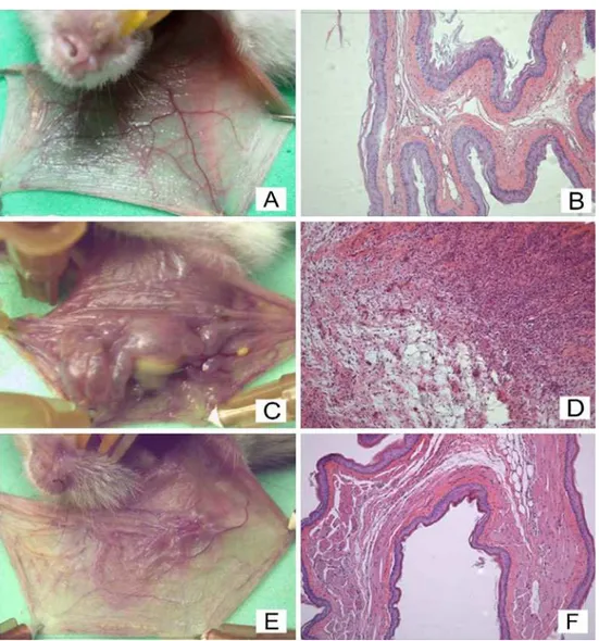
The i.p. administration of 5-FU, followed by mechan-ical trauma of the cheek pouch, caused significant lesions (p<0.05), reflected by accentuated erythema, hemorrhage, extensive ulcers, and abscesses (Fig.3cand Table 1) com-pared with the N group (Fig.3aand Table1) and MT group (Table 1). The treatment of the animals with 5 mg/kg LP prevented 5-FU-induced oral damage, reflected by reduced erythema and the absence of ulcerations and abscesses on day 10 (p<0.05; Fig.3e, g, respectively; Table1).
The histopathology of the cheek pouch of the animals subjected to 5-FU-induced oral mucositis revealed ingurgi-tation and accentuated vascular dilaingurgi-tation, intense cellular infiltration with neutrophil prevalence, hemorrhagic areas, edema, abscesses, and extensive ulcers (Fig.3dand Table1) compared with the normal cheek pouches of hamsters not subjected to oral mucositis (Fig.3band Table1) and the MT group (Table1). The treatment with LP (Fig.3fand Table1) significantly (p<0.05) reduced 5-FU-induced inflammatory cell infiltration, edema, and hemorrhage and prevented the formation of ulcers and abscesses.
The cheek pouches of the hamsters subjected to 5-FU-induced oral mucositis (C group) showed marked and signif-icant immunostaining of TNF-α(p<0.001), IL-1β(p<0.001), iNOS (p<0.05), and COX-2 (p<0.05) on inflamed conjunc-tive tissue (Table2and Figs.4d, e and5d, e) compared with the cheek pouches of the N group (Table2and Figs.4b, c and
5b, c). In epithelial tissue, only iNOS immunostaining was significantly (p<0.05) different from the N group (Fig.5e). LP (5 mg/kg) significantly decreased TNF-α (p<0.05), IL-1β
(p<0.01), iNOS (p<0.05), and COX-2 (p<0.01) immunos-taining in conjunctive tissue compared with the C group (Table 2 and Figs. 4d, e and 5d, e). In epithelial tissue, 5 mg/kg LP inhibited only COX-2 (p< 0.05) and iNOS (p< 0.05) immunostaining compared with the C group (Table 2 and Fig. 5f, g).
Discussion
The present study evaluated whether LP protects against 5-FU-induced oral mucositis. We also investigated the possi-ble modulating effect on the expression of pro-inflammatory mediators. An important aspect of our study was the use of an animal model that mimics clinical oral mucositis. We demonstrated that LP significantly and dose-dependently reduced macroscopic and microscopic lesions induced by 5-FU in the oral mucosa. Oral mucositis is characterized by an intense inflammatory reaction caused by chemotherapeu-tic agents on mucosa lamina propria cells (Sonis et al.
2004a). We verified that LP treatment decreased inflamma-tory cell infiltration, edema, hemorrhage, ulcers, and ab-scesses. This effect of LP is consistent with several studies that reported an anti-inflammatory effect of this product of
C. procera in different animal models (Ramos et al.2009; Arya and Kumar2005). Additionally, the pioneer study with the isolated latex protein fraction assessed the protective effect of LP against ifosfamide-induced hemorrhagic cystitis,
Table 2 Effect ofC. procera(LP) upon immunohistochemistry analysis for TNF-α, COX-2, IL-1β, and iNOS expression in the cheek pouches of hamsters subjected to 5-fluorouracil-induced oral mucositis
Tissue TNF-α COX-2 IL-1β iNOS
Ep Cj Ep Cj Ep Cj Ep Cj
N (0–1) 0.5 (0–1) 0.5 (0–0) 0 (0–0) 0 (1–4) 1.5 (0–4) 2.5 (0–4) 2 (0–1) 0.5 MT (1–2) 1.5 (1–1) 1 (0–2) 1 (1–2) 1 (1–2) 2 (1–3) 1 (2–3) 3 (1–3) 2 C (1–3) 2 (3–4) 4**** (1–2) 2 (4–4) 4**** (0–4) 2 (3–4) 4*** (3–4) 4*** (3–4) 4*** LP (0–1) 1 (1–2) 1* (0–1) 0* (1–2) 1** (0–1) 0.5 (0–1) 0.5* (0–3) 2* (0–2) 0.5**
another clinically related animal model. In that animal model, the authors found a marked protective effect (Alencar et al.
2004).
In the inflammatory response, neutrophil migration is an important event that occurs at the microvascular level (Carlos and Harlan1994). It is a consequence mainly of the release of neutrophil chemotactic factors by resident cells, inducing roll-ing and adhesion of neutrophils on endothelial cells, followed
by their transmigration to the extravascular space (Guo and Ward2002; Lindbom and Werr2002). In the present study, we observed a dose-dependent inhibitory effect on MPO activity, indicating less neutrophil infiltration into the lesion site. Previous study reported that intravascular administration of LP and more purified LP-fractions significantly re-duced neutrophil influx at the site of inflammation by inhibit-ing the rollinhibit-ing and adherence of neutrophils reflected by
Fig. 4 Representative photomicrography of the effect ofC. proceraon the
intravital microscopy (Ramos et al.2009). Several mecha-nisms might be involved in this anti-inflammatory phenome-non. A possible mode of action involves the direct effect on selectins found on leukocyte or endothelial cell membranes (Assreuy et al.1999). Another possibility is a direct effect on neutrophil migration through the downregulation of chemo-kine receptors (McColl and Clark-Lewis1999). Some authors have suggested that neutrophil migration induced by IL-1β
and TNF-αis not caused by a direct effect on neutrophils but occurs via the release of chemotactic factors from resident macrophages (Faccioli et al.1990). Despite the modulatory activity of LP on different pro-inflammatory mediators, as reported here and in preceding studies (Kumar et al.2011; Lima-Filho et al.2010; Alencar et al. 2004), the pathway underlying such an effect is still an aim of investigation. The ability of LP to inhibit inflammation even when
Fig. 5 Representative photomicrography of the effect ofC. proceraon
given by different route of administration is worth of note, and it is likely signalizing events involving LP recruit a complex web of chemokines.
In a model of intestinal mucositis, Tsuji et al. (2003) found that 5-FU delayed the elevation of acute inflammatory cytokines and increased portal endotoxin content, simulta-neous gut mucosal injury, bacterial translocation to the mesenteric lymph nodes, and sepsis-like symptoms. Lima-Filho et al. (2010) reported a protective effect of LP in the control of systemic murine infection. This effect was asso-ciated with a modulating effect on pro-inflammatory cyto-kine expression rather than with increased bactericidal capacity. To test the hypothesis that LP modulates the in-flammatory process by controlling pro-inin-flammatory medi-ators, we investigated the expression of TNF-α, IL-1β, COX-2, and iNOS. We found markedly reduced expression of these mediators after LP injection.
Agents known to attenuate the expression of cyto-kines have demonstrated the efficacy in the prevention of oral mucositis. Previous studies, including one of our own, demonstrated that inhibition of TNF-α production along with other cytokines is effective in preventing the development of mucositis (Lima et al. 2005). Pretreat-ment with the immunomodulating drugs thalidomide and pentoxifiline prevented mucositis in animals as well as atorvastatin, a statin that has many pleiotropic actions, including anti-inflammatory and anti-oxidant effects (Medeiros et al. 2011).
Previous studies found that COX-2 (Sonis2002; Sonis et al. 2004b; Logan et al. 2007) and iNOS (Caballero et al.
1985) play important roles in mucosal injury and inflamma-tory events that lead to oral mucositis. In addition to its pro-inflammatory effects, COX-2 also plays an important role in angiogenesis (Vane et al. 1998). Furthermore, iNOS has been reported to be associated with several human malig-nant tumors, including breast, brain, lung, prostate, colorec-tal, and pancreatic carcinomas, Kaposi's sarcoma, and melanoma (reviewed by Singh and Gupta2011). Proteins ofC. proceralatex were also reported to exhibit in vivo anti-cancer activity against sarcoma 180 (Oliveira et al.2010). Combining pharmacotherapeutics with anti-inflammatory effects as an adjuvant to cancer treatment is desirable and might have important clinical implications for the manage-ment of mucositis. The use ofC. proceralatex proteins may be promising.
We demonstrated that the protein fraction of Calotropis proceralatex protects against 5-FU-induced oral mucositis. We also verified an inhibitory effect on the expression of pro-inflammatory mediators. In conclusion, we showed that the protein fraction of the latex fromC. procerawas effective in the prevention of oral mucositis in a hamster model. However, well-designed clinical studies are needed to confirm the clin-ical efficacy of this treatment in humans. Despite that LP
represents a mix of latex proteins, a series of manuscripts has described different biochemical aspects and enzymatic profile of these samples, and no acute or chronic toxicity has been associated to this protein fraction (for review, see previ-ous studies of Ramos et al.).
Acknowledgments The authors thank Maria Silvandira França Pin-heiro, Department of Physiology and Pharmacology, Faculty of Medi-cine, Federal University of Ceará, Brazil, for technical assistance and Michael Arends for the English correction. This work was supported by the Brazilian Agency for Scientific and Technological Development (CNPq) and Fundação Cearense de Apoio ao Desenvolvimento Cientí-fico e Tecnológico.
Conflicts of interest statement The authors declare that there is no conflict of interest, and the funding source(s) had no involvement in the research design, writing, or other aspects of this manuscript.
References
Alencar NM, Fiqueiredo IS, Vale MR, Bitencurt FS, Oliveira JS, Ribeiro RA, Ramos MV (2004) Anti-inflammatory effect of the latex from Calotropis procera in three different experimental models: peritonitis, paw edema and hemorrhagic cystitis. Planta Med 70:1144–1149
Altmann GG (1974) Changes in the mucosa of the small intestine fol-lowing methotrexate administration or abdominal X-irradiation. Am J Anat 140:263–279
Arya S, Kumar VL (2005) Anti-inflammatory efficacy of extracts of latex ofCalotropis proceraagainst different mediators of inflam-mation. Mediators Inflamm 2005:228–232
Assreuy AMS, Martins GJ, Moreira MEF, Brito GAC, Cavada BS, Ribeiro RA, Flores CA (1999) Prevention of cyclophosphamide-induced hemorrhagic cystitis by glucose-mannose binding plant lectins. J Urol 161:1988–1993
Balis FM, Savitch JL, Bleyer WA, Reuman GH, Poplack DG (1985) Remission induction of meningeal leukemia with high-dose intra-venous methotrexate. J Clin Oncol 3:485–489
Bishop JF, Joshua DE, Lowenthal RM, Kronenberg H, Whiteside MG, Cobcroft R, Dodds A, Wolf M, Manoraran A (1986) A phase I-II study of cytosine arabinose, daunorubicin, and VP16-213 in adult patients with acute non-lymphocytic leukemia. Aust N Z J Med 16:48–51
Bradley PP, Christensen RD, Rothstein G (1982) Cellular and extra-cellular myeloperoxidase in pyogenic inflammation. Blood 60:618–622
Caballero GA, Ausman RK, Quebbeman EJ (1985) Long-term, ambu-latory, continuous IV infusion of 5-FU for the treatment of ad-vanced adenocarcinomas. Cancer Treatment Rep 69:13–15 Carlos TM, Harlan JM (1994) Leukocyte-endothelial adhesion
mole-cules. Blood 84:2068–2101
Faccioli LH, Souza GE, Cunha FQ, Poole S, Ferreira SH (1990) Recombinant interleukin-1 and tumor necrosis factor induce neu-trophil migration “in vivo” by indirect mechanisms. Agents Actions 30:344–349
Freitas CDT, Oliveira JS, Miranda MR, Macedo MS, Sales MP, Villas-Boas LA, Ramos MV (2007) Enzymatic activities and protein profile of latex fromCalotropis procera. Plant Physiol Biochem 45:781–789
Haagen J, Krohn H, Röllig S, Schmidt M, Wolfram K, Dörr W (2009) Effect of selective inhibitors of inflammation on oral mucositis: preclinical studies. Radiother Oncol 92:472–476
Keefe DM, Schubert MM, Elting LS, Sonis ST, Epstein JB, Raber-Durlacher JE, Migliorati CA, McGuire DB, Hutchins RD, Peterson DE (2007) Updated clinical practice guidelines for the prevention and treatment of mucositis. Cancer 109:820–831
Kismann KG, Groth D (1999) Plantas infestantes e nocivas, 2nd edn. BASF, São Paulo
Kumar VL, Basu N (1994) Anti-inflammatory activity of the latex of Calotropis procera. J Ethnopharmacol 44:123–125
Kumar S, Dewan S, Sangraula H, Kumar VL (2001) Anti-diarrhoeal activity of the latex of Calotropis procera. J Ethnopharmacol 76:115–118
Kumar VL, Chaudhary P, Ramos MV, Mohan M, Matos MPV (2011) Protective effect of proteins derived from the latex ofCalotropis proceraagainst inflammatory hyperalgesia in monoarthritic rats. Phytother Res 25:1336–1341
Leitão RFC, Ribeiro RA, Bellaguarda EAL, Macedo FDB, Silva LR, Oriá RB, Vale ML, Cunha FQ, Brito GAC (2007) Role of nitric oxide on pathogenesis of 5-fluorouracil induced experimental oral mucositis in hamster. Cancer Chemother Pharmacol 59:603–612 Leitão RFC, Ribeiro RA, Lira AMS, Silva LR, Bellaguarda EAL,
Macedo FDB, Sousa RB, Brito GAC (2008) Glutamine and alanyl-glutamine accelerate the recovery from 5-fluorouracil-induced experimental oral mucositis in hamster. Cancer Chemo-ther Pharmacol 61:215–222
Lima V, Brito GA, Cunha FQ, Rebouças CG, Falcão BA, Augusto RF, Souza ML, Leitão BT, Ribeiro RA (2005) Effects of the tumour necrosis factor-α inhibitors pentoxifylline and thalidomide in short-term experimental oral mucositis in hamsters. Eur J Oral Sci 113:210–217
Lima-Filho JV, Patriota JM, Silva AF, Filho NT, Oliveira RS, Alencar NM, Ramos MV (2010) Proteins from latex ofCalotropis procera prevent septic shock due to lethal infection bySalmonella Enter-icaserovar Typhimurium. J Ethnopharmacol 129:327–334 Lindbom L, Werr J (2002) Integrin-dependent neutrophil migration in
extravascular tissue. Semin Immunol 14:115–121
Logan RM, Gibson RJ, Sonis ST, Keefe DMK (2007) Nuclear factor-κB (NF-κB) and cyclooxygenase-2 (COX-2) expression in the oral mucosa following cancer chemotherapy. Oral Oncol 43:395–401 Lorenzi H, Matos FJA (2002) Plantas medicinais no Brasil: nativas e
exóticas. Instituto Plantarum, São Paulo
McColl SR, Clark-Lewis I (1999) Inhibition of murine neutrophil recruitment in vivo by CXC chemokine receptor antagonists. J Immunol 163:2829–2835
Medeiros CACX, Leitão RFC, Macedo RN, Barboza DRMM, Gomes AS, Nogueira NAP, Alencar NMN, Ribeiro RA, Brito GAC (2011) Effect of atorvastatin on 5-fluorouracil-induced experimental oral mucositis. Cancer Chemother Pharmacol 67:1085–1100
Oliveira JS, Bezerra PD, de Freitas CDT, Marinho-Filho JDB, de Moraes OM, Pessoa C, Costa-Lotufo LV, Ramos MV (2007) In vitro cytotoxicity against different humar cancer cell lines of laticifer proteins of Calotropis procera (Ait.)R.Br. Toxicol In Vitro 21:1563–1573
Oliveira JS, Costa-Lotufo LV, Bezerra DP, Alencar NMN, Marinho-Filho JDB, Figueiredo IST, Moraes OM, Pessoa C, Alves APNN, Ramos MV (2010) In vivo growth inhibition of sarcoma 180 by latex proteins fromCalotropis procera. Naunyn Schmiedebergs Arch Pharmacol 382:139–149
Oliveira RSB, Figueiredo IST, Freitas LB, Pinheiro RS, Brito GA, Alencar NMN, Ramos MV, Ralph MT, Lima-Filho JV (2012) Inflammation induced by phytomodulatory proteins from the latex of Calotropis procera (Asclepiadaceae) protects against Salmonella infection in a murine model of typhoid fever. Inflamm Res. doi:10.1007/s00011-012-0460-8
Peterson DE, Bensadoun RJ, Roila F, ESMO Guidelines Working Group (2010) Management of oral and gastrointestinal muco-sitis: ESMO Clinical Practice Guidelines. Ann Oncol 21:261– 265
Ramos MV, Bandeira GP, Freitas CDT, Nogueira NAP, Alencar NMN, Sousa PAS, Carvalho AFFU (2006) Latex constituents of Calo-tropis procera(R. Br.) display toxicity upon egg hatching and larvae ofAedes aegypti(Linn). Mem Inst Oswaldo Cruz 101:503– 510
Ramos MV, Aguiar VC, Melo VMM, Mesquita RO, Silvestre PP, Oliveira JS, Oliveira RSB, Macedo NMR, Alencar NMN (2007) Immunological and allergenic responses induced by latex frac-tions of Calotropis procera (Ait.) R.Br. J Ethnopharmacol 111:115–122
Ramos MV, Oliveira JS, Figueiredo JG, Figueiredo IST, Kumar VL, Bitencourt FS, Cunha FQ, Oliveira RSB, Bomfim LR, Lima-Filho JV, Alencar NMN (2009) Involvement of NO in the inhibitory effect ofCalotropis proceralatex protein fractions on leukocyte rolling, adhesion and infiltration in rat peritonitis model. J Ethno-pharmacol 125:387–392
Ramos MV, Viana CA, Silva AF, Freitas CDT, Figueiredo IST, Oli-veira RSB, Alencar NMN, Lima-Filho JV, Kumar VL (2012) Proteins derived from latex ofC. proceramaintain coagulation homeostasis in septic mice and exhibit thrombin- and plasmin-like activities. Naunyn Schmiedebergs Arch Pharmacol 385:455–463 Roth BJ, Sledge GW, Williams SD, Meyer SC, Ansari R, Fisher WB
(1991) Methotrexate, vinblastine, doxorubicin, and cisplatin in metastatic breast cancer: a phase II trial of the Hoosier Oncology Group. Cancer 68:248–252
Rubenstein EB, Peterson DE, Schubert M et al (2004) Clinical practice guidelines for the prevention and treatment of cancer therapy-induced oral and gastrointestinal mucositis. Cancer 100:2026– 2046
Sangraula H, Dewans S, Kumar VL (2002) Evaluation of anti-inflamatory activity of the latex ofCalotropis procerain differ-ent models of inflammation. Inflammopharmocol 9:257–264 Singh S, Gupta AK (2011) Nitric oxide: role in tumour biology and
iNOS/NO-based anticancer therapies. Cancer Chemother Pharma-col 67:1211–1224
Soares PM, Lima SR, Matos SG, Andrade MM, Patrocinio MC, de Freitas CD, Ramos MV, Criddle DN, Cardi BA, Carvalho KM, Assreuy AM, Vasconcelos SM (2005) Anti-nociceptive activity of Calotropis proceralatex in mice. J Ethnopharmacol 99:125–129 Sonis ST (2002) The biologic role for nuclear factor-kappa B in disease
and its potential involvement in mucosal injury associated with anti-neoplastic therapy. Crit Rev Oral Biol Med 13:380–389 Sonis ST, Fey EG (2002) Oral complications of cancer therapy.
On-cology 16:680–686
Sonis ST, Tracey C, Shklar G, Jenson J, Florine D (1990) An animal model for mucositis induced by cancer chemotherapy. Oral Surg Oral Med Oral Pathol 69:437–443
Sonis ST, Elting LS, Keefe D, Peterson DE, Schubert M, Hauer-Jensen M, Bekele BN, Raber-Durlacher J, Donnelly JP, Rubenstein EB (2004a) Perspectives on cancer therapy-induced mucosal injury: pathogenesis, measurement, epidemiology, and consequences for patients. Cancer 100(9 Suppl):1995–2025
Sonis ST, O’Donnell KE, Popat R, Bragdon C, Phelan S, Cocks D, Epstein JB (2004b) The relationship between mucosal cyclooxygenase-2 (COX-2) expression and experimental radiation-induced mucositis. Oral Oncol 40:170–176
Tsuji E, Hiki N, Nomura S, Fukushima R, Kojima J, Ogawa T, Mafune K, Mimura Y, Kaminishi M (2003) Simultaneous onset of acute inflammatory response, sepsis-like symptoms and intestinal mu-cosal injury after cancer chemotherapy. Int J Cancer 107:303–308 Vane JR, Bakhle YS, Botting RM (1998) Cyclooxygenases 1 and 2.