Azilsartan Reduced TNF-
α
and IL-1
β
Levels,
Increased IL-10 Levels and Upregulated
VEGF, FGF, KGF, and TGF-
α
in an Oral
Mucositis Model
Aurigena Antunes de Araújo1
*‡, Hugo Varela2‡, Caroline Addison Carvalho Xavier de
Medeiros3, Gerly Anne de Castro Brito4, Kênio Costa de Lima5, Ligia Moreno de Moura6,
Raimundo Fernandes de Araújo Júnior7
1Postgraduate Programs in Public Health and Pharmaceutical Science, Department of Biophysics and Pharmacology, Federal University of Rio Grande Norte (UFRN), Natal, RN, Brazil,2Postgraduate Program in Public Health, UFRN, Natal, RN, Brazil,3Department of Biophysics and Pharmacology, UFRN; and Postgraduate Program in Health and Society, State University of Rio Grande Norte (UERN), Natal, RN, Brazil,4Postgraduate Program in Pharmacology and Morphology, Department of Morphology, Federal University of Ceará (UFC), Fortaleza, CE, Brazil,5Postgraduate Program in Public Health and Health Science, Department of Dentistry, UFRN, Natal, RN, Brazil,6Postgraduate Program in Public Health, UFRN; and University Potiguar (UnP), Natal, RN, Brazil,7Postgraduate Program in Functional & Structural Biology and Health Science, Department of Morphology, UFRN, Natal, RN, Brazil
‡These authors contributed equally to this work.
Abstract
Oral mucositis (OM) is a common complication of treatments for head and neck cancer, par-ticularly radiotherapy with or without chemotherapy. OM is characterised by oral erythema, ulceration, and pain. The aim of this study was to evaluate the effect of azilsartan (AZT), an angiotensin II receptor antagonist, on 5-fluorouracil (5-FU)-induced oral mucositis (OM) in Syrian hamsters. OM was induced by the intraperitoneal administration of 5-FU on experi-mental days 1 (60mg/Kg) and 2 (40mg/Kg). Animals were pretreated with oral AZT (1, 5, or 10 mg/kg) or vehicle 30 min before 5-FU injection and daily until day 10. Experimental treatment protocols were approved by the Animal Ethics Committee Use/CEUA (Number 28/2012) of the UFRN. Macroscopic analysis and cheek pouch samples were removed for histopathologic analysis. Myeloperoxidase (MPO), Malonyldialdehyde (MDA), interleukin-1 beta (IL-1β), interleukin-10 (IL-10), and tumour necrosis factor-alpha (TNF-α) were ana-lysed by Enzyme Linked Immuno Sorbent Assay (ELISA). Vascular endothelial growth factor (VEGF), fibroblast growth factor (FGF), keratinocyte growth factor (KGF), and transforming growth factor (TGF)-αwere measured by immunohistochemistry. Analysis of variance followed by Bonferroni’s test was used to calculate the means of intergroup differ-ences (p0.05). Treatment with 1 mg/kg AZT reduced levels MPO (p<0.01), MDA (p<0.5) and histological inflammatory cell infiltration, and increased the presence of granulation tis-sue. AZT treatment at 1 mg/kg reduced the TNF-α(p<0.05) and IL-1β(p<0.05) levels, in-creased the cheek pouch levels of IL-10 (p<0.01), and upregulated VEGF, FGF, KGF, and TGF-α. Administration of AZT at higher doses (5 and 10 mg/kg) did not significantly reverse
OPEN ACCESS
Citation:de Araújo AA, Varela H, de Medeiros CACX, de Castro Brito GA, de Lima KC, de Moura
LM, et al. (2015) Azilsartan Reduced TNF-αand
IL-1βLevels, Increased IL-10 Levels and Upregulated
VEGF, FGF, KGF, and TGF-αin an Oral Mucositis
Model. PLoS ONE 10(2): e0116799. doi:10.1371/ journal.pone.0116799
Academic Editor:Chunxue Bai, Zhongshan Hospital Fudan University, CHINA
Received:September 29, 2014
Accepted:December 14, 2014
Published:February 17, 2015
Copyright:© 2015 de Araújo et al. This is an open access article distributed under the terms of the
Creative Commons Attribution License, which permits unrestricted use, distribution, and reproduction in any medium, provided the original author and source are credited.
Data Availability Statement:All relevant data are within the paper.
Funding:The authors have no support or funding to report.
the OM. AZT at a dose of 1 mg/kg prevented the mucosal damage and inflammation associ-ated with 5-FU-induced OM, increasing granulation and tissue repair.
Introduction
Oral mucositis (OM) is a common complication of treatments for head and neck cancer, par-ticularly radiotherapy with or without chemotherapy. OM is characterised by oral erythema, ulceration, and pain. The condition can predispose patients with neutropenia to septicaemia [1,2].
There are five phases in OM pathogenesis. The initiation phase involves the initial injury to cells by radiotherapy and/or chemotherapy. This injury may be induced directly via DNA dam-age or (more commonly) indirectly via reactive oxygen species. The consequent activation of various enzymes and transcription factors eventually leads to the upregulation of genes coding for inflammatory cytokines, such as tumour necrosis factor (TNF)-α, interleukin (IL)-1β, and IL-6, which target the submucosa and basal epithelium. The resulting inflammation and tissue damage lead to ulceration and subsequent bacterial colonisation, further feeding a vicious cycle of inflammatory cytokine-mediated damage. The final healing phase involves signalling via the extracellular matrix, resulting in epithelial proliferation, epithelialisation, and reestablishment of the mucosal barrier [3].
Different therapeutic approaches for cancer treatment-induced OM have been reported, in-cluding intensive oral hygiene care [4], antimicrobial agents [5], anti-inflammatory agents [6], cytokines, growth factors [7], and topical agents, such as laser therapy [8,9] or medicinal plants [10,11]. To date, however, no single intervention has been able to prevent and treat OM; com-binations of treatments acting on different phases of OM must be used. Moreover, it is still un-clear which strategies reduce OM, as no evidence supports any treatment as having superior efficiency and efficacy [12].
The mucosal immune response, including tolerogenic prevention of inflammatory reactions and the secretion of antigen-nonspecific suppressor cytokines (e.g., IL-10) [13]. Various signal-ling pathways have the ability to increase keratinocyte migration and proliferation. These path-ways include epidermal growth factor (EGF) family members, such as transforming growth factor-alpha (TGF-α) [14]. In addition, other growth factors are involved in granulation tissue formation, such as vascular endothelial growth factor (VEGF) [15] and fibroblast growth factor (FGF)-2 [16].
Our group has studied angiotensin receptor blockers (ARBs) because these drugs have been shown to interfere with pathways that mediate inflammation in an experimental animal model [17–20]. The purpose of the study reported in this paper was to investigate the anti-inflamma-tory activity of azilsartan (AZT) in an experimental model of OM.
Material and Methods
Animals
acclimatised for 7 days and fasted for 12 h before the experiments. All efforts were made to minimize the number of animals used and their suffering degree.
Induction of experimental OM
Hamsters were divided into six groups (n = 6/group). On days 1 and 2 of the experiment, 5-FU was administered intraperitoneally (i.p.) at 60 and 40 mg/kg, respectively, in accordance with an experimental OM model described by Medeiroset al. [21]. On day 4, animals were anaes-thetised with 2% xylazine (10 mg/kg) and 10% ketamine (70 mg/kg). The cheek pouch mucosa was irritated by superficial scratching to potentiate OM. Irritation was performed twice, by dragging the tip of an 18-gauge needle across the everted cheek pouch in a linear manner, to re-produce the clinical signs of chronic irritation and to create a condition favourable for OM by mechanical trauma (MT). Animals were sacrificiedon day 10 by an overdose of anaesthesia with 2% thiopental (80 mg/kg, i.p.). After death a cardiac puncture was performed.
Experimental groups
Hamsters in the three experimental OM groups were treated with oral AZT (EDARBI, USA) at a dosage of 1, 5, or 10 mg/kg (groups AZT1/5-FU, AZT5/5-FU, and AZT10/5-FU, respective-ly). Oral AZT was administered 30 min before 5-FU injection, and then daily until the ham-sters were sacrificied on day 10. Hamham-sters in the three control groups (Normal, MT, and 5-FU/ Saline groups) were treated with saline instead of AZT, with or without 5-FU and/or MT treat-ment. The Normal group was not treated with 5-FU or MT; the MT group was not treated with 5-FU, but received MT on day 4; and the 5-FU/Saline group received i.p. 5-FU on days 1 and 2 (60 and 40 mg/kg, respectively), and MT of the cheek pouches on day 4.
Macroscopic analysis of cheek pouch
Photographs were used for scoring lesions. For macroscopic analysis, inflammatory aspects, such as erythema, erosion, vasodilatation, epithelial ulcerations, and abscesses, were evaluated in a single-blind fashion and graded as follows[21]: Score 0, completely healthy cheek pouch with no erosion or vasodilatation; Score 1, presence of erythema, but no evidence of erosion in the cheek pouch; Score 2, severe erythema, vasodilation, and surface erosion; Score 3, forma-tion of ulcers in one or more faces of the mucosa, but not affecting more than 25% of the sur-face area of the cheek pouch, as well as severe erythema and vasodilatation; Score 4, cumulative formation of ulcers of about 50% of the surface area of the cheek pouch; and Score 5, virtually complete ulceration of the cheek pouch mucosa, in which the fibrosis makes oral mucosa exposure difficult.
Histopathologic analysis
Specimens were fixed in 10% neutral buffered formalin, dehydrated, and embedded in paraffin. Sections measuring 5-μm thick were obtained for haematoxylin-eosin staining and were
exam-ined by light microscopy (×40 magnification). Inflammatory cell infiltration, vasodilatation, presence of haemorrhagic areas, oedema, ulcerations, and abscesses were determined in a sin-gle-blinded fashion and graded as follows [21]: Score 1, normal epithelium and connective tis-sue without vasodilatation, absence of or discreet cellular infiltration, and absence of
presence of haemorrhagic areas, oedema, and eventual ulceration, and absence of abscesses; and Score 4, severe vasodilatation, and inflammatory infiltration with neutrophil.
Immunohistochemical analysis of VEGF, FGF, keratinocyte growth
factor (KGF), and TGF-α
Tissue was processed as described previously [21]. Using a microtome, six 4-μm-thick sections
of check pouch tissue were obtained from each of the Normal, 5-FU/Saline, AZT1/5-FU, and AZT5/5-FU groups. Samples were transferred to gelatine-coated slides. Each tissue section was deparaffinised, rehydrated, washed with 0.3% Triton X-100 in phosphate buffer, and quenched with endogenous peroxidase (3% hydrogen peroxide). Tissue sections were incubated over-night at 4°C with primary antibodies (Santa Cruz Biotechnology, INTERPRISE, Brazil) against VEGF, FGF, KGF, and TGF-α(all at 1:400 dilution). Slices were washed with phosphate buffer and incubated with a streptavidin/horseradish peroxidase (HRP)-conjugated secondary anti-body (Biocare Medical, Concord, CA, USA) for 30 min. Immunoreactivity to TGF, KGF, FGF, and VEGF was visualised by a colorimetric-based detection kit following the manufacturer’s protocol (TrekAvidin-HRP Label + Kit from Biocare Medical, Dako, USA).
Myeloperoxidase (MPO) assay
The extent of neutrophil accumulation in Check pouch tissue samples was measured by assay-ing MPO activity. Cheek pouch mucosa (6 samples per group) was harvested as described above and stored at -70°C until required for assay. After homogenisation and centrifugation (2000 ×gfor 20 min), the MPO activity in these samples (in units of MPO/mg tissue) was de-termined by a previously described colorimetric method [22].
Malonyldialdehyde (MDA) assay
Malonyldialdehyde (MDA) is an end product of lipid peroxidation. To quantify the increase in free radicals in check pouch tissue sample, MDA content was measured via the assay described by Esterbauer and Cheeseman [23]. Check pouch tissue samples (6 samples per group) were suspended in buffer Tris HCl 1:5 (w/v) and minced with scissors for 15 sec on an ice-cold plate. The resulting suspension was homogenised for 2 min with an automatic Potter homogenizer and centrifuged at 2500 × g at 4°C for 10 min. The supernatants were assayed to determine MDA content. The results are expressed as nanomoles of MDA per gram of tissue.
IL-1β, IL-10 and TNF-α
assay
Biochemical, white blood cell (WBC) and bacteraemia analyses
On day 10, animals were killed by an overdose of anaesthesia with 2% thiopental (80 mg/kg, i.p.). Blood samples were collected by heart puncture for subsequent biochemical, WBC, and bacteraemia analyses.
For biochemical analyses, serum was obtained by centrifuging total blood without anticoag-ulants at 2,500 rpm for 15 min. Serum levels of alanine amino transferase (ALT), aspartate amino transferase (AST), creatinine, and urea were determined by using standardised diagnos-tic kits (LABTEST) and spectrophotometry.
For WBC analysis, 20μl of total blood were added to 380μl of Turk solution. Total and
dif-ferential counts of leukocytes (Number of leukocytes/mm³) were determined by standard man-ual procedures using light microscopy [26]. For bacteraemia analysis, 10μl of total blood were
diluted tenfold in brain-heart infusion media (BHI). Bacterial growth was analysed after 24 to 48 h at 37°C by visual analysis of the turbidity of the culture medium. A turbid medium indi-cates bacteraemia (+), and a non-turbid medium suggests absence of bacteria in the blood. Or-ganisms that grew on BHI were streaked for isolation on 5% sheep blood agar plates,
MacConkey agar plates, and mannitol-salt-agar plates. Organisms that grew were identified by standard methods, including Gram stain morphology, DNAse, catalase, coagulase, motility, and sugar metabolism methods.
Statistical analysis
Data are presented as the mean ± standard error of the mean or as the median (range), when appropriate. Analysis of variance (ANOVA) followed by Bonferroni’s test was used to calculate the means. The Kruskal-Wallis test followed by Dunn’s test was used to compare medians (GraphPad Prism 5.0 Software, La Jolla, CA, USA). A p-value<0.05 indicated a statistically
significant difference. Analysis of MPO, MDA, IL-1β, IL-10 and TNF-αassay were performed in triplicate.
Results
Effects of AZT on OM
Treatment of hamsters with 5-FU followed by MT of the cheek pouch (5-FU/Saline group) caused lesions within 10 days, as evidenced by erythema, hyperaemia, haemorrhagic areas, and extensive ulceration. Clinical dates were confirmed by histopathologic analysis, which revealed severe vascular ingurgitation and vasodilatation, accentuated inflammatory infiltration with the prevalence of neutrophils, oedema, and extensive ulceration and abscesses (Figs.1and2,
Table 1). On day 10, the median (range) for the macroscopic score was 5 (4.5–5) and for the histologic score was 4 (4–4).
Compared to the 5-FU/Saline group, the MT group evidenced less severe clinical lesions, with discrete erythema but no ulcers. Clinical dates were confirmed by histopathologic analysis, which showed reduced levels of cellular inflammation, oedema, and haemorrhage, although granulation tissue was still observed (Figs.1and2,Table 1). On day 10, the median (range) for the macroscopic score was 2 (1.5–3) and for the histologic score was 2 (2–2) (#p<0.5 and
##p<0.01 vs. 5-FU/Saline group).
Compared to the 5-FU/Saline group, the AZT1/5-FU group showed less severe clinical le-sions, with discrete erythema but no ulcers. Histopathologic analysis revealed reduced cellular inflammation, oedema, and haemorrhage, but granulation tissue was still present (Figs.1and
Compared to the 5-FU/Saline group, the AZT5/5FU and AZT10/5FU groups did not evi-dence significant reductions in the severity of the clinical lesions. These groups presented ery-thema, hyperaemia, haemorrhagic areas, and extensive ulceration. Histopathologic analysis showed cellular inflammation, with prevalence of neutrophils, oedema, ulcers, and abscesses (Figs.1and2,Table 1). Medians (ranges) for the macroscopic scores were 4 (4–4) and 3 (3–3), respectively, and for the histologic scores were 3 (2.25–3) and 2 (3–3.75), respectively, on day 10 (all p>0.05 vs. 5-FU/Saline group). Differences in immunohistochemistry markers on day
10 were observed among the Normal, 5-FU/Saline, AZT1/5-FU, and AZT5/5-FU groups (Fig. 3). Treatment with AZT at 1 mg/kg (AZT1/5FU) caused a considerable increase in immu-nostaining for VEGF, FGF, KGF, and TGF-αin the check pouch tissues when compared to ani-mals in the 5-FU/Saline group.
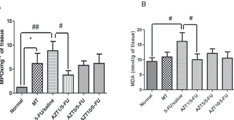
MPO and MDA levels
The MPO activity was measured in the cheek pouch as an indicator of neutrophil infiltration. Levels of MPO in the Normal and AZT1/5-FU groups were decreased (p<0.01 and p<0.05,
respectively) compared to levels in the 5-FU/Saline group (Fig. 4A). Levels of MPO in the MT group were increased (p<0.05) compared to levels in the Normal group (Fig. 4A). Levels of
MDA in the Normal and AZT1/5-FU groups were decreased (p<0.05) compared to levels in
the 5-FU/Saline group (Fig. 4B).
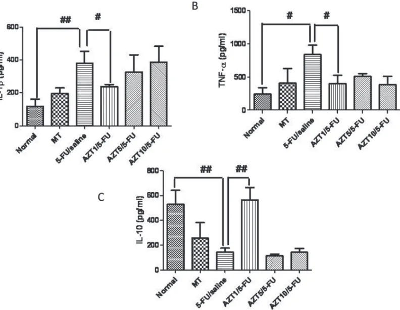
IL-1β, IL-10, and TNF-α
levels
Levels of IL-1βand TNF-αin the Normal group were decreased compared to levels in the 5-FU/Saline group (p<0.01,Fig. 4Aand p<0.05,Fig. 4B, respectively). Levels of IL-10 in the
Normal group were decreased compared to levels in the 5-FU/Saline group (p<0.01,Fig. 4C).
Treatment with AZT at 1 mg/kg (AZT1/5-FU) significantly blocked this elevation in TNF-α (p<0.05) and IL-1β(p<0.05) levels in the cheek pouch (Fig. 5A and 5B, respectively), while
significantly increasing the levels of anti-inflammatory cytokine IL-10 compared to the 5-FU/ Saline group (p<0.01,Fig. 5C).
Effects of AZT on systemic changes: Biochemical, WBC and
bacteraemia analyses
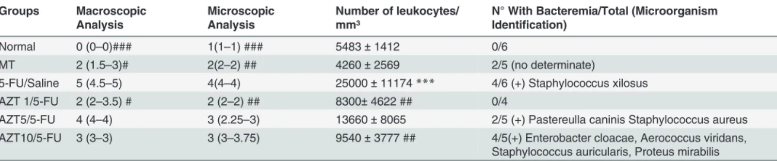
To determine the effect of AZT on systemic changes, biochemical, WBC, and bacteraemia anal-yses were performed. Levels of hepatic enzymes (ALT and AST) and markers of renal function (urea and creatinine) did not show a significant difference between the groups (Table 2). On day 10, animals in the 5-FU/Saline group presented significantly increased leukocyte levels compared to the Normal control group (p<0.001;Table 1).Table 1shows the results of the
bacteraemia analysis for animals in the various groups on day 10. Animals in the Normal con-trol group did not exhibit bacteraemia (-). The 5-FU/Saline group had the highest proportion of animals with bacteraemia (Staphylococcus xylosuswas found in 4/6 samples). The AZT1/5-FU group did not present any sample with bacteraemia. Bacteraemia (+) samples were found in the AZT5/5-FU and AZT10/5-FU groups, and different bacterial species were identified. Fig 1. Macroscopic aspects of hamster cheek pouches.Representative cheek pouches are shown for animals subjected to: A: saline (Normal Group); B: mechanical trauma (MT Group); C: i.p. 5-FU on days 1 and 2 (60 mg/kg and 40 mg/kg, respectively), and MT of the cheek pouches on day 4 (5-FU/Saline Group); D: daily oral AZT (1 mg/kg), i.p. 5-FU on days 1 and 2 (60 mg/kg and 40 mg/kg, respectively), and MT of the cheek pouches on day 4 (AZT1/5-FU Group); E: daily oral AZT (5 mg/kg), i.p. 5-FU on days 1 and 2 (60 mg/kg and 40 mg/kg, respectively), and MT of the cheek pouches on day 4 (AZT5/5-FU Group); F: daily oral AZT (10 mg/kg), i.p. 5-FU on days 1 and 2 (60 mg/kg and 40 mg/kg, respectively), and MT of the cheek pouches on day 4 (AZT10/5-FU Group).
Discussion
AZT is an ARB that was approved for use in 2011. At its maximal dose, AZT has superior effi-cacy to both olmesartan and valsartan at their maximal, approved doses, without increasing ad-verse events [27]. Studies have shown that AZT exhibits anti-inflammatory activity [18], confirming reports regarding the anti-inflammatory activities of ARBs [17,28,29].
The 5-FU/Saline group in this study showed a median macroscopic score of 5, with nearly complete ulceration of the cheek pouch mucosa and fibrosis that made exposure of the oral mucosa difficult. These findings differed significantly from those of the AZT1/5-FU group (median macroscopic score = 2), which evidenced erythema and vasodilation but no ulcera-tion. The results were confirmed by histopathologic analysis, which revealed reduced infiltra-tion of inflammatory cells and oedema. Treatment of animals with 5 mg/kg AZT (AZT5/5-FU group) or 10 mg/kg AZT (AZT10/5-FU group) did not reduce the 5-FU–induced lesions in the cheek pouch, as indicated by the macroscopic and histologic scores.
The histopathologic results confirmed the presence of granulation tissue in the AZT1/5-FU group. The macrophages in granulation tissue control the cellularity of wounds by inducing ap-optosis and phagocytising various wound cells. Macrophages target neutrophils during the in-flammatory phase of repair, as well as fibroblasts and endothelial cells (ECs) during the resolution of this phase [30]. Neutrophils become less prevalent as the wound matures, and macrophages emerge as the predominant inflammatory cell. Neutrophils undergo apoptosis in the wound and are recognised and ingested by macrophages [31]. We found that the AZT1/5-FU group showed a significant reduction in the number of neutrophils, as confirmed by the sig-nificant reduction of level MPO.
Wound healing is an evolutionarily conserved, complex, multicellular process that, in skin, aims at barrier restoration. This process involves the coordinated efforts of several cell types, including keratinocytes, fibroblasts, ECs, macrophages, and platelets. The migration, infiltra-tion, proliferainfiltra-tion, and differentiation of these cells culminate in inflammainfiltra-tion, new tissue for-mation, and, ultimately, wound closure. This complex process is executed and regulated by an Fig 2. Microscopic aspects of hamsters cheek pouches.Animals subjected to without oral mucositis that received saline (Normal) (A, E, I); Animals subjected to Trauma in oral mucosa that received Saline (MT) (B, F, J); Animals subjected to 5-FU/saline that received Saline (C, G, K); Animals subjected that received AZT1/5-FU (D). Animals subjected that received AZT5/5-FU (H), Animals subjected that received AZT10/5-FU.
doi:10.1371/journal.pone.0116799.g002
Table 1. Macroscopic, microscopic, Number of leucocytes/mm3and N° With Bacteremia/Total(Microorganism Identification) analysis of hamsters check pouches subjected of experimental oral mucositis, Natal, RN, 2014.
Groups Macroscopic Analysis
Microscopic Analysis
Number of leukocytes/ mm³
N° With Bacteremia/Total (Microorganism Identification)
Normal 0 (0–0)### 1(1–1) ### 5483±1412 0/6
MT 2 (1.5–3)# 2(2–2) ## 4260±2569 2/5 (no determinate)
5-FU/Saline 5 (4.5–5) 4(4–4) 25000±11174*** 4/6 (+) Staphylococcus xilosus AZT 1/5-FU 2 (2–3.5) # 2 (2–2) ## 8300±4622 ## 0/4
AZT5/5-FU 4 (4–4) 3 (2.25–3) 13660±8065 2/5 (+) Pastereulla caninis Staphylococcus aureus AZT10/5-FU 3 (3–3) 3 (3–3.75) 9540±3777 ## 4/5(+) Enterobacter cloacae, Aerococcus viridans,
Staphylococcus auricularis, Proteus mirabilis
(#p<0.05, ##p<0.01, ###p<0.001 compared groups with 5-FU/saline;***p<0.001 compared groups with Normal Group)
equally complex signalling network involving numerous growth factors, cytokines, and chemo-kines [32].
OM is characterised by an intense inflammatory reaction, caused by the effects of chemo-therapeutic agents on the mucosa lamina propria cells, which results in the production of proinflammatory cytokines (e.g., IL-1β, IL-6, and TNFα) [33]. We demonstrated that oral treatment with 1 mg/kg AZT significantly (P<0.05) reduced TNF-αand IL-1βlevels in an
ex-perimental model of OM in hamsters. In agreement with our data, various articles have shown that AZT decreases IL-1βproduction [18].
IL-10 is the important cytokine with anti-inflammatory properties. In monocytes and mac-rophages, IL-10 diminishes the production of inflammatory mediators and inhibits antigen presentation, although it enhances their uptake of antigens [34]. In our study, the AZT1/5-FU group showed significantly increased levels of IL-10 and TGF-αcompared to 5FU/saline group. Increased tissue levels of TGF-αcan indicate macrophage migration to granulation tis-sue [35]. Macrophages initiate granulation tissue development and release various growth fac-tors, including FGF, EGF, TGF, and platelet-derived growth factor (PDGF) [32]. Macrophages provide an ongoing source of cytokines to modulate inflammatory cell adhesion, cell migra-tion, and fibroblast proliferation. Within hours of injury, re-epithelialisation is initiated, and Fig 3. Photomicrographs of hamsters cheek pouches showing immunoreactivity to TGF-α, FGF, KGF and VEGF.Hamsters subjected to Normal (A, D, G, J, M, P, S); Hamsters subjected to 5- FU/saline (B, E, H, K, N, Q, T); Hamsters subjected to treated with AZT1/5-FU (C, F, I, L, O, R, U) and Hamsters subjected to treated with AZT5/5-FU (C, F, I, L, O, R, U). Images are shown at 40× magnification. Bar = 100μm. Arrow indicates high or moderate labeling in the oral mucosa. Asterisk indicates mild or moderate labeling in the oral mucosa. Triangle and asterisk indicate oral mucosa. Triangle and arrow indicate high labeling of oral mucosa. Asterisk and triangle indicate mild labeling of oral mucosa.
doi:10.1371/journal.pone.0116799.g003
Fig 4. MPO (A) and MDA (B) in Normal, MT, 5-FU/saline and groups treated with AZT1/5-FU, AZT5/5-FU and AZT10/5-FU (#p<0.05, ##p<0.01,
compared groups with 5-FUT/saline Group;*p<0.05, compared groups with Normal Group).
Fig 5. Levels of IL-1β(A), TNF-α(B) and IL-10 (C), and, Normal, MT, 5-FU/saline and groups AZT1/5-FU, AZT5/5-FU and AZT10/5-FU (#p<0.05,
##p<0.01, compared group with 5-FU/saline).
doi:10.1371/journal.pone.0116799.g005
Table 2. Determination of the alanine amino transferase (ALT) and aspartate amino transferase (AST), creatinine and urea parameters Biochemistry of experimental oral mucositis, Natal, RN, 2014.
Groups ALT AST Urea Creatinine
Normal 92.8±4.9 56.2±19.3 47. 4±7.7 0.2±0.08
MT 92.0±17.1 44.4±16.7 50.8±15.9 0.18±0.08
5-FU/Saline 115±49.4 78.5±58.7 38.9±10.0 0.28±0.1
AZT 1/5-FU 87.3±22.6 50.5±12.7 42±4.8 0.3±0.0
AZT5/5-FU 68.4±13.1 57±37.7 38±5.6 0.27±0.5
AZT10/5-FU 86.8±15.9 45±12.4 37±5.6 0.25±0.05
the release of EGF, TGF-α, and FGF acts to stimulate epithelial cell migration and proliferation [36].
TGF-αhas the ability to increase keratinocyte migration and proliferation [14]. FGFs are produced by keratinocytes, fibroblasts, ECs, smooth muscle cells, chondrocytes, and mast cells. FGF-2 or FGF is increased in the acute wound and plays a role in granulation tissue remodel-ling [16]. FGF-7 or KGF-1, which may be important during the late stages of neovascularisa-tion, upregulates VEGF [37], stimulates the proliferation and migration of keratinocytes, and plays an important role in re-epithelialisation [36]. In the present investigation, the AZT1/5-FU group evidenced increased tissue expression of growth factors (i.e., VEGF, FGF, KGF, and TGF-α) involved in the healing process and, especially, in the activation of keratinocytes and neovascularisation with collagen scaffold formation.
The use of AZT was tested in three doses; however, only the 1 mg/kg dose (AZT1/5-FU group) showed efficacy. At this dose, the inflammatory process was reduced, with a significant reduction in MPO levels, notable presence of granulation tissue, and increased levels of IL-10, VEGF, FGF, KGF, and TGF-α. The use of AZT showed a dose-independent effect, with prefer-ence for a lower dose in order to achieve the best clinical results by accelerating the healing pro-cess. The effect of AZT on systemic changes was tested through biochemical, WBC, and bacteraemia analyses. AZT proved to be a very safe drug; even associated with 5-FU (used to induce OM), AZT did not cause changes in the biochemical parameters. With the WBC and bacteraemia analyses, bacteraemia was found in four of six samples in the 5-FU/Saline group, which also showed significant leukocytosis. Although 5-FU treatment is expected to result in leukopenia, the observed leukocytosis was probably in response to bacterial lipopolysaccharide or host proinflammatory cytokines as a result of secondary infection. The AZT1/5-FU group no showed the presence of bacteria in samples. These findings strengthen the evidence for a protective role of AZT in 5-FU–induced OM.
A limitation of the present study is the use of an experimental model of oral mucositis that was established in hamsters. Given that major genetic differences exist between rodents and humans, the present results need to be confirmed in primates prior to the clinical application of azilsartan to humans.
In the present study, we show that azilsartan mediates a protective effect towards the inflam-matory parameters that characterize 5-FU-induced OM. Many of the functional effects demon-strated are dependent on two key factors related to azilsartan as a molecule: 1) it has a high affinity and slow dissociation rate from AT1R, and 2) it exhibits inverse agonistic properties. These properties suggest that azilsartan represents a unique therapeutic agent for the potential treatment of a wide range of angiotensin II-dependent cardiometabolic diseases. These diseases include cardiac hypertrophy, unstable atherosclerotic plaques, cardiac fibrosis, insulin resis-tance, renoprotection and antiinflammatory effects [38,39]. In addition, the results obtained from a number of clinical studies indicate that angiotensin II (AII) receptor type 1 (ATR1)-blocking drugs (ARBs) mediate anti-inflammatory effects [40–42].
hypertensive cancer patients will benefit from an anti-inflammatory effect mediated by azilsar-tan-based chemotherapy
Acknowledgments
Tarciso Bruno Montenegro Sampaio, Biologist. Universidade Potiguar, Natal, RN, Brazil.
Author Contributions
Conceived and designed the experiments: AAA RFAJ GACB CACXM. Performed the experi-ments: AAA RFAJ HV. Analyzed the data: AAA RFAJ HV. Contributed reagents/materials/ analysis tools: AAA RFAJ HV GACB CACXM LMM KCL. Wrote the paper: AAA RFAJ HV GACB CACXM.
References
1. Scully C, Epstein J, Sonis S (2003) Oral mucositis: a challenging complication of radiotherapy, chemo-therapy, and radiochemotherapy: part 1, pathogenesis and prophylaxis of mucositis. Head Neck 25: 1057–1070. PMID:14648865
2. Rimulo AL, Ferreira MC, Abreu MH, Aguirre-Neto JC, Paiva SM (2011) Chemotherapy-induced oral mucositis in a patient with acute lymphoblastic leukaemia. Eur Arch Paediatr Dent 12: 124–127. PMID: 21473846
3. Georgiou M, Patapatiou G, Domoxoudis S, Pistevou-Gompaki K, Papanikolaou A (2012) Oral Mucosi-tis: understanding the pathology and management. Hippokratia 16: 215–216. PMID:23935285
4. Hasenau C, Clasen BP, Roettger D (1988) [Use of standardized oral hygiene in the prevention and therapy of mucositis in patients treated with radiochemotherapy of head and neck neoplasms]. Laryngol Rhinol Otol (Stuttg) 67: 576–579. PMID:3236992
5. Donnelly JP, Bellm LA, Epstein JB, Sonis ST, Symonds RP (2003) Antimicrobial therapy to prevent or treat oral mucositis. Lancet Infect Dis 3: 405–412. PMID:12837345
6. Nicolatou-Galitis O, Sarri T, Bowen J, Di Palma M, Kouloulias VE, et al. (2013) Systematic review of anti-inflammatory agents for the management of oral mucositis in cancer patients. Supportive Care in Cancer 21: 3179–3189. doi:10.1007/s00520-013-1847-yPMID:23702538
7. von Bultzingslowen I, Brennan MT, Spijkervet FKL, Logan R, Stringer A, et al. (2006) Growth factors and cytokines in the prevention and treatment of oral and gastrointestinal mucositis. Supportive Care in Cancer 14: 519–527. PMID:16775647
8. de Castro JF, Abreu EG, Correia AV, Brasil CD, da Cruz Perez DE, et al. (2013) Low-Level Laser in Prevention and Treatment of Oral Mucositis in Pediatric Patients with Acute Lymphoblastic Leukemia. Photomed Laser Surg.
9. Lopez TC, Martins MD, Pavesi VC, Ferreira LS, Bussadori SK, et al. (2013) Effect of laser phototherapy in the prevention and treatment of chemo-induced mucositis in hamsters. Braz Oral Res 27: 342–348. doi:10.1590/S1806-83242013005000019PMID:23752482
10. Elad S, Meidan I, Sellam G, Simaan S, Zeevi I, et al. (2013) Topical curcumin for the prevention of oral mucositis in pediatric patients: case series. Altern Ther Health Med 19: 21–24. PMID:23981369
11. Tanideh N, Tavakoli P, Saghiri MA, Garcia-Godoy F, Amanat D, et al. (2013) Healing acceleration in hamsters of oral mucositis induced by 5-fluorouracil with topical Calendula officinalis. Oral Surg Oral Med Oral Pathol Oral Radiol 115: 332–338. doi:10.1016/j.oooo.2012.08.450PMID:23182376
12. Rodriguez-Caballero A, Torres-Lagares D, Robles-Garcia M, Pachon-Ibanez J, Gonzalez-Padilla D, et al. (2012) Cancer treatment-induced oral mucositis: a critical review. Int J Oral Maxillofac Surg 41: 225–238. doi:10.1016/j.ijom.2011.10.011PMID:22071451
13. Strober W, Fuss IJ, Blumberg RS (2002) The immunology of mucosal models of inflammation. Annu Rev Immunol 20: 495–549. PMID:11861611
14. Li Y, Fan J, Chen M, Li W, Woodley DT (2006) Transforming growth factor-alpha: a major human serum factor that promotes human keratinocyte migration. J Invest Dermatol 126: 2096–2105. PMID: 16691197
16. Powers CJ, McLeskey SW, Wellstein A (2000) Fibroblast growth factors, their receptors and signaling. Endocrine-Related Cancer 7: 165–197. PMID:11021964
17. Nakano A, Hattori Y, Aoki C, Jojima T, Kasai K (2009) Telmisartan inhibits cytokine-induced nuclear factor-kappaB activation independently of the peroxisome proliferator-activated receptor-gamma. Hypertens Res 32: 765–769. doi:10.1038/hr.2009.95PMID:19590508
18. Araujo AA, Varela H, Brito GA, Medeiros CA, Araujo Lde S, et al. (2014) Azilsartan Increases Levels of IL-10, Down-Regulates MMP-2, MMP-9, RANKL/RANK, Cathepsin K and Up-Regulates OPG in an Ex-perimental Periodontitis Model. PLoS One 9: e96750. doi:10.1371/journal.pone.0096750PMID: 24819928
19. Araujo AA, Lopes de Souza G, Souza TO, de Castro Brito GA, Saboia Aragao K, et al. (2013) Olmesar-tan decreases IL-1beta and TNF-alpha levels; downregulates MMP-2, MMP-9, COX-2, and RANKL; and upregulates OPG in experimental periodontitis. Naunyn Schmiedebergs Arch Pharmacol 386: 875–884. doi:10.1007/s00210-013-0886-8PMID:23775504
20. Araujo AA, Souza TO, Moura LM, Brito GA, Aragao KS, et al. (2013) Effect of telmisartan on levels of IL-1, TNF-alpha, down-regulated COX-2, MMP-2, MMP-9 and RANKL/RANK in an experimental peri-odontitis model. J Clin Periodontol 40: 1104–1111. doi:10.1111/jcpe.12160PMID:24118063
21. Medeiros CA, Leitao RF, Macedo RN, Barboza DR, Gomes AS, et al. (2011) Effect of atorvastatin on 5-fluorouracil-induced experimental oral mucositis. Cancer Chemother Pharmacol 67: 1085–1100. doi: 10.1007/s00280-010-1409-7PMID:20661736
22. Souza MH, Troncon LE, Cunha FQ, Oliveira RB (2003) Decreased gastric tone and delayed gastric emptying precede neutrophil infiltration and mucosal lesion formation in indomethacin-induced gastric damage in rats. Brazilian Journal of Medical and Biological Research 36: 1383–1390. PMID: 14502371
23. Esterbauer H, Cheeseman KH (1990) Determination of aldehydic lipid peroxidation products: malonal-dehyde and 4-hydroxynonenal. Methods Enzymol 186: 407–421. PMID:2233308
24. Safieh-Garabedian B, Poole S, Allchorne A, Winter J, Woolf CJ (1995) Contribution of interleukin-1 beta to the inflammation-induced increase in nerve growth factor levels and inflammatory hyperalgesia. Br J Pharmacol 115: 1265–1275. PMID:7582555
25. Kendall C, Ionescu-Matiu I, Dreesman GR (1983) Utilization of the biotin/avidin system to amplify the sensitivity of the enzyme-linked immunosorbent assay (ELISA). J Immunol Methods 56: 329–339. PMID:6833765
26. de Souza GE, Ferreira SH (1985) Blockade by antimacrophage serum of the migration of PMN neutro-phils into the inflamed peritoneal cavity. Agents Actions 17: 97–103. PMID:4083185
27. White WB, Weber MA, Sica D, Bakris GL, Perez A, et al. (2011) Effects of the angiotensin receptor blocker azilsartan medoxomil versus olmesartan and valsartan on ambulatory and clinic blood pressure in patients with stages 1 and 2 hypertension. Hypertension 57: 413–420. doi:10.1161/
HYPERTENSIONAHA.110.163402PMID:21282560
28. Saavedra JM (2012) Angiotensin II AT(1) receptor blockers as treatments for inflammatory brain disor-ders. Clin Sci (Lond) 123: 567–590. doi:10.1042/CS20120078PMID:22827472
29. Nagib MM, Tadros MG, ELSayed MI, Khalifa AE (2013) Anti-inflammatory and anti-oxidant activities of olmesartan medoxomil ameliorate experimental colitis in rats. Toxicology and Applied Pharmacology 271: 106–113. doi:10.1016/j.taap.2013.04.026PMID:23665423
30. Brancato SK, Albina JE (2011) Wound Macrophages as Key Regulators of Repair Origin, Phenotype, and Function. American Journal of Pathology 178: 19–25. doi:10.1016/j.ajpath.2010.08.003PMID: 21224038
31. Meszaros AJ, Reichner JS, Albina JE (1999) Macrophage phagocytosis of wound neutrophils. J Leu-koc Biol 65: 35–42. PMID:9886244
32. Barrientos S, Stojadinovic O, Golinko MS, Brem H, Tomic-Canic M (2008) Growth factors and cytokines in wound healing. Wound Repair and Regeneration 16: 585–601. doi:10.1111/j.1524-475X.2008. 00410.xPMID:19128254
33. Sonis ST, Elting LS, Keefe D, Peterson DE, Schubert M, et al. (2004) Perspectives on cancer therapy-induced mucosal injury: pathogenesis, measurement, epidemiology, and consequences for patients. Cancer 100: 1995–2025. PMID:15108222
34. Sabat R, Grutz G, Warszawska K, Kirsch S, Witte E, et al. (2010) Biology of interleukin-10. Cytokine Growth Factor Rev 21: 331–344. doi:10.1016/j.cytogfr.2010.09.002PMID:21115385
35. Rappolee DA, Mark D, Banda MJ, Werb Z (1988) Wound macrophages express TGF-alpha and other growth factors in vivo: analysis by mRNA phenotyping. Science 241: 708–712. PMID:3041594
37. Niu J, Chang Z, Peng B, Xia Q, Lu W, et al. (2007) Keratinocyte growth factor/fibroblast growth factor-7-regulated cell migration and invasion through activation of NF-kappaB transcription factors. J Biol Chem 282: 6001–6011. PMID:17200110
38. Kajiya T, Ho C, Wang J, Vilardi R, Kurtz TW (2011) Molecular and cellular effects of azilsartan: a new generation angiotensin II receptor blocker. J Hypertens 29: 2476–2483. doi:10.1097/HJH. 0b013e32834c46fdPMID:21986624
39. Ojima M, Igata H, Tanaka M, Sakamoto H, Kuroita T, et al. (2011) In vitro antagonistic properties of a new angiotensin type 1 receptor blocker, azilsartan, in receptor binding and function studies. J Pharma-col Exp Ther 336: 801–808. doi:10.1124/jpet.110.176636PMID:21123673
40. Clancy P, Koblar SA, Golledge J (2014) Angiotensin receptor 1 blockade reduces secretion of inflam-mation associated cytokines from cultured human carotid atheroma and vascular cells in association with reduced extracellular signal regulated kinase expression and activation. Atherosclerosis 236: 108–115. doi:10.1016/j.atherosclerosis.2014.06.011PMID:25016365
41. Taguchi I, Toyoda S, Takano K, Arikawa T, Kikuchi M, et al. (2013) Irbesartan, an angiotensin receptor blocker, exhibits metabolic, anti-inflammatory and antioxidative effects in patients with high-risk hyper-tension. Hypertens Res 36: 608–613. doi:10.1038/hr.2013.3PMID:23425956