Analysis of dimensions, activation and median frequency
of cervical flexor muscles in young women with migraine
or tension-type headache
Débora Wanderley1, Alberto G. Moura Filho2, Joaquim J. S. Costa Neto1, Gisela R. Siqueira2, Daniella A. de Oliveira2
ABSTRACT | Background: Central and peripheral mechanisms may be involved in migraine and tension-type headache pathogenesis, however the role of muscle disorders in their pathophysiological mechanisms remains unclear. Objectives: To assess the association between the presence of migraine or tension-type headache and changes in longus colli muscle dimensions and sternocleidomastoid muscle activity. Method: An observational study with 48 women comparing the following groups: migraine (n=21), tension-type headache (n=16), and control (n=11). The cross-sectional area, lateral and anteroposterior dimensions, and shape ratio of the longus colli muscle were measured using ultrasound. The activation of the sternocleidomastoid muscle was assessed by signal amplitude and the decline in median frequency using surface electromyographic analysis. Results: The dimensions of the longus colli muscle did not differ between groups (p>0.05). Post-test analysis showed lower sternocleidomastoid muscle activation on both sides, at the onset of contraction, in the group with tension-type headache when compared to the control group {right sternocleidomastoid [tension-type headache: 0.39 (0.30-0.49); control: 0.58 (0.42-0.76); p=0.026] and left sternocleidomastoid [tension-type headache: 0.39 (0.31-0.48); control: 0.60 (0.42-0.79); p=0.039], Tukey’s post hoc test}. There was no difference between the three groups in sternocleidomastoid muscle activation, on both sides, at the end of contraction (p>0.05). Intergroup analysis showed no difference in the rate of decline in median frequency (p>0.05). Conclusion: The group with tension-type headache exhibited less activation at the onset of sternocleidomastoid muscle contraction. No association was observed between the presence of headache and alterations in longus colli muscle dimensions, median frequency, and sternocleidomastoid muscle activation at the end of contraction.
Keywords: migraine disorders; tension-type headache; neck muscles; ultrasonography; electromyography; movement.
HOW TO CITE THIS ARTICLE
Wanderley D, Moura Filho AG, Costa Neto JJS, Siqueira GR, de Oliveira DA. Analysis of dimensions, activation and median frequency
of cervical lexor muscles in young women with migraine or tension-type headache. Braz J Phys Ther. 2015 May-June; 19(3):243-250.
http://dx.doi.org/10.1590/bjpt-rbf.2014.0093
1Programa de Pós-graduação em Neuropsiquiatria e Ciências do Comportamento, Universidade Federal de Pernambuco (UFPE), Recife, PE, Brazil 2Departamento de Fisioterapia, UFPE, Recife, PE, Brazil
Received: Aug. 26, 2014 Revised: Nov. 16, 2014 Accepted: Dec. 16, 2014
Introduction
Migraine and tension-type headache are the most common source of pain in young workers and they
have also been characterized as signiicant public health
issues, with great socioeconomic impact1-4. These
are primary headaches, and central and peripheral mechanisms may be involved in their pathogenesis5,6.
A number of authors6-10 have sought to elucidate
the involvement of peripheral nociceptive stimuli in the pathogenesis of migraine and tension-type headache. However, the role of muscle alterations in the activation of the physiopathological process of these types of headache remains unclear8-10.
A possible reason for the association between changes in craniocervical muscles in patients with
migraine or tension-type headache is the activation of trigemino-cervical nucleus during migraine or tension-type headache attacks. In this nucleus, there is a convergence of cervical and trigeminal nociceptive afferents in the trigeminocervical complex, releasing vasoactive algogenic substances, which facilitate pain transmission to the brain and promote awareness of the central nervous system4-5,11.
This is supported by other indings suggesting that
individuals with migraine or tension-type headache are more likely to self-report neck pain and to report cervical muscles disorders, such as limited range of motion11, changes in head and cervical posture11,
fatigue and activation during contractions7,15. Several
methods have been used to verify this association, including photogrammetry11, electromyography7,15-17,
and ultrassonography12-14.
Prior studies7,15-17 have used electromyography
to assess the relationship between headache and
alterations in the supericial cervical activity of the sternocleidomastoid, splenius, and trapezius muscles.
The results suggest that changes in activation7,16,17 and
fatigability7,15 are more frequent in individuals with
headache. However, previous assessments of neck muscle dimensions only included patients with chronic neck pain and did not include subjects with migraine.
Patients with chronic neck pain exhibited lower activation in deep neck muscles, such as the longus capitis and longus colli muscles12-14, accompanied by
increased sternocleidomastoid muscle activity12 as
a compensatory mechanism. In two studies13,14, the
ultrasonographic images of these muscles showed a reduction in thickness13, cross-sectional area13, and
anteroposterior dimension in individuals with chronic
neck pain. Other indings suggest the presence of
atrophy and alterations in muscle recruitment14.
In this respect, deep lexor muscles in the cervical
region are essential to cervical spine postural control.
Thus, imbalances between these muscles and supericial
cervical muscles make the cervical spine less stable and more vulnerable to other forces involved in maintaining posture, causing overload in other muscles18.
Despite the importance of deep and supericial cervical lexor muscles in stabilizing and maintaining
cervical lordosis and indications that migraine and tension-type headache are associated with changes in neck muscles, there is still a lack of studies7,15-17 that
use non-invasive techniques to assess those muscles in individuals with migraine or tension-type headache not related to chronic neck pain.
Thus, the aim of the present study was to assess the association between the presence of migraine or tension-type headache and changes in the longus colli muscle dimensions and sternocleidomastoid muscle activity, using ultrasonography and surface electromyography.
Method
This is an observational, cross-sectional type study
comparing three groups. The sample size was not
calculated because this is a pilot study. The study was approved by the Human Research Ethics Committee of the Health Sciences Center of Universidade Federal
de Pernambuco (UFPE), Recife, PE, Brazil (CAAE
02219412.5.0000.5208). All participants gave their informed consent.
Data were collected from October/2012 to December/2013 in the Physical Therapy Department of UFPE.
Participants
The sample was composed of young adult women, aged between 20 and 30 years, to avoid biases due to the presence of muscle changes associated with biological aging. Only nulliparous women were included in order to prevent biases related to the relationship between hormones and the presence of headache19.
Clinical diagnosis, established by a neurologist based
on criteria proposed by The International Classiication
of Headache Disorders20, was used to divide the
sample into migraine, tension-type headache, and control groups.
The migraine group was composed of women diagnosed with episodic migraine (less than 15 days with headache per month), with following characteristics: pure migraine (with aura, without aura or both) or probable migraine (with aura and without aura or both). Participants who had probable migraine associated with tension-type headache or migraine associated with tension-type headache were also allocated to the migraine group20. The tension-type
headache group was formed by women with episodic tension-type headache (less than 15 days with headache per month)20. The control group was composed of
participants who had intermittent headache crises over their lifetime that were not associated with the characteristics of primary headaches and those who did not meet the diagnostic criteria of migraine or tension-type headache20.
The exclusion criteria were as follows: 1) body
mass index ≥30 as obesity can increase the risk of
chronic migraine19; 2) chronic migraine, chronic
tension-type headache or chronic neck pain due to the association between muscle alterations and chronification of headache and neck pain7,16,17;
3) diseases or dysfunctions such as myopathies,
ibromyalgia, abnormalities, fractures or history of
cervical spine or thoracic surgery, symptomatic spinal disc herniation, rheumatoid arthritis, and history of
spinal tumors; 4) score ≥15 in the neck disability
index21, score ≥36 in the Beck depression inventory
or score ≥30 in the Beck anxiety inventory22 as neck
The neck disability index is a questionnaire adapted
and validated for the Brazilian population that provides
information about how neck pain affects the ability to perform activities of daily living. It consists of
10 sections, scored from 0 to 5 each. Scores ≥15 indicate
moderate neck dysfunction21.
The Beck depression and anxiety inventories are instruments for research of depressive and anxiety
symptoms, translated and validated for the Brazilian
population, and consists of 21 items. In the Beck depression inventory, the severity of symptoms varies from 0 to 3, with a maximum score of 63. Scores
≥36 indicate severe depression. In the Beck anxiety
inventory, the sum of the items results in a total score
that can range from 0 to 63, and scores ≥30 indicate
severe anxiety22.
Data collection procedure
An examiner, blinded to the diagnosis of headache, performed an ultrasonographic assessment of the longus colli muscle and surface electromyographic evaluation of the sternocleidomastoid muscle. During data collection, participants could not be menstruating or taking medication such as muscle relaxants,
analgesics or anti-inlammatories, in the 48 hours
before the examination.
To avoid measurement bias, the examiner was trained to perform the evaluations.
Ultrasonographic assessment of the longus
colli muscle
B-mode ultrasonography (Aloka 1500 with
7.5 MHz linear transducer) was used to measure the
cross-sectional area (cm2), as well as the lateral (cm),
anteroposterior (cm), and shape ratio dimensions of the muscle.
The cross-sectional area was considered the greatest distance between the inner edges of the margins of the muscle image, without including fascial contours13. The lateral and anteroposterior
dimensions were considered the greatest distance between one margin and another of the image, in the lateral and anteroposterior direction, respectively13.
The shape ratio was obtained by dividing lateral and anteroposterior dimension values13.
Participants were positioned in dorsal decubitus,
knees lexed, arms along the body and head in midline
position14. The transducer was placed longitudinally in
the anterior region of the neck, parallel to the trachea,
approximately ive centimeters from the midline at the
level of vertebrae C5 and C6. The longus colli muscle
is bordered inferiorly and medially by the vertebral body, laterally by the carotid artery, and superiorly by the retropharyngeal space13.
Three measurements of the muscle dimensions were taken at rest and during contraction, with a one-minute interval between them. In contraction,
subjects were instructed to perform a cervical lexion,
without removing their head and shoulders from the table, sustaining it for ten seconds14. The average of
the three results for each measurement was used to compare intergroup dimensions.
Surface electromyography of the
sternocleidomastoid muscle
The EMG device (model 410C, EMG System of
Brazil Ltda.) was connected to a portable computer. The equipment has four channels, with pre-ampliiers, sample frequency per channel of 2000 Hz, Butterworth band-pass ilter between 20 and 500 Hz, ampliied
2000 times (common-mode rejection >120 dB),
digitized with a frequency of 2 KHz per channel
and amplitude range between –5 and +5 volts. After
collection, the signal was digitized, converted to .txt format and analyzed using the software of the
device itself.
Signal capture followed ISEK (International Society of Electrophysiology and Kinesiology) Standards for
Reporting EMG Data23. Electromyographic signals
were detected using two rectangular surface electrodes (Ag/AgCl, self-adhesive, bipolar) on the muscle, twenty millimeters apart24.
Participants were placed in dorsal decubitus, knees
lexed and feet resting on the table, arms lexed and
hands above their head and resting on the table, in order to reduce the action of trunk and scapular waist muscles25. The electrodes were placed on the midpoint
of the muscle belly, along the length of the muscle
ibers17, from a reference line running between the
lower point of the mastoid process and the center of
the sternal furcula. The reference electrode was ixed
to the lateral epicondyle of the left humerus.
In the irst ive seconds of collection, participants
remained at rest with their head at maximum rotation amplitude to the opposite side to that assessed and
leaning toward the same side. Cervical lexion was then
performed, up to maximum amplitude25, maintaining
the position in isometric contraction for twenty-ive
seconds, subjects not raising their shoulders from
the table, followed by ive seconds of rest. The
sternocleidomastoid muscle was assessed unilaterally
The electrical activities analyzed were median frequency (Hz) and signal amplitude (v), through a time
window manually driven by the software of the device itself. One-second samples of the electromyographic
recording were analyzed at the start of the rest period
(between 2 and 3 seconds), at the onset of contraction (between 6 and 7 seconds), and at the end of contraction (between 28 and 29 seconds).
Median frequency was measured at the start and
end of muscle contraction in order to analyze the
rate of decline at the end of contraction15. Muscle
activation was determined from the amplitude of
the electromyographic signal quantiied by root mean square (RMS) values. Data were normalized
by subtracting RMS values at the start and end of contraction from baseline values27. The average of
median frequency and normalized RMS values was
used for intergroup comparison.
Processing and data analysis
The data bank was digitized into Microsoft
Excel (2007) spreadsheets and exported to SPSS 20.0 for
analysis. Data were presented as mean (conidence
interval) and percentage. All variables exhibited
normal distribution in the Kolmogorov-Smirnov test.
To verify the association between variables, paired t-test, one-way ANOVA (for intergroup comparisons separately), repeated measures two-way ANOVA (for intra and intergroup comparisons performed simultaneously) and Tukey’s post hoc test were
performed. Statistical signiicance was set at the 95% conidence level (p<0.05).
Results
A total of 52 participants were recruited, 4 of whom refused to take part in the study. Forty-eight women, aged on average 22.67 years (CI: 22.1-23.23), were allocated to the migraine (n=21), tension-type (n=16), and control (n=11) groups.
Of the participants with headache (n=37), 43% (n=16/37) suffered from episodic tension-type headache and 57% (n=21/37) migraine. Considering only the migraine group (n=21), we found the following frequency of subtypes: 67% (n=14/21) had migraine without aura; 14% (n=3/21) migraine without aura associated with episodic tension-type headache; 9% (n=2/21) migraine with aura; 5% (n=1/21) migraine with aura associated with episodic tension-type headache; and 5% (n=1/21) migraine with aura and without aura.
There was no intergroup difference (p>0.05) in age, height, weight or body mass index. The scores for the
Neck disability index were statistically signiicant in
the intergroup analysis (p=0.01; one-way ANOVA). Post-test analysis showed higher scores in the migraine
group than in the control group (p<0.01); and higher
scores in tension-type headache than in the control group (p=0.08). The Beck depression inventory scores and the Beck anxiety inventory scores did not differ between groups (p>0.05; Table 1).
Regarding participants with headache in Table 1, there was no difference between groups in the time
Table 1. General characteristics of sample.
Groups
Control (n=11) Migraine (n=21) TTH (n=16) p*
Age (years) 21.64 (21.02-22.26) 23.1 (22.12-24.07) 22.81 (21.73-23.9) 0.123
Weight (kg) 57.26 (53.03-61.49) 58.67 (55.22-62.12) 57.34 (54.06-60.63) 0.794
Height (m) 1.64 (1.61-1.66) 1.64 (1.62-1.67) 1.62 (1.6-1.64) 0.263
BMI (kg/m2) 21.31 (19.38-23.23) 21.43 (20.24-22.63) 21.86 (20.69-23.03) 0.824
NDI 3.18 (1.72-4.88) 8.86 (6.06-10.23) 6.4 (4.87-9.25) 0.01†
BDI 4.80 (1.88-7.72) 7.71 (3.96-11.47) 4.0 (2.37-5.63) 0.16
BAI 6.50 (1.61-11.39) 6.52 (3.56-9.48) 5.50 (2.79-8.21) 0.88
Time with headache (years) - 8.71 (6.80-10.63) 8.0 (6.03-9.97) 0.59
Headache frequency (days/6 months)
- 24.48 (11.75-37.20) 6.50 (2.68-10.32) 0.01**
Headache duration (hours) - 6.62 (5.03-8.21) 7.44 (1.31-13.57) 0.21
with headache (p=0.59) and headache duration (p=0.21). However, headache frequency was higher in the migraine group than in the tension-type headache group (p=0.01).
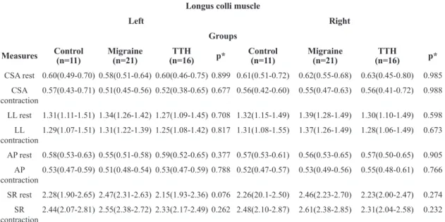
Ultrasonographic analysis of the left and right longus colli muscle during rest and contraction revealed no intergroup difference in cross-sectional area or lateral, anteroposterior, and shape ratio dimensions (Table 2).
There was a statistically signiicant intergroup
difference in right and left sternocleidomastoid muscle
activation at the onset of contraction (p<0.05; repeated
measures two-way ANOVA). Post-test analysis showed less sternocleidomastoid muscle activation on both sides at the start of contraction in the group with tension-type
headache when compared to the control group {right sternocleidomastoid [tension-type headache: 0.39 (0.30-0.49); control: 0.58 (0.42-0.76); p=0.026] and left sternocleidomastoid [tension-type headache: 0.39 (0.31-0.48); control: 0.60 (0.42-0.79); p=0.039], Tukey’s post-hoc test}. There was no difference between the three groups in sternocleidomastoid muscle activation, on either side, at the end of contraction (p>0.05; repeated measures two-way ANOVA; Table 3).
In intragroup analysis, there was a signiicant difference (p<0.05; repeated measures two-way
ANOVA) in median frequency variation between the start and end of sternocleidomastoid muscle contraction, indicating a decline in median frequency at the end of
Table 2. Intergroup analysis of ultrasonography of the longus colli muscle during rest and contraction.
Longus colli muscle
Left Right
Groups
Measures Control (n=11) Migraine (n=21) (n=16)TTH p* Control (n=11) Migraine (n=21) (n=16)TTH p*
CSA rest 0.60(0.49-0.70) 0.58(0.51-0.64) 0.60(0.46-0.75) 0.899 0.61(0.51-0.72) 0.62(0.55-0.68) 0.63(0.45-0.80) 0.985
CSA contraction
0.57(0.43-0.71) 0.51(0.45-0.56) 0.52(0.38-0.65) 0.677 0.56(0.42-0.60) 0.55(0.47-0.63) 0.56(0.41-0.72) 0.988
LL rest 1.31(1.11-1.51) 1.34(1.26-1.42) 1.27(1.09-1.45) 0.708 1.32(1.15-1.49) 1.39(1.28-1.49) 1.30(1.10-1.49) 0.598
LL
contraction
1.29(1.07-1.51) 1.31(1.22-1.39) 1.25(1.08-1.42) 0.817 1.31(1.08-1.55) 1.37(1.26-1.49) 1.28(1.06-1.49) 0.673
AP rest 0.58(0.53-0.63) 0.55(0.51-0.58) 0.59(0.52-0.65) 0.377 0.57(0.53-0.61) 0.56(0.53-0.65) 0.57(0.50-0.65) 0.905
AP contraction
0.53(0.47-0.59) 0.51(0.48-0.54) 0.53(0.47-0.59) 0.788 0.52(0.47-0.57) 0.53(0.49-0.56) 0.55(0.48-0.61) 0.766
SR rest 2.28(1.90-2.65) 2.47(2.31-2.63) 2.15(1.93-2.36) 0.076 2.26(20.1-2.50) 2.46(2.23-2.70) 2.23(2.00-2.47) 0.274
SR contraction
2.44(2.07-2.81) 2.55(2.38-2.72) 2.33(2.17-2.49) 0.262 2.48(2.10-2.87) 2.61(2.38-2.85) 2.31(2.04-2.58) 0.232
*One-way ANOVA. Data are shown as mean and conidence interval. TTH: tension-type Headache; n: number; CSA: cross sectional area, cm2; LL: lateral measure, cm; AP: anteroposterior measure, cm; SR: shape ratio.
Table 3. Intergroup analysis of the means of normalized root mean square values of the sternocleidomastoid muscle.
Root mean square Sternocleidomastoid muscle
Left Right
Groups Beginning of
contraction (6-7s)
End of contraction (28-29s)
Beginning of contraction (6-7s)
End of contraction (28-29s)
Migraine (n=21)
0.53 (0.44-0.63)
0.42 (0.35-0.48)
0.46 (0.39-0.53)
0.39 (0.34-0.44)
Control (n=11)
0.60 (0.42-0.79)
0.49 (0.37-0.62)
0.58 (0.42-0.76)
0.44 (0.32-0.58)
p†=0.039 p†=0.026
TTH (n=16)
0.39 (0.31-0.48)
0.38 (0.30-0.47)
0.39 (0.30-0.49)
0.35 (0.27-0.45)
p* 0.038** 0.188 0.033** 0.369
contraction in all groups. However, the difference in the rate of median frequency decline, from the start to the end of sternocleidomastoid muscle contraction,
was not signiicant between groups (p=0.092 on the left
side and p=0.97 on the right side; repeated measures two-way ANOVA; Table 4).
Discussion
The hypothesis that women with migraine or tension-type headache have a shorter longus colli muscle and increased sternocleidomastoid muscle activity, when compared to women without headache,
was not conirmed. On the other hand, the present study
is innovative in that it assesses muscles with important
stabilization and postural alignment functions in the
cervical region in a young population with migraine or episodic tension-type headache.
Since muscle alterations may be more associated
with biological aging and chroniication of headache
and neck pain7,16,17 than with episodic headache in
a younger population (15 to 24 years), the present study avoided a confusion factor in the association
between muscle modiications and the emergence of
headaches. Therefore, differences between our results and those reported in the literature7,16,17 may be due
to the different characteristics and age groups of the
populations analyzed.
In this study, there was less activation at the onset of sternocleidomastoid muscle contraction in the group with tension-type headache, when compared to participants without headache. Corroborating
our indings, other authors have also observed less
activation and more prolonged sternocleidomastoid muscle relaxation in participants with chronic neck pain associated with headache16.
On the other hand, studies that showed greater activation and fatigability in cervical muscles, such as the sternocleidomastoid, splenius17, frontal
and temporal7, assessed participants with chronic
tension-type headache7,17. Thus, the increase in muscle
activation and fatigability may be a consequence of
the motor control reorganization strategy in subjects
with chronic headache17, generating muscle overload
and fatigue, increased nociception17, and changes in
the type of muscle iber7.
Therefore, the reduced muscle activation values observed in the group with tension-type headache may be related to the increased basal tonus in the sternocleidomastoid muscle and smaller amount of fast
ibers. The reduction in type II ibers can be associated
with lower speed of contraction, reducing the muscle activation in the tension-type group7. Similarly, the
absence of a difference in sternocleidomastoid muscle activation in the group with migraine suggests that these
individuals may have another type of muscle iber.
The neck disability index scores were higher in the migraine and tension-type headache groups than in the control group, suggesting that neck pain adds
signiicantly to the overall disability of individuals with
migraine or tension-type headache. Neck pain-related disability can be associated with changes in body posture28 and seems to increase as the frequency
of migraine attacks increases29. Furthermore, this
Table 4. Intra and inter-group analysis of the variation in median frequency between the beginning and the end of the contraction of the sternocleidomastoid muscle.
Median frequency Sternocleidomastoid muscle
Left Right
Groups
Beginning of contraction
(6-7s)
End of contraction
(28-29s)
p*
Beginning of contraction
(6-7s)
End of contraction
(28-29s)
p*
Control (n=11) 75.99 (69.31-82.66)
66.40 (58.88-73.91)
0.010 76.70
(70.24-83.15)
64.98 (55.42-74.53)
0.008
Migraine (n=21) 87.23 (80.69-93.77)
75.70 (70.09-81.30)
<0.001 85.74 (78.71-92.78)
74.77 (67.81-81.73)
<0.001
TTH (n=16)
82.75 (76.57-88.94)
66.40 (60.71-72.08)
<0.001 78.85 (71.28-86.42)
67.86 (61.84-73.89)
<0.001
p† 0.092 0.974
disability may be a functional consequence of changes in craniocervical posture in migraine patients29.
With respect to the analysis of deep cervical muscles, one study10 showed that people with chronic
neck pain exhibited imbalance between deep and
supericial cervical lexor muscles, such as the longus
colli and sternocleidomastoid muscles. In our study, similar results were expected in groups with migraine or tension-type headache. However, given that they are deep muscles in the cervical region, alterations in this structure may only be perceived when there is prolonged accumulation of nociceptive stimuli, as in chronic neck pain12,14.
Moreover, there are also reports of reduced thickness14, cross-sectional area, and anteroposterior
dimension13 in the longus colli muscle of individuals
with chronic neck pain. However, it is important to underscore that chronic neck pain may be linked to muscle atrophy and alterations in muscle dimension and recruitment14, justifying the discrepancies in the
indings of the present study. Thus, alterations in
cervical muscles are most likely correlated to pain
chroniication, as occurs in the aging process or in
conditions such as chronic migraine, cervicogenic headache and chronic neck pain, than to the pathogenesis of headache7,16,17,19.
One of the limitations of the present study was that it is not a longitudinal study and does not involve a long-term follow-up of participants to establish a more accurate association between alterations in cervical muscles and the presence of headache. Additionally, the need to control some factors that possibly trigger the headache and the higher prevalence of migraine compared with tension-type headache and women without primary headache limited our ability to obtain a larger sample, especially for the control group.
The clinical importance of our study is that the knowledge of changes in muscle function will be important to guide physical therapy treatment in patients with headache. The decreased activation in the sternocleidomastoid muscle at the onset of contraction in the tension-type headache group suggests the importance of more muscle assessments in patients with headache. Furthermore, patients with headache
beneited from the Neck disability index evaluation,
because in our study they were more likely to report neck disability than the control group, suggesting that the prevalence of neck pain is higher in patients with headache.
Conclusion
In the present study, the group with tension-type headache showed less sternocleidomastoid muscle activation at the onset of contraction. No association was observed between the presence of headache and alterations in the dimensions of the longus colli muscle, median frequency, and sternocleidomastoid muscle activation at the end of contraction.
References
1. Morelli JGS, Rebelatto JR. The effectiveness of manual therapy in individuals with headaches, with and without cervical degeneration: analysis of six cases. Rev Bras Fisioter. 2007;11(4):285-9. http://dx.doi.org/10.1590/ S1413-35552007000400013.
2. Winter AC, Berger K, Buring JE, Kurth T. Associations of socioeconomic status with migraine and non-migraine headache. Cephalalgia. 2012;32(2):159-70. http://dx.doi. org/10.1177/0333102411430854. PMid:22174348 3. Reynales H, Aycardi E, Valencia D. [Migraine: implications
for work, disability and request for health services in Colombia]. Rev Neurol. 2001;32(11):1001-5. PMid:11562818. 4. Fumal A, Schoenen J. Tension-type headache: current research
and clinical management. Lancet Neurol. 2008;7(1):70-83. http://dx.doi.org/10.1016/S1474-4422(07)70325-3. PMid:18093564
5. Burstein R. Deconstructing migraine headache into peripheral
and central sensitization. Pain. 2001;89(2-3):107-10. http:// dx.doi.org/10.1016/S0304-3959(00)00478-4. PMid:11166465 6. Sohn J-H, Choi H-C, Jun A-Y. Differential patterns of muscle
modification in women with episodic and chronic tension-type headache revealed using surface electromyographic analysis. J Electromyogr Kinesiol. 2013;23(1):110-7. http:// dx.doi.org/10.1016/j.jelekin.2012.08.001. PMid:22947196 7. Jensen R, Fuglsang-Frederiksen A, Olesen J. Quantitative
surface EMG of pericranial muscles in headache. A population study. Electroencephalogr Clin Neurophysiol. 1994;93(5):335-44. http://dx.doi.org/10.1016/0168-5597(94)90121-X. PMid:7525241
8. Rollnik JD, Karst M, Fink M, Dengler R. Botulinum toxin type A and EMG: a key to the understanding of chronic tension-type headaches? Headache. 2001;41(10):985-9. http:// dx.doi.org/10.1046/j.1526-4610.2001.01193.x. PMid:11903527 9. Bartsch T. Migraine and the neck: new insights from basic data. Curr Pain Headache Rep. 2005;9(3):191-6. http://dx.doi. org/10.1007/s11916-005-0061-0. PMid:15907257 10. Bartsch T, Goadsby PJ. Increased responses in trigeminocervical
nociceptive neurons to cervical input after stimulation of the dura mater. Brain. 2003;126(Pt 8):1801-13. http://dx.doi. org/10.1093/brain/awg190. PMid:12821523
12. Falla DL, Jull GA, Hodges PW. Patients with neck pain demonstrate reduced electromyographic activity of the deep cervical flexor muscles during performance of the craniocervical flexion test. Spine. 2004;29(19):2108-14. http://dx.doi.org/10.1097/01.brs.0000141170.89317.0e. PMid:15454700
13. Javanshir K, Rezasoltani A, Mohseni-Bandpei MA, Amiri M, Ortega-Santiago R, Fernández-de-Las-Peñas C. Ultrasound assessment of bilateral longus colli muscles in subjects with chronic bilateral neck pain. Am J Phys Med Rehabil. 2011;90(4):293-301. http://dx.doi.org/10.1097/ PHM.0b013e31820173e5. PMid:21173685
14. Jesus-Moraleida FR, Ferreira PH, Pereira LSM, Vasconcelos CM, Ferreira ML. Ultrasonographic analysis of the neck flexor muscles in patients with chronic neck pain and
changes after cervical spine mobilization. J Manipulative Physiol Ther. 2011;34(8):514-24. http://dx.doi.org/10.1016/j. jmpt.2011.08.006. PMid:21978544
15. Oksanen A, Pöyhönen T, Metsähonkala L, Anttila P, Hiekkanen H, LaimiK, et al. Neck flexor muscle fatigue in adolescents with headache: an electromyographic study. Eur J Pain. 2007;11(7):764-72. http://dx.doi.org/10.1016/j. ejpain.2006.12.003. PMid:17291797
16. Barton PM, Hayes KC. Neck flexor muscle strength, efficiency, and relaxation times in normal subjects and subjects with unilateral neck pain and headache. Arch Phys Med Rehabil. 1996;77(7):680-7. http://dx.doi.org/10.1016/ S0003-9993(96)90008-8. PMid:8669995
17. Fernández-de-las-Peñas C, Falla D, Arendt-Nielsen L, Farina D. Cervical muscle co-activation in isometric contractions is enhanced in chronic tension-type headache patients. Cephalalgia. 2008;28(7):744-51. http://dx.doi.org/10.1111/ j.1468-2982.2008.01584.x. PMid:18460003
18. Mayoux-Benhamou MA, Revel M, Vallée C, Roudier R, Barbet JP, Bargy F. Longus colli has a postural function on cervical curvature. Surg Radiol Anat. 1994;16(4):367-71. http://dx.doi.org/10.1007/BF01627655. PMid:7725191 19. Peres MF, Sanchez del Rio M, Seabra ML, Tufik S, Abucham
J, Cipolla-Neto J, et al. Hypothalamic involvement in chronic migraine. J Neurol Neurosurg Psychiatry. 2001;71(6):747-51. http://dx.doi.org/10.1136/jnnp.71.6.747. PMid:11723194 20. Headache Classification Subcommittee of the International
Headache Society. The international classification of headache disorders. Cephalalgia. 2004;24(1 Suppl 1):9-160. PMid:14979299.
21. Cook C, Richardson JK, Braga L, Menezes A, Soler X, Kume P, et al. Cross-cultural adaptation and validation of the
Brazilian Portuguese version of the Neck Disability Index
and Neck Pain and Disability Scale. Spine. 2006;31(14):1621-7. http://dx.doi.org/10.1097/01.brs.0000221989.53069.16. PMid:16778699
22. Stulz N, Crits-Christoph P. Distinguishing anxiety and depression in self-report: purification of the beck anxiety inventory and beck depression inventory-II. J Clin Psychol. 2010;66(9):927-40. PMid:20694959.
23. Merletti R, Di Torino P. Standards for reporting EMG data.
J Electromyogr Kinesiol. 1999;9(1):3-4.
24. Hermens HJ, Freriks B, Disselhorst-Klug C, Rau G. Development of recommendations for SEMG sensors and sensor placement procedures. J Electromyogr Kinesiol. 2000;10(5):361-74. http://dx.doi.org/10.1016/S1050-6411(00)00027-4. PMid:11018445
25. Kendall HO, Kendall FP, Wadsworth GE. Músculos, pruebas y funciones. Barcelona: JIMS; 1974.
26. Basmajian JV. Electrofisiología de la acción muscular. Buenos Aires: Medica Panamericana; 1976.
27. Madill SJ, McLeanL. Quantification of abdominal and pelvic floor muscle synergies in response to voluntary pelvic floor muscle contractions. J Electromyogr Kinesiol. 2008;18(6):955-64. http://dx.doi.org/10.1016/j.jelekin.2007.05.001. PMid:17646112
28. Gonçalves MC, Florencio LL, Chaves TC, Speciali JG, Bigal ME, Bevilaqua-Grossi D. Do women with migraine have higher prevalence of temporomandibular disorders? Braz J Phys Ther. 2013;17(1):64-8. http://dx.doi.org/10.1590/ S1413-35552012005000054. PMid:23117652
29. Florencio LL, Chaves TC, Carvalho GF, Gonçalves MC, Casimiro EC, Dach F, et al. Neck pain disability is related to the frequency of migraine attacks: a cross-sectional study. Headache. 2014;54(7):1203-10. http://dx.doi.org/10.1111/ head.12393. PMid:24863346
Correspondence
Daniella Araújo de Oliveira
Departamento de Fisioterapia
Programa de Pós-Graduação em Fisioterapia Universidade Federal de Pernambuco
Avenida Jornalista Aníbal Fernandes, s/n, Cidade Universitária
CEP 50740-560, Recife, PE, Brazil