cop
yr
ight
© ABE&M todos os dir
eitos r
eser
v
ados
perspectiva
GEORGE S. EISENBARTH
JOY JEFFREY
Barbara Davis Center for Childhood Diabetes, University of Colorado at Denver and Health Sciences Center, Aurora, CO, USA.
Recebido em 13/02/2008 Aceito em 18/02/2008
ABSTRACT
We can now predict the development of Type 1A (Immune Mediated) diabe-tes primarily through the determination of four biochemically characterized islet autoantibodies [insulin, GAD65, IA-2 (ICA512) and (Znt8)]. Prediction is possible because beta-cell destruction is chronically progressive and very slow in most, but not all individuals. We can also prevent type 1A diabetes in animal models and a major goal is the prevention of type 1A diabetes in man with multiple clinical trials underway. (Arq Bras Endocrinol Metab 2008; 52/2:146-155)
Keywords: Type 1 diabetes; Anti-islet autoimmunity; Autoantibodies; HLA, Genetic
RESUMO A História do Diabetes Melito Tipo 1.
Atualmente o desenvolvimento do diabetes melito tipo 1 A( imune mediado) pode ser predito através da determinação de quatro auto-anticorpos antiilho-tas [antiinsulina, anti-GAD65, anti-IA2 (ICA512) e (anti-Znt8)] caracterizados bioquimicamente. A predição dessa doença é possível devido a destruição das células-beta, não em todos os indivíduos mas na sua maioria, ser crônica e lentamente progressiva. Também é possível prevenir o DM1 A em modelos animais e o objetivo maior é a prevenção dessa doença em humanos, para os quais vários protocolos clínicos estão em andamento. (Arq Bras Endocrinol Metab 2008;52/2:146-155)
Descritores: Diabetes do tipo 1; Auto-imunidade antiilhota; Auto-anticorpos; HLA; Genética
INTRODUCTION
cop
yr
ight
© ABE&M todos os dir
eitos r
eser
v
ados
GENETICS OF TYPE 1A DIABETES
Type 1A diabetes can develop in the setting of rare “monogenic” disorders such as the IPEX syndrome (Immune Dysfunction, Polyendocrinopathy, Enterop-athy, X-linked) (8) and APS-1 (Autoimmune Polyen-docrine Syndrome Type 1) (also known as APECED [Autoimmune Polyendocrinopathy-Candidiasis-Ecto-dermal Dystrophy]) (9) . The IPEX syndrome devel-ops as a result of mutations of the FoxP3 gene that controls the development of regulatory T cells (10). In the absence of such regulatory T cells, which turn off pathogenic T cells, approximately 80% of IPEX children develop type 1 diabetes. Children with the syndrome can develop diabetes as early as at birth, and often develop diabetes as neonates. These children ex-press GAD65 and insulin autoantibodies which aids in their diagnosis. Children with the severest form of the disorder can benefit from bone marrow transplanta-tion. The APS-1 syndrome develops secondary to mu-tations of a gene (AIRE: Autoimmune Regulator) that controls expression of “peripheral” antigens in the thymus, such as insulin (11). Over time these
chil-dren develop multiple disorders, such as mucocutane-ous candidiasis, hypoparathyroidism, Addison’s disease and type 1A diabetes.
Despite the existence of these monogenic disor-ders, type 1A diabetes usually develops in a polygenic manner with genes within and linked to HLA (Figure 2) (12). By far HLA DR and DQ alleles are the major determinant of the disease (13), followed by polymor-phisms of the insulin gene (14) and thirdly a lym-phocyte specific phosphatase (PTPN22) (15). A number of additional genes have been implicated, such as CTLA-4 (16) but their effects on diabetes risk are relatively small and currently play no role in the predic-tion of diseases (Figure 2) (16).
The highest risk genotype for type 1A diabetes has the HLA alleles DR3/4-DQ8 (DQ8 is DQA1*0301, DQB1*0302) (See www.barbaradaviscenter.org book on Immunology of Diabetes for explanation of HLA nomenclature and Teaching Slides). Children with this genotype comprise 2.4% of newborns in Denver, Colo-rado and more than 30% of children developing type 1A diabetes. With additional typing for DP alleles we
Figure 1. Stages in development of type 1A diabetes (7).
Age
B
e
ta
c
e
ll
m
a
s
s
Normal insulin release
Progressive loss insulin release
Glucose normal
C- peptide present
No C - peptide
cop
yr
ight
© ABE&M todos os dir
eitos r
eser
v
ados
can now predict risk of 20% for development of anti-islet autoimmunity at birth in the general population and 80% for siblings of patients with this genotype who share identical by descent these alleles (17-19). The im-proved ability to genetically predict now allows design of trials for the prevention of diabetes prior to the de-tection of islet autoimmunity, with the caveat that trials in genetically at risk individuals by necessity should uti-lize very safe agents, given the remaining uncertainty as to subsequent activation of autoimmunity.
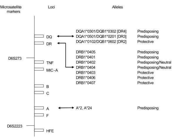
The loci in the HLA region encoding DR and DQ molecules display the strongest association for both dia-betes susceptibility and protection (Figure 3) (20). The HLA-DQ locus, the locus most strongly associated with diabetes susceptibility, encodes for multiple variants of the molecule, a heterodimer consisting of two chains (α and β) which are involved in immune recognition and antigen presentation to CD4 T cells. Alleles in this locus can either be predisposing or protective, the degree to which is influenced by the DR allele with which they are in linkage disequilibrium. While the DR-3-DQ2
mole-cules (DQB1*0201) and DR4-DQ8 (DQB1*0302) are associated with susceptibility, the DQB1*0602 allele is associated with dominant protection (19). Two additio-nal haplotypes, which are strongly protective, are DRB*1401, DQA1*0101, DQB1* 0503 and DRB1* 0701, DQA1*0201, DQB1*0303 (21).
Recently Baschal has found that he absence of the reportedly protective alleles DPB1*0402 and/or DRB1*0403 in DR3-DQB1*0201/DR4-DQB1*0302 individuals confers a 55% risk of persistently expressing anti-islet autoantibodies for relatives (children with a parent or sibling with type 1 diabetes) according to survival curve analysis as compared to 0% for those with either protective allele and a 20% risk for developing islet immunity in children without a type 1 diabetes relative (18).
Additional MHC and MHC-linked loci contribute to diabetes risk (22-26).Siblings are known to have a higher diabetes risk than the offspring of a parent with diabetes, even though siblings and offspring share ap-proximately half of their genome with their diabetic
pro-Figure 2. Odds ratios for the susceptibility allele for the ten independent T1D-associated genes or regions according to SNPs
marking each gene or region. The HLA class II SNP (rs3129934) was the marker with the highest association with T1D in the MHC region (positions 25–35 Mb on chromosome 6) in the WTCCC study(12).
1 2 3 4 5 6 7
HLA cla
ss II INS PTPN
22
CD25 12q2 4
ERB B3e
PTPN 2
KIAA
0350 CTLA4 CD25
O
d
d
s
R
a
ti
o
Susceptibility alleles for T1D-associated genes or regions
cop
yr
ight
© ABE&M todos os dir
eitos r
eser
v
ados
band. Siblings can share both HLA haplotypes which are identical to their proband, whereasoffspring inherit only one haplotype from their single diabeticparent. Further-more the same designated haplotype (i.e. the highest risk DR3/DR4-DQ8) can be identical-by-descent from pa-rent of origin between siblings or not. Therefore sharing of multiple genetic polymorphismsof DR, DQ genes and non-DR, DQ genes linked to the MHC regionon both copies of chromosome 6 could cause the increase siblingrisk, as high as 80% (Figure 4) (27).
“Ancestral” MHC haplotypes extend over 1 million nucleotides so that a series of polymorphisms and gene loci are remarkably conserved in almost total linkage disequilibrium (28-30). This has been confirmed by throughput SNA analysis and extensive resequencing of the MHC region.
AUTOIMMUNITY
The islet beta-cell zinc cation efflux transporter Znt8 (Scl30A8) is a major newly defined (31). This transporter
was discovered as an autoantigen because it is specifically expressed in islet beta–cells, where it is associated with the regulated pathway of insulin secretion. Znt8 facilitates the transportation of Zn2+ from the cytoplasm into the insulin
secretory granule and the concentration of Zn2+ within
the granule lumen where the zinc cation binds to insulin hexamers. Fluid phase radioassays have already been vali-dated for autoantibodies to this autoantigen in the most recent CDC affiliated DASP workshop and approximately 60% of new onset patients have autoantibodies reacting with the zinc transporter. These radioassays follow the earlier development of radioassays for autoantibodies re-acting with insulin, GAD65 (Glutamic Acid Decarboxy-lase) and IA-2 (Insulinoma Associated). Insulin autoantibodies develop within weeks of the starting of subcutaneous injection of insulin, and, thus, after insulin therapy measurement of insulin autoantibodies is not use-ful. Assays for each of the above 3 autoantibodies can be set at the 99th percentile of controls and approximately
90% of children with new onset diabetes express either one or the other autoantibody (Figure 5).
Figure 3. HLA Region and IDDM Susceptibility. Schematic representation of the HLA region on Chromosome 6 showing
microsat-ellite markers, loci, and alleles associated with IDDM susceptibility. Distances between loci are grossly approximated (20). DQ
C B TNF
A
F
HFE DR
D6S2223 D6S273
MIC- A
DQA1*0501/DQB1*0201 [DR3] Predisposing DQA1*0102/DQB1*0602 [DR2] Protective
DRB1*0405 Predisposing
DRB1*0401 Predisposing
DRB1*0402 Predisposing/Neutral DRB1*0404 Predisposing/Neutral
DRB1*0403 Protective
DRB1*0406 Protective
DRB1*0407 Protective
A*2, A*24 Predisposing
cop
yr
ight
© ABE&M todos os dir
eitos r
eser
v
ados
Figure 4. Extreme risk for
dia-betes autoimmunity. Life table analysis of DR3/4-DQ2/8 sib-lings of patients with type 1 di-abetes in the DAISY study followed from birth for the de-velopment of anti-islet autoan-tibodies. These relatives with the highest risk DR3/4-DQ2/8 HLA genotype were subdivid-ed by the number of HLA hap-lotypes inherited identical by descent to their proband dia-betic sibling. High risk cohort are DR3/4-DQ8 siblings that share both MHC haplotypes identical-by-descent with their proband, N=29. Low risk cohort are DR3/4-DQ8 siblings that do not share both MHC haplo-types identical-by-descent with their proband, N=19 (27).
Figure 5. Overlapping prevalence of
ZnT8A, GADA, IA2A, and IAA at onset. (A) Seropositive individuals evaluated with three-autoantibody standard or with Zn-T8A substituted for GADA, IA2A, or IAA. The ZnT8A assay incorporates both C-ter-minal and N/C assays in the one mea-surement. (B) Seropositive individuals evaluated with four-autoantibody stan-dard. ZnT8 antibodies(ZnTA) were found in 26% of T1D subjects classified as au-toantibody-negativeon the basis of exist-ing markers [glutamate decarboxylase (GADA),protein tyrosine phosphatase IA2 (IA2A), antibodies to insulin(IAA), and islet cytoplasmic autoantibodies (ICA)]. The combined measurement ofZnT8A, GADA, IA2A, and IAA raised autoimmunity de-tection ratesto 98% at disease onset, a level that approaches that neededto detect prediabetes in a general pediat-ric population (32).
0.0 2.5 5.0 7.5 10.0 12.5 15.0
0 10 20 30 40 50 60 70 80 90 100
Siblings share 2 MHC haplotypes
Siblings share 0 or 1 haplotype
Age (years) A u to a n tib o d y p o s iti v e ( % ) ZnT 4 9 11 17 55 6 5 14 12 13 16 29 10
IA2+INS = 9 GAD
Zn
T+GAD = 9
INS IA2
B
A
IA2 IA2 IA2ZnT ZnT ZnT
INS
INS GAD INS GAD
GAD 21 26 68 9 20 21 45 13 13 28 19 16 8 34 19 21 21 20 61 39 18 10 19 23 71 41 25 29 9 26 15 84
Total = 223
No Ab = 4
≥1 Ab 94.2%
≥2 Ab 71.3%
≥1 Ab 95.5%
≥2 Ab 70.0%
≥1 Ab 92.4% ≥1 Ab 96.0%
≥2 Ab 63.2%
≥1 Ab 98.2%
≥2 Ab 79.4%
≥2 Ab 65.0%
Aly et al. Extreme genetic risk for type 1A diabetes. PNAS 2006. 103:14074.
cop
yr
ight
© ABE&M todos os dir
eitos r
eser
v
ados
utilize binding of GAD65 to antigen captured by plate bound anti-GAD antibodies can perform as well as the fluid phase radioassays and kits for such assays are now available (4). Despite excellent assays, a subset of chil-dren with new onset diabetes are still negative for all anti-islet autoantibodies (33).
Given genetic susceptibility, the first islet autoanti-body to appear during the first five years of life is usu-ally autoantibodies to insulin (30). Subsequently GAD65 autoantibodies may be the first to appear and insulin autoantibodies become less common, such that if onset of diabetes is after age 12 the majority of chil-dren do not express insulin autoantibodies (34). GAD65 autoantibodies are the most common in adults with Latent Autoimmune Diabetes of Adults (LADA) (35). We believe LADA is type 1A diabetes developing in an adult, diagnosed prior to development of ketoaci-dosis and a severe insulin deficiency.
ally diabetes (36). Using a competitive radiobinding-assay Achenbach et al measured IAA affinity in sequential IAA-positive samples from children who are followed by birth in the BABYDIAB cohort in Europe. All high-affinity IAAs required conservation of human insulin A chain residues 8-13 and were reactive with proinsulin. High affinity was associated with HLA DRB1*04, young age of IAA appearance, and subse-quent progression to multiple islet autoantibodies or type 1 diabetes and thus identifying children at high risk (Figure 7). Of note the data were consistent with the early and sustained presentation of proinsulin in the context of the highest risk allele HLA DR4. The same group followed autoantibodies to GAD (GADAs) for heterogeneity in affinity and epitope recognition in the BABYDIAB cohort of children (37). Affinity was hi-gher in multiple islet autoantibody-positive children and in children who carried the HLA DR3 haplotype.
Figure 6. Progression to type 1 diabetes
of relatives of patients with type 1 diabe-tes subdivided by number of “biochemi-cal” anti-islet autoantibodies for GAD, ICA512 (IA-2) and insulin (31).
0 20 40 60 80 100
0 2.5 5 7.5 10 12.5 15
3 A bs
2 A bs
1 A b
Percent not Diabetic
Years of Follow-up
3 Ab n = 41 17 8 1
2 Abs n = 44 27 15 4 2 1
1 Abs n = 93 23 14 10 6 4
From Verge et. al. Prediction of type I diabetes in first-degree relatives using a combination of insulin, GAD, and ICA512bdc/IA-2 autoantibodies. Diabetes. 1996;45:926-33.
cop
yr
ight
© ABE&M todos os dir
eitos r
eser
v
ados
At present we know relatively little concerning the pancreatic pathology of the prediabetic process in man, but a recent collaborative study, Network for Pancreatic Organ Donors with Diabetes (nPOD), sponsored by the JDRF is seeking to address this lack. In particular pan-creases from cadaveric donors expressing islet autoanti-bodies have been analyzed and their histology made available on a web page (
www.jdrfnPOD.org
). Initial studies suggest that individuals expressing multiple islet autoantibodies are likely to have insulitis (38,39), but it is likely that there will be several different pathologic processes leading to beta-cell destruction. Insulitis is not uniform and the same pancreas will have normal islets, pseudoatrophic islets (islets lacking all insulin producing cells) that have no insulitis, and islets with insulitis (40). The insulitis of man, concordant with the chronic nature of the development of type 1A diabetes, is usually relati-vely mild in terms of number of islet involved at any given time. A major question is whether islet insulin-producing cells remain in patients with longstanding type 1A diabe-tes. Current evidence suggests that for most individuals less than 1% of islet beta-cell mass remains, but there are individuals with long-term type 1 diabetes with remai-ning islet beta-cells (41).Though islet autoantibodies are measured to aid the diagnosis and prediction of type 1A diabetes the
disease is most likely T-cell mediated. (Even T-cell me-diated disorders can be dependent upon B lymphocytes, particularly for antigen presentation, and a trial of anti-CD20 antibodies in new onset patients by TrialNet is now fully enrolled.) The measurement of T cells targe-ting islet autoantigens, and in particular T cell assays with the ability to distinguish patients with type 1 dia-betes from controls, has been particularly difficult. Re-cent assays measuring response of memory T cells in contrast to naïve T cells (naïve T cells of controls res-pond to islet autoantigens) holds promise for the deve-lopment of predictive T cell assays (42,43).
METABOLIC PROGRESSION
Given the recognition of the chronic nature of the de-velopment of type 1A diabetes and the existence of ex-cellent islet autoantibody assays, it has been possible to define multiple metabolic parameters that detect ab-normalities prior to the onset of diabetes. Loss of first phase insulin secretion following intravenous glucose develops in most individuals months to years prior to the onset of diabetes (44). Similarly, abnormalities on oral glucose tolerance testing, especially at the two-hour time point, usually precede diabetes (45,46). In children followed to the onset of diabetes a chronic rise
Figure 7. Mature high-affinity immune responses to (pro)insulin anticipate the autoimmune
cas-cade that leads to type 1 diabetes (30).
"F alse Positive" Hig h Risk
0 20 40 60 80 100
Affin >10(9) Mu ltip e Ab s f/u Diab etes Proin su lin
Percent Binding
cop
yr
ight
© ABE&M todos os dir
eitos r
eser
v
ados
OVERT DIABETES
It is clear that beta-cells of the mouse can regenerate with recovery from acute destruction and severe hyper-glycemia (47). Islet beta-cells appear to arise primarily from replication of existing beta-cells in the mouse. (48) As NOD mice progress to diabetes beta-cell repli-cation increases, apparently slowing progression to dia-betes, with diabetes occurring when approximately 20% of beta-cells have been destroyed. With immunothera-py at the onset of overt diabetes recovery of beta-cell function can be demonstrated in the NOD mouse (49). Evidence for beta-cell replication has been presented for man at onset of diabetes (50) and functioning beta-cells can remain in long-term patients. Unfortunately beta-cell mass appears to be severely compromised and potential for replication in man is unknown (51). Pa-tients with more than fifty years of type 1 diabetes are being studied at the Joslin Diabetes Center (50-year Medalist Study) with a subset of patients still expressing limited amounts of C-peptide (52). Understanding the limits of islet beta-cell replication in man and the path-way from islet stem cell to mature islet beta-cell is an important avenue to achieve beta-cell replacement therapies(53).
CONCLUSION
At present we still lack the tools for in vivo imaging of
either beta-cell mass or insulitis in man. In animal mod-els a number of techniques have shown promise (54,55), but to date we either lack sufficient data in man to assess utility or the studies have been inconclu-sive. It is likely that methods that have been utilized to image insulitis in the NOD mouse model (vascular leakage imaged with iron nanoparticles) will be difficult to apply to man if our current understanding of histolo-gy of new onset pancreas is accurate. There is relatively little insulitis in man and the insulitis is non-synchro-nous. With the lack of ability to image the pancreas, our understanding of the natural history of the disease comes primarily from indirect measurements of insulin secre-tion and the ability to detect anti-islet autoimmunity
al understanding of the pathology of the disorder at all stages, including the “prediabetic” phase, and this will provide histologic data to help guide development of imaging modalities. In the absence of firm knowledge that would be provided with imaging modalities, we believe that type 1A diabetes develops for most indi-viduals as a result of chronic progressive beta-cell de-struction and that the process once initiated (e.g. expression of ≥2 biochemical autoantibodies) very few individuals escape from almost complete beta- cell de-struction. We believe a concentred effort to find indi-viduals who escape progression to diabetes should be undertaken to both better define the natural history of the disease and to search for factors that might natu-rally abrogate the pathologic process leading to type 1 diabetes.
REFERENCES
1. Eisenbarth GS. Update in type 1 diabetes. J Clin Endocrinol Metab. 2007;92(7):2403-7.
2. Diagnosis and classification of diabetes mellitus. Diabetes Care. 2008;31 Suppl 1:S55-S60.
3. Genome-wide association study of 14,000 cases of seven common diseases and 3,000 shared controls. Nature. 2007;447(7145):661-78.
4. Liu E, Eisenbarth GS. Accepting clocks that tell time poorly: fluid-phase versus standard ELISA autoantibody assays. Clin Immunol. 2007;125(2):120-6.
5. Yang Y, Santamaria P. Lessons on autoimmune diabetes from animal models. Clin Sci (Lond). 2006;110(6):627-39.
6. Norris JM, Yin X, Lamb MM, Barriga K, Seifert J, Hoffman M et al. Omega-3 polyunsaturated fatty acid intake and islet au-toimmunity in children at increased risk for type 1 diabetes. JAMA. 2007;298(12):1420-8.
7. Eisenbarth GS. Type I diabetes mellitus. A chronic autoimmu-ne disease. N Engl J Med. 1986;314:1360-8.
8. Wildin RS, Freitas A. IPEX and FOXP3: Clinical and research perspectives. J Autoimmun. 2005;25 Suppl:56-62.
9. Su MA, Anderson MS. Aire: an update. Curr Opin Immunol. 2004;16(6):746-52.
10. Fontenot JD, Gavin MA, Rudensky AY. Foxp3 programs the development and function of CD4+CD25+ regulatory T cells. Nat Immunol. 2003;4(4):330-6.
11. Mathis D, Benoist C. A decade of AIRE. Nat Rev Immunol. 2007;7(8):645-50.
cop
yr
ight
© ABE&M todos os dir
eitos r
eser
v
ados
13. Noble JA, Valdes AM, Cook M, Klitz W, Thomson G, Erlich HA. The role of HLA class II genes in insulin-dependent diabetes mellitus: Molecular analysis of 180 Caucasian, multiplex fami-lies. Am J Hum Genet. 1996;59(5):1134-48.
14. Pugliese A, Zeller M, Fernandez A, Zalcberg LJ, Bartlett RJ, Ricordi C et al. The insulin gene is transcribed in the human thymus and transcription levels correlate with allelic variation at the INS VNTR-IDDM2 susceptibility locus for type I diabetes. Nat Genet. 1997;15(3):293-7.
15. Bottini N, Musumeci L, Alonso A, Rahmouni S, Nika K, Ro-stamkhani M et al. A functional variant of lymphoid tyrosine phosphatase is associated with type I diabetes. Nat Genet. 2004;36(4):337-8.
16. Marron MP, Raffel LJ, Garchon HJ, Jacob CO, Serrano-Rios M, Martinez LM et al. Insulin-dependent diabetes mellitus (IDDM) is associated with CTLA4 polymorphisms in multiple ethnic groups. Hum Mol Genet. 1997;6(8):1275-82.
17. Aly TA, Baschal EE, Jahromi MM, Fernando MS, Babu SR, Fin-gerlin TE et al. Analysis of SNPs Identifies Major Type 1A Dia-betes Locus Telomeric of the MHC. DiaDia-betes. 2007;.
18. Baschal EE, Aly TA, Babu SR, Fernando MS, Yu L, Miao D et al. HLA-DPB1*0402 Protects Against Type 1A Diabetic Autoim-munity in the Highest Risk DR3-DQB1*0201/DR4-DQB1*0302 DAISY Population. diab. 2007;56(Epub ahead of print):2405-9. 19. Baisch JM, Weeks T, Giles R, Hoover M, Stastny P, Capra JD.
Analysis of HLA-DQ genotypes and susceptibility in insulin-de-pendent diabetes mellitus. N Engl J Med. 1990;322(26):1836-41. 20. Steck AK, Pugliese A, Eisenbarth GS. Prediction of Type IA
Dia-betes: The Natural History of the Prediabetic Period. In: Eisen-barth GS, editor. Type 1 Diabetes: Molecular, Cellular and Clinical Immunology. 2008.
21. Redondo MJ, Kawasaki E, Mulgrew CL, Noble JA, Erlich HA, Freed BM et al. DR and DQ associated protection from type 1 diabetes: comparison of DRB1*1401 and DQA1*0102-DQB1*0602. J Clin Endocrinol Metab. 2000;85(10):3793-7. 22. Tarn AC, Thomas JM, Dean BM, Ingram D, Schwarz G,
Botta-zzo GF et al. Predicting insulin-dependent diabetes. Lancet. 1988;I(8590):845-50.
23. Blomhoff A, Lie BA, Myhre AG, Kemp EH, Weetman AP, Aksel-sen HE et al. Polymorphisms in the cytotoxic T lymphocyte antigen-4 gene region confer susceptibility to Addison's dise-ase. J Clin Endocrinol Metab. 2004;89(7):3474-6.
24. Hanifi MP, de Knijf P, Roep BO, van der AB, Naipal A, Gorus F et al. Genetic structure of IDDM1: two separate regions in the major histocompatibility complex contribute to susceptibility or protec-tion. Belgian Diabetes Registry. Diabetes. 1998;47(2):263-9. 25. Inoue K, Ikegami H, Fujisawa T, Noso S, Nojima K, Babaya N et
al. Allelic variation in class I K gene as candidate for a second component of MHC-linked susceptibility to type 1 diabetes in non-obese diabetic mice. Diabetologia. 2004;47(4):739-47. 26. Hattori M, Yamato E, Itoh N, Senpuku H, Fujisawa T, Yoshino
M et al. Cutting edge: homologous recombination of the MHC class I K region defines new MHC-linked diabetogenic suscep-tibility gene(s) in non-obese diabetic mice. J Immunol. 1999;163(4):1721-4.
27. Aly TA, Ide A, Jahromi MM, Barker JM, Fernando MS, Babu SR et al. Extreme Genetic Risk for Type 1A Diabetes. Proc Natl Acad Sci USA. 2006;103(38):14074-9.
28. Barker JM, Goehrig SH, Barriga K, Hoffman M, Slover R, Eisenbarth GS et al. Clinical characteristics of children diag-nosed with type 1 diabetes through intensive screening and follow-up. Diabetes Care. 2004;27(6):1399-404.
29. Barker JM, Barriga K, Yu L, Miao D, Erlich H, Norris JN et al. Prediction of autoantibody positivity and progression to type 1 diabetes: Diabetes Autoimmunity Study in the Young (DAI-SY). J Clin Endocrinol Metab. 2004;89:3896-902.
30. Achenbach P, Koczwara K, Knopff A, Naserke H, Ziegler AG, Bonifacio E. Mature high-affinity immune responses to (pro) insulin anticipate the autoimmune cascade that leads to type 1 diabetes. J Clin Invest. 2004;114(4):589-97.
31. Verge CF, Gianani R, Kawasaki E, Yu L, Pietropaolo M, Jackson RA et al. Prediction of type I diabetes in first-degree relatives using a combination of insulin, GAD, and ICA512bdc/IA-2 au-toantibodies. Diabetes. 1996;45(7):926-93.
32. Wenzlau JM, Juhl K, Yu L, Moua O, Sarkar SA, Gottlieb P et al. The cation efflux transporter ZnT8 (Slc30A8) is a major auto-antigen in human type 1 diabetes. Proc Natl Acad Sci U S A. 2007;104(43):17040-5.
33. Wang J, Miao D, Babu S, Yu J, Barker J, Klingensmith G et al. Prevalence of autoantibody-negative diabetes is not rare at all ages and increases with older age and obesity. J Clin Endocri-nol Metab. 2007;92(1):88-92.
34. Vardi P, Ziegler AG, Matthews JH, Dib S, Keller RJ, Ricker AT et al. Concentration of insulin autoantibodies at onset of type I diabetes. Inverse log-linear correlation with age. Diabetes Care. 1988;11(9):736-9.
35. Palmer JP, Hampe CS, Chiu H, Goel A, Brooks-Worrell BM. Is latent autoimmune diabetes in adults distinct from type 1 dia-betes or just type 1 diadia-betes at an older age? Diadia-betes. 2005;54 Suppl 2:S62-7.:S62-S67.
36. Achenbach P, Bonifacio E, Williams AJ, Ziegler AG, Gale EA, Bingley PJ. Autoantibodies to IA-2beta improve diabetes risk assessment in high-risk relatives. Diabetologia. 2008;51:488-92 37. Mayr A, Schlosser M, Grober N, Kenk H, Ziegler AG, Bonifacio
E et al. GAD autoantibody affinity and epitope specificity iden-tify distinct immunization profiles in children at risk for type 1 diabetes. Diabetes. 2007;56(6):1527-33.
38. In't VP, Lievens D, De Grijse J, Ling Z, van der AB, Pipeleers-Marichal M et al. Screening for insulitis in adult autoantibody-positive organ donors. Diabetes. 2007;56(9):2400-4.
39. Gianani R, Putnam A, Still T, Yu L, Miao D, Gill RG et al. Initial results of screening of non - diabetic organ donors for expres-sion of islet autoantibodies. J Clin Endocrinol Metab. 2006;91:1855-61.
40. Foulis AK, McGill M, Farquharson MA. Insulitis in type 1 (insu-lin-dependent) diabetes mellitus in man - macrophages, lym-phocytes, and interferon-gamma containing cells. J Pathol. 1991;165(2):97-103.
41. Butler AE, Galasso R, Meier JJ, Basu R, Rizza RA, Butler PC. Modestly increased cell apoptosis but no increased beta-cell replication in recent-onset type 1 diabetic patients who died of diabetic ketoacidosis. Diabetologia. 2007;50(11):2323-31. 42. Monti P, Scirpoli M, Rigamonti A, Mayr A, Jaeger A, Bonfanti
R et al. Evidence forin vivo primed and expanded autoreactive T cells as a specific feature of patients with type 1 diabetes. J Immunol. 2007;179(9):5785-92.
43. Danke NA, Yang J, Greenbaum C, Kwok WW. Comparative study of GAD65-specific CD4+ T cells in healthy and type 1 diabetic subjects. J Autoimmun. 2005;25(4):303-11.
44. Barker JM, McFann K, Harrison LC, Fourlanos S, Krischer J, Cuthbertson D et al. Pre-type 1 diabetes dysmetabolism: maxi-mal sensitivity achieved with both oral and intravenous gluco-se tolerance testing. J Pediatr. 2007;150(1):31-6.
cop
yr
ight
© ABE&M todos os dir
eitos r
eser
v
ados
47. Nir T, Melton DA, Dor Y. Recovery from diabetes in mice by beta-cell regeneration. J Clin Invest. 2007;117(9):2553-61. 48. Dor Y, Brown J, Martinez OI, Melton DA. Adult pancreatic
beta-cells are formed by self-duplication rather than stem-cell diffe-rentiation. Nature. 2004;429(6987):41-6.
49. Ablamunits V, Sherry NA, Kushner JA, Herold KC. Autoimmu-nity and beta-cell regeneration in mouse and human type 1 diabetes: the peace is not enough. Ann N Y Acad Sci. 2007;1103:19-32. Epub@2007 Mar 21:19-32.
50. Redondo MJ, Yu L, Hawa M, Mackenzie T, Pyke DA, Eisenbarth GS et al. Heterogeneity of type I diabetes: analysis of mono-zygotic twins in Great Britain and the United States. Diabeto-logia. 2001;44(3):354-62.
51. Nielsen CH, El Fassi D, Hasselbalch HC, Bendtzen K, Hegedus L. B-cell depletion with rituximab in the treatment of autoimmune diseases. Graves' ophthalmopathy the latest addition to an ex-panding family. Expert Opin Biol Ther. 2007;7(7):1061-78.
Opin Endocrinol Diabetes Obes. 2007;14(4):277-82.
54. Turvey SE, Swart E, Denis MC, Mahmood U, Benoist C, Weissle-der R et al. Noninvasive imaging of pancreatic inflammation and its reversal in type 1 diabetes. J Clin Invest. 2005;115(9):2454-61. 55. Harris PE, Ferrara C, Barba P, Polito T, Freeby M, Maffei A.
VMAT2 gene expression and function as it applies to imaging beta-cell mass. J Mol Med. 2008;86(1):5-16.
Address correspondence:
George S. Eisenbarth
Barbara Davis Center for Childhood Diabetes, University of Colorado 303-724-6847, USA