cop
yr
ight
© ABE&M todos os direitos reser
v
ados
clinical case report
JULIANA GABRIEL R. DE ANDRADE
MARA SANCHES GUARAGNA
FERNANDA CAROLINE SOARDI
GIL GUERRA-JÚNIOR
MARICILDA PALANDIDE MELLO
ANDRÉA TREVAS MACIEL-GUERRA
Grupo Interdisciplinar de Estudos da Determinação e Diferenciação do Sexo (GIEDDS), Faculdade de Ciências Médicas, Universidade de Campinas (Unicamp) (JGRA, GGJ, ATMG); Centro de Biologia Molecular e Engenharia Genética (CBMEG), Unicamp (MSG, FCS, MPM); Campinas, SP, Brasil.
Received in 25/8/2008 Accepted in 14/10/2008
ABSTRACT
Aim: To present phenotypic variability of WT1-related disorders. Methods: De-scription of clinical and genetic features of fi ve 46,XY patients with WT1 anom-alies. Results: Patient 1: newborn with genital ambiguity; he developed Wilms tumor (WT) and chronic renal disease and died at the age of 10 months; the heterozygous 1186G>A mutation compatible with Denys-Drash syndrome was detected in this child. Patients 2 and 3: adolescents with chronic renal dis-ease, primary amenorrhea and hypergonadotrophic hypogonadism; patient 2 had a gonadoblastoma. The heterozygous IVS9+4, C>T mutation, compatible with Frasier syndrome was detected. Patient 4: 9-year-old boy with aniridia, genital ambiguity, dysmorphisms and mental defi ciency; a heterozygous 11p deletion, compatible with WAGR syndrome was detected. Patient 5: 2 months old, same diagnosis of patient 4; he developed WT at the age of 8 months. Conclusions: Constitutional abnormalities of WT1 cause gonadal and renal anomalies and predisposition to neoplasia and must be investigated in patients with ambiguous genitalia, chronic renal disease and(or) Wilms tumors; primary amenorrhea with chronic renal disease; and aniridia, genital ambiguity and dysmorphisms. (Arq Bras Endocrinol Metab 2008; 52/8:1236-1243)
Keywords: Sex differentiation; WT1 gene; Denys-Drash syndrome; Frasier syndrome; WAGR syndrome
RESUMO
Achados Clínicos e Genéticos de Cinco Pacientes com Anomalias Relacionadas ao Gene WT1.
Objetivo: Descrever a variabilidade fenotípica das anomalias relacionadas ao WT1. Métodos: Descrição das características clínicas e genéticas de cinco pa-cientes 46,XY com anomalias no WT1. Resultados: Paciente 1: Recém-nascido com ambigüidade genital desenvolveu tumor de Wilms (TW) e insufi ciência renal crônica (IRC), com óbito aos 10 meses. Detectada a mutação 1186G>A em heterozigose, compatível com síndrome de Denys-Drash. Pacientes 2 e 3: Adolescentes com IRC, amenorréia primária e hipogonadismo hipergonado-trófi co; a paciente 2 apresentava gonadoblastoma. Ambas apresentavam mutação IVS9+4, C>T em heterozigose, característica da síndrome de Frasier. Paciente 4: Idade 9 anos, aniridia, ambigüidade genital, dismorfi smos e defi -ciência mental; deleção 11p, compatível com síndrome WAGR foi encontrada em heterozigose. Paciente 5: Dois meses, mesmo diagnóstico do paciente 4, desenvolveu TW aos 8 meses. Conclusões: Alterações constitucionais do WT1 determinam anomalias gonadais, renais e predisposição a neoplasias; devem ser pesquisadas em casos de ambigüidade genital associada a IRC e(ou) TW; de amenorréia primária com IRC; e aniridia, ambigüidade genital e dismorfi smos. (Arq Bras Endocrinol Metab 2008; 52/8:1236-1243)
cop
yr
ight
© ABE&M todos os direitos reser
v
ados
sex ambiguity as a result of dysgenetic testis, diffuse mesangial sclerosis with chronic renal disease and high incidence of Wilms tumour (WT) (13-14). The clinical picture of FS includes dysgenetic gonads with male-to-female sex reversal in 46,XY subjects and pubertal delay in both sexes, nephrotic syndrome and focal segmental glomerulosclerosis leading to chronic renal disease , and high incidence of gonadoblastoma but not of WT. Mutations in FS affect the splice site in intron 9, with
WT1(+KTS) isoforms (15-16) losses.
WAGR (WT, aniridia, genitourinary malformations, mental retardation) is a contiguous gene syndrome ari-sing from deletions of chromosome 11p13 which en-compass at least both the PAX6 and WT1 genes (17-19).
We report fi ve patients followed in a reference ser-vice for disorders of sex development which illustrate the broad spectrum of presentation of WT1-associated disorders.
SUBJECTS AND METHODS
Patients
The fi ve patients were evaluated by the Interdisciplina-ry Study Group of Disorders of Sex Development (GIEDDS) of the State University of Campinas, São Paulo. The protocol was approved by the local Ethics Committee (N. 434/06) and informed consent was obtained from the parents of the children included in the study.
Laboratory assays
LH, FSH, testosterone were measured by electroche-miluminescence (BM/Hitachi Elecsys 2010, Roche Diagnostics, Boehringer, Mannheim, Germany).
Karyotype
Chromosome analysis of peripheral blood lymphocytes was performed by G-banding at 500-600 bands resolu-tion using standard procedures.
Genomic DNA extraction, amplifi cation and sequencing
Genomic DNA from peripheral blood leukocytes was purifi ed by Proteinase K digestion and phenol/chloro-form extraction followed by ethanol precipitation using standard techniques (20).
INTRODUCTION
S
ex determination is a complex and yet not fully elu-cidated process which depends on a complex ne-twork of interrelated genes. Gonadal development starts by the end of the 5th week of gestation with themigration of primordial germ cells from the yolk sac to the gonadal anlage. Formation of the primordial gona-ds, which have no apparent sexual differences up to 8 weeks, depend on the expression of many genes, inclu-ding SF-1 (steroidogenic factor 1) (1), DAX1 (dosage-sensitive sex reversal, adrenal hypoplasia critical region, on chromosome X, gene 1) (2) and WT1 (Wilms tu-mour 1) (3).
WT1 (OMIM 607102) is located at 11p13 and en-codes a zinc fi nger motif-containing transcription factor involved in regulation of growth and differentiation (4). Beyond its role in the genesis of Wilms tumour (5-6), it regulates early gonad and kidney development (7).
Alternative splicing generates four major WT1 iso-forms: an alternative splice site in intron 9 allows the addition of three amino acids (KTS) between zinc fi n-gers 3 and 4 and the fi fth exon, encoding 17 aminoaci-ds, may or may not be present. These isoforms are highly conserved among different species and play a crucial role in normal gene function. Gene action de-pends on the predominant isoform: WT1 (-KTS) va-riants act as transcriptional regulators, while WT1
(+KTS) participates on the regulation of certain genes at the post-transcriptional level (8-10).
WT1 (-KTS) isoforms act in association with the product of SF-1 to promote expression of anti-mülle-rian hormone (AMH), responsible for regression of the müllerian ducts in male embryos. The product of DAX1 can repress the synergistic action of WT1 and SF-1, re-sulting in down-regulation of AMH (11). In vitro ex-periments suggested that WT1 (-KTS) variants are also responsible for transcriptional activation of SRY, which activates the male differentiation pathway (10).
Mutations in WT1 are found in a variety of syndro-mes, including Denys-Drash (DDS, OMIM 194080), Frasier (FS, OMIM 136680) and WAGR (OMIM 194072) syndromes. Both DDS and FS are characterized by gonadal and renal anomalies and predisposition to ne-oplasia associated with “de novo” constitutional WT1
cop
yr
ight
© ABE&M todos os direitos reser
v
ados
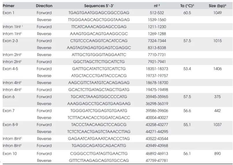
The 10 exons and the exon-intron junction regions of the WT1 gene were PCR amplifi ed from genomic DNA with primers described in Table 1. Genomic se-quence of the WT1 gene was obtained in the published sequence ENSG00000184937 (www.ensembl.org).
The fi nal volume of all reactions was 50 uL and con-tained 10X Taq DNA polymerase buffer (Invitrogen, CA, USA), 1.0-1.5 mM MgCl2, 2 mM of each dNTP, 20 pmol of each primer, 300-500 ng genomic DNA templates 2 units recombinant Taq DNA polymerase (Invitrogen), 5% DMSO used only for exon 1. After a fi rst denaturation step (5 min, 94°C), the cycling profi le was: 94°C, 1 min; 53,5°C – 63,5°C, 1 min; 72°C 1-6 min (30 cylcles), followed by 5 min at 72°C (fi nal exten-sion). The size of the PCR products was verifi ed in 1% agarose gel electrophoresis stained with ethidium bromi-de. Before sequencing, purifi cation of PCR products was performed using the Wizard® SV Gel and PCR clean-up
system (Promega, Madison, WI, USA). Further direct sequencing using ABI PRISM Big Dye Terminator v3.1 Cycle Sequencing Kit (ABI PRISM/PE Biosystems, Foster City, CA, USA) was carried out in two separate reactions for each exon, except for exon 1 which requi-red four reactions, using sense and antisense primers (Table 1). The sequences were obtained in an automatic sequencer ABI PRISM 3700 DNA Analyzer (ABI PRISM/PE Biosystems). Free softwares Chromas Lite and CLC Sequence Viewer v.5.0.1 were used to analyze and compare sequences with the published WT1 sequen-ce at Ensembl database.
RESULTS
Case 1
A 1-month-old child was referred to us due to sex am-biguity. He was born at term after an uneventful
preg-Table 1. Primers designed for WT1 coding sequence amplifi cation.
Primer Direction Sequences 5’-3’ nt 2 Ta (°C)3 Size (bp)4
Exon 1 Forward TGAGTGAATGGAGCGGCCGAG 512-532 60.5 1049
Reverse TTGGGAAGCAGCTGGGTAAGAG 1539-1560 Intron 1Int 1 Forward TTCATCAAACAGGAGCCGAG 1211-1230
Intorn 1Int1 Reverse AAAGTGGACAGTGAAGGCGC 1269-1288
Exon 2-3 Forward CTGTCCCAAGGTCACATCCAG 7324-7344 57.5 1015
Reverse AAGTAGTAGAGTGGAGTCGAGGC 8313-8338 Intorn 2Int1 Reverse ATTTGCTGTGGGTTAGGAATTC 7710-7731
Intron 2Int1 Forward GGCTTAGCTTCTTGCATTCTG 7921-7941
Exon 4-5 Forward GATTTGCATATTCTGTCATTCTG 18351-18373 53.4 1406
Reverse ATGCTACCCTGATTACCCACG 19737-19757
Intron 4Int1 Reverse AAGCGTTCTAATGTCACAGAGAG 18678-18700
Intron 4Int1 Forward GCACTCTTGATAGCTAGCTTGATG 19475-19498
Exon 6 Forward TGCATCTAAAGTGGCCCCATG 35945-35965 57.5 375
Reverse AAAGGAGCCTGCAGTGAAGAAG 36298-36319
Exon 7 Forward TGGGGATCTGGAGTGTGAATG 39586-39606 56.6 442
Reverse TCTTTACAACACCTGGATCAGACC 40004-40027
Exon 8-9 Forward TACCCTAACAAGCTCCAGCG 43258-43277 55,1 1037
Reverse TCTCTCAACTGAGTCTAAACCTTAG 44271-44295 Intorn 8Int1 Reverse GAGAATCATGAAATCAACCCTAG 43522-43544
Intron 8Int1 Forward TGAGGCAGATGCAGACATTG 43949-43968
Exon 10 Forward CGGGCCTTGATAGTTGAACTTG 46892-46913 56.1 890
Reverse GTTTCTTAAGAGCAGTGTGCCAG 47759-47781
cop
yr
ight
© ABE&M todos os direitos reser
v
ados
nancy with a birth weight of 3,655 g and length 50 cm. He was the second child of unrelated parents and fami-ly history was unremarkable. He had a 2-cm phallus with chordee, penoscrotal hypospadias, shawl scrotum, bilateral cryptorchidism, and there was no dysmorphic picture associated to sex ambiguity. Sonography revea-led no mullerian derivatives while genitography showed a urogenital sinus.
His karyotype was 46,XY, and there were normal basal levels of LH (5.4 U/L), FSH (3.8 U/L), free (5.4 pg/mL) and total (157 ng/dL) testosterone (T). When the child was 6 months old, a hCG stimulation test was performed (three intramuscular injections of Chorionic Gonadotropin (Profasi®, 1,000 IU, on
suc-cessive days), and testosterone levels increased from 157 to 395 ng/dL. He developed unilateral WT and chronic renal disease at 8 months, and died 2 months later as a result of septicemia.
An 1186G>A heterozygous mutation was detected in exon 9 and confi rmed the diagnosis of DDS; this case was fi rst reported by Tagliarini and cols. (21).
Case 2
A 17.3-year-old girl was evaluated for primary amenor-rhea and hypergonadotrophic hypogonadism. She was born at term after an uneventful pregnancy, with a bir-th weight of 3,560g and lengbir-th 46cm. She was bir-the se-cond child of unrelated parents, and family history was unremarkable. She was subject to renal transplantation at 11 years as a consequence of chronic renal disease; at the same age, bilateral inguinal hernia repair was per-formed.
She referred spontaneous pubertal onset. On phy-sical examination, she had normal external genitalia and pubertal development was on Tanner stage B3P2.
There was no dysmorphic picture. Ultrasound revealed a 2.8cm3 uterus, and gonads could not be found.
Her karyotype was 46,XY, and there were high le-vels of FSH (188 U/L) and LH (46 U/L) and low estradiol (11pg/mL). Bilateral gonadectomy was per-formed, and histology revealed a right dysgenetic go-nad with mesonephric remnants and a gogo-nadoblastoma on the left. Female hormonal replacement therapy was initiated later on. Molecular analysis revealed an IVS9+4, C>T heterozygous mutation in intron 9 (Fi-gure 1), thus confi rming the diagnosis of Frasier syn-drome.
Case 3
A 18-year-old girl was referred with a suspected diag-nosis of FS to molecular analysis. She was the second child of unrelated parents, and family history was unre-markable. The girl had chronic renal disease treated with peritoneal dialysis, and primary amenorrhea was investigated when she was 15 years old – cytogenetic investigation revealed a 46,XY karyotype, bilateral go-nadectomy was performed and histology revealed dys-genetic gonads. She has been on HRT since then.
On physical examination, she had no dysmorphic picture, external genitalia were normal, and breast de-velopment was incomplete.
Molecular analysis revealed the same mutation of case 2, thus confi rming the diagnosis of FS.
Case 4
A 9-year-old boy presented with a history of sex ambi-guity, aniridia, mental and motor retardation and dys-morphic features. He was born at term by cesarian section for breech presentation, after an uneventful pregnancy, with a birth weight of 2,550g. He was the
cop
yr
ight
© ABE&M todos os direitos reser
v
ados
fi rst child in a sibship of three; his parents were not re-lated, and family history was unremarkable.
Physical examination revealed fl at occiput, small and dysmorphic ears, short upslanted palpebral fi ssures, aniridia, nystagmus, short nose with high nasal bridge, clinodactyly of the V fi ngers, bilateral single transverse palmar crease, predominance of arches on the fi nger-tips, fusiform fi ngers, nail hypoplasia, increased inter-mamillary distance and diastasis recti. He had a 4.5cm-phallus, bifi d scrotum, penoscrotal hypospadia and nonpalpable gonads.
Ophthalmologic evaluation revealed macular hypo-plasia. The testes were not seen on pelvic ultrasound and genitography did not reveal a urogenital sinus.
He had prepubertal levels of LH (<0.2 U/L), FSH (0.4 U/L), total (<20 ng/dL) and free (1 pg/mL) tes-tosterone. An hCG stimulation test was performed, and testosterone levels increased from <20 to 396 ng/ dL. Cytogenetic investigation revealed a de novo
46,XY,del(11p) karyotype, thus leading to the diagno-sis of WAGR syndrome.
Case 5
A 3-month-old boy was evaluated for aniridia and dysmorphic features. He was born in the 38th week
of gestation by cesarian section, and intrauterine growth retardation was noted at the 7th month. Birth
weight was 2,650g and length 44cm. He was the only child of unrelated parents, and family history was unremarkable.
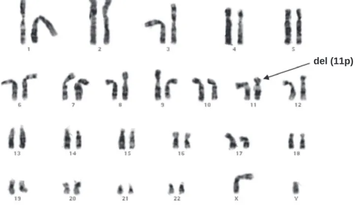
On physical examination, he presented high fo-rehead, low anterior hairline, dysmorphic ears, ante-verted nostrils, notched alae nasi, long and fl at philtrum, thin upper lip, retrognathism, short neck, single transverse palmar crease and hypoplastic nails. He had a 3.5-cm phallus, bilateral cryptorchidism and hypoplastic scrotum. Ophthalmologic evaluation re-vealed photophobia, nystagmus, remnants of pupilla-ry membrane and peripheral iris and mottled retinal pigment epithelium.
He had normal levels of LH (9.5 U/L), FSH (8.8 U/L), and total testosterone (669 ng/dL) for age. His karyotype was 46,XY,del (11p) de novo (Figure 2), lea-ding to the diagnosis of WAGR syndrome. When he was 8 months old, a unilateral Wilms tumour was de-tected by sonography. He was subject to nephrectomy, chemotherapy and radiotherapy. There was no tumor relapse until the age of 4 years, and renal function re-mained normal. Data on these fi ve cases are summari-zed in Table 2.
Figure 2. Karyotype of patient 5.
cop
yr
ight
© ABE&M todos os direitos reser
v
ados
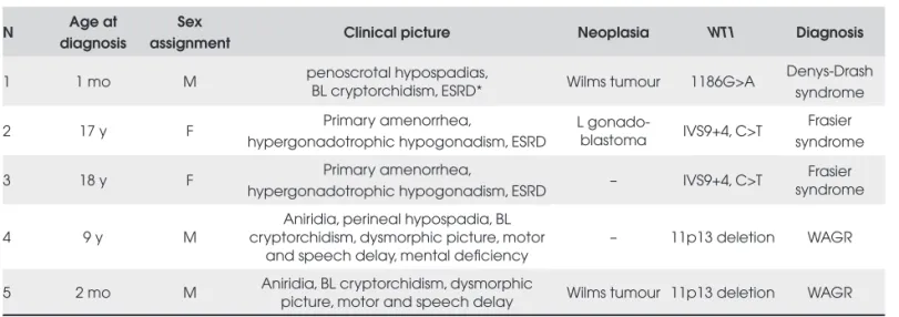
Table 2. Description of fi ve 46,XY patients with WT1-related disorders.
N Age at diagnosis
Sex
assignment Clinical picture Neoplasia WT1 Diagnosis
1 1 mo M penoscrotal hypospadias,
BL cryptorchidism, ESRD* Wilms tumour 1186G>A
Denys-Drash syndrome
2 17 y F Primary amenorrhea,
hypergonadotrophic hypogonadism, ESRD
L
gonado-blastoma IVS9+4, C>T
Frasier syndrome
3 18 y F Primary amenorrhea,
hypergonadotrophic hypogonadism, ESRD – IVS9+4, C>T
Frasier syndrome
4 9 y M
Aniridia, perineal hypospadia, BL cryptorchidism, dysmorphic picture, motor
and speech delay, mental defi ciency
– 11p13 deletion WAGR
5 2 mo M Aniridia, BL cryptorchidism, dysmorphic
picture, motor and speech delay Wilms tumour 11p13 deletion WAGR
BL = bilateral; ESRD = end stage renal disease; F = female; L = left; M = male; NB = newborn; *deceased (10 months)
DISCUSSION
Disorders of gonadal development (DGD) are a highly heterogeneous group of disorders of sex development (DSD) and include individuals with dysgenetic gonads (streaks), dysgenetic or rudimentary testes and true hermaphroditism or ovotesticular DSD. Some 46,XY individuals with DGD are born with sex ambiguity, and thus may be evaluated in infancy. However, those with female internal and external genitalia (male-to-female sex reversal) may be diagnosed only in adolescence be-cause of pubertal delay. The latter are of great concern because of the risk of neoplastic transformation of dys-genetic gonads, which is signifi cantly elevated after adolescence (22). Hormonal activity of gonadoblasto-ma gonadoblasto-may be found in some patients (23); in case 2, for instance, there was spontaneous breast development which may be due to an estrogen-producing gonado-blastoma.
Among DGD, WT1-related disorders are characte-rized by the association of gonadal and renal anomalies. As a consequence, screening for mutations in WT1
should be considered in 46,XY patients with ambiguous genitalia associated with chronic renal disease and (or) WT and in 46,XY females with hypergonadotrophic hypogonadism and history of chronic renal disease, thus allowing the diagnosis of DDS and FS.
In addition, all newborns with aniridia who do not have a family history of this ocular anomaly must be subject to high-resolution cytogenetic testing, which detects deletions involving 11p13 in up to 20% of
indi-viduals (24). FISH testing with probes spanning PAX6,
WT1, the regions fl anking PAX6, and the intervening sequence between PAX6 and WT1 can also be used to detect cryptic deletions in individuals with other clinical features of WAGR and normal cytogenetic studies (24).
Genotype-phenotype correlations in WT1-related disorders are well established. Mutations in DDS pa-tients inactivate DNA binding by the zinc fi ngers, le-ading to early and severe impairment of renal function, dysgenetic testes and high incidence of WT, while in FS mutations in the donor splice site of intron 9 of the WT1 gene lead typically to dysgenetic gonads, end-stage renal failure in the second decade and gonadoblastoma. In turn, the reduced haploinsuffi -ciency of WT1 in 11p13 deletion has a less pronounced effect on development, especially on that of the renal system.
However, there are some reports of atypical pre-sentations, including a 46,XY child with sex ambiguity, nephrotic syndrome, gonadal tumour and normal tes-tosterone production despite high levels of gonadotro-pins, who had a mutation associated with FS (25), In another study, a 46,XY child with sex ambiguity, nor-mal testosterone production, aortic coarctation and no renal disease was found to have a P181S mutation in
WT1 inherited from the mother (26).
cop
yr
ight
© ABE&M todos os direitos reser
v
ados
cases of steroid-resistant nephrotic syndrome (SRNS) (27-28). Routine evaluation of patients with this syn-drome would allow early diagnosis of both DDS and FS in both sexes.
The heterozygous 1186G>A (D396N) mutation in exon 9 of patient 1 leads to an aspartic acid-aspara-gine substitution changing the structural organization of the third zinc fi nger of the WT1 protein. It was described in 1991 (13) and is a frequent fi nding in DDS; the most frequent mutation is 1180C>T (R394W) (39.6%) (29). The apparent dominant-ne-gative nature of DDS mutations results from the ac-tion of altered WT1 in blocking the normal activity of the wildtype protein (12).
The heterozygous mutation in intron 9 found in both patients with FS (IVS9+4, C>T) is the most fre-quent mutation identifi ed in these patients (52%) (15,25). This and the other four different mutations described in intron 9 of WT1 in patients with FS lead to reversal of the (+KTS)/(-KTS) ratio from 2:1 to 1:2 (15-16). Most of the patients with FS show the +4 C>T and +5G>A mutations; this hotspot is probably a con-sequence of the potential to deaminate 5-methylcytosi-ne at the +4/+5 CpG dinucleotide (16).
Recurrence risk of WT1-related disorders varies ac-cording to each specifi c situation. DDS and FS usually arise as a consequence of de novo mutations, while 11p deletion in WAGR syndrome may be de novo or may result from transmission by a parent with a balanced chromosome rearrangement (24).
In conclusion, constitutional abnormalities of WT1 should be considered in patients with ambiguous geni-talia and renal disease (chronic renal disease or Wilms tumors), primary amenorrhea with chronic renal disea-se, and those with aniridia, genital ambiguity and dys-morphic picture with or without WT.
Acknowledments: We are grateful to the Main Clinical tory of the University Hospital and to the Cytogenetics Labora-tory of the Department of Medical Genetics of State University of Campinas (Unicamp). No potencial confl ict of interest rele-vant to this article was reported.
REFERENCES
1. Luo X, Ikeda, Y, Parker, KL. A cell-specifi c nuclear receptor is essential for adrenal and gonadal development and sexual di-fferentiation. Cell. 1994;77:481-90.
2. Swain A, Zanaria E, Hacker A, Lovell-Badge R, Camerino G. Mouse Dax1 expression is consistent with a role in sex deter-mination as well as in adrenal and hypothalamus function. Nature Genet. 1996;12:404-9.
3. Pritchard-Jones K, Fleming S, Davidson D, Bickmore W, Por-teous D, Gosden C, et al. The candidate Wilms’ tumour gene is involved in genitourinary development. Nature. 1990;346: 194-7.
4. Rose EA. Glaser T, Jones C, Smith CL, Lewis WH, Call, KM, et al. Complete physical map of the WAGR region of 11p13 loca-lizes a candidate Wilms’ tumour gene. Cell. 1990;60:405-508. 5. Call KM, Glaser T, Ito CY, Buckler AJ, Pelletier J, Haber DA, et
al. Isolation and characterization of a zinc fi nger polypeptide gene at the human chromosome 11 Wilms’ tumour locus. Cell. 1990;60:509-20.
6. Gessler M, Konig A, Bruns GAP. Homozygous deletion in Wil-ms tumours of a zinc-fi nger gene identifi ed by chromosome jumping. Nature. 1990;343:774-8.
7. Pritchard-Jones K, Fleming S, Davidson D, Bickmore W, Por-teous D, Gosden C, et al. The candidate Wilms’ tumour gene is involved in genitourinary development. Nature. 1990;346: 194-7.
8. Haber DA, Sohn RL, Buckler AJ, Pelletier J, Call KM, Housman DE. Alternative splicing and genomic structure of the Wilms tumour gene WT1. Proc Nat Acad Sci. 1991;88:9618-22. 9. Laity JH, Chung J, Dyson HJ, Wright PE. Alternative splicing of
Wilms’ tumour suppressor protein modulates DNA binding activity through isoform-specifi c DNA-induced conformatio-nal changes. Biochemistry. 2000;39:5341-8.
10. Hossain A, Saunders GF. The human sex-determining gene SRY is a direct target of WT1. J Biol Chem. 2001;276:16817-23. 11. Nachtigal MW, Hirokawa Y, Enyeart-VanHouten DL, Flanagan
JN, Hammer GD, Ingraham HA. Wilms’ tumour 1 and Dax-1 modulate the orphan nuclear receptor SF-1 in sex-specifi c gene expression. Cell. 1998;93:445-54.
12. Moffett P, Bruening W, Nakagama H, Bardeesy N, Housman D, Housman DE, Pelletier J. Antagonism of WT1 activity by protein self-association. Proc Natl Acad Sci USA. 1995;92:11105-9. 13. Pelletier J, Bruening W, Kashtan CE, Mauer SM, Manivel JC,
Striegel JE, et al. Germline mutations in the Wilms’ tumor su-ppressor gene are associated with abnormal urogenital deve-lopment in Denys-Drash syndrome. Cell. 1991;67(2):437-47. 14. Patek CE, Little MH, Fleming S, Miles C, Charlieu JP, Clarke AR,
et al. A zinc fi nger truncation of murine WT1 results in the characteristic urogenital abnormalities of Denys-Drash syn-drome. Proc Natl Acad Sci USA. 1999;96(6):2931-6.
15. Barbaux S, Niaudet P, Gubler M-C, Grunfeld J-P, Jaubert F, Kut-tenn F, et al. Donor splice-site mutations in WT1 are responsi-ble for Frasier syndrome. Nature Genet. 1997;17:467-70. 16. Klamt B, Koziell A, Poulat F, Wieacker P, Scambler P, Berta P,
Gessler M. Frasier syndrome is caused by defective alternati-ve splicing of WT1 leading to an altered ratio of WT1 +/-KTS splice isoforms. Hum Mol Genet. 1998;7(4):709-14.
17. Miller RW, Fraumeni Jr. JF, Manning MD. Association of Wil-ms’ tumour with aniridia, hemihypertrophy and other conge-nital malformations. New Eng J Med. 1964;270:922-7. 18. Schmickel RD. Chromosomal deletions and enzyme defi
cien-cies. J Pediat. 1986;108:244-6.
cop
yr
ight
© ABE&M todos os direitos reser
v
ados
20. Sambrook J, Fritsch EF, Maniatis TE. Molecular cloning, a la-boratory manual. Cold Spring Harbor, New York. 1989. 21. Tagliarini EB, Assumpção JG, Scolfaro MR, Mello MP,
Maciel-Guerra AT, Maciel-Guerra Júnior G, et al. Mutations in SRY and WT1 genes required for gonadal development are not responsible for XY partial gonadal dysgenesis. Braz J Med Biol Res. 2005;38(1):17-25.
22. Verp MS, Simpson JL. Abnormal sexual differentiation and neoplasia. Cancer Genet Cytogenet. 1987;25:191-218.
23. Hoepffner W, Horn LC, Simon E, Sauerbrei G, Schröder H, Thamm-Mücke B, et al. Gonadoblastoma in 5 patients with 46,XY gonadal dysgenesis. Exp Clin Endocrinol Diabetes. 2005;113(4):231-5.
24. Hingorani M, Moore A. Aniridia. In: GeneReviews at Gene-Tests: Medical Genetics Information Resource (database on the internet). Seattle: University of Washington, 2002 [Upda-ted 2008 July 12; ci[Upda-ted 2008 August 19]. Available from: http:// www.genetests.org.
25. Melo KF, Martin RM, Costa EM, Carvalho FM, Jorge AA, Ar-nhold IJ, et al. An unusual phenotype of Frasier syndrome due to IVS9 +4C>T mutation in the WT1 gene: predominantly male ambiguous genitalia and absence of gonadal dysgenesis. J Clin Endocrinol Metab. 2002;87(6):2500-5.
26. Köhler B, Pienkowski C, Audran F, Delsol M, Tauber M, Paris F, et al. An N-terminal WT1 mutation (P181S) in an XY patient with ambiguous genitalia, normal testosterone production, absence of kidney disease and associated heart defect: enlar-ging the phenotypic spectrum of WT1 defects. Eur J Endocri-nol. 2004;150(6):825-30.
27. Cho HY, Lee JH, Choi HJ, Lee BH, Ha IS, Choir Y, et al. WT1 and NPHS2 mutations in Korean children with steroid-resistant ne-phrotic syndrome. Pediatr Nephrol. 2008;23(1):63-70. 28. Gbadegesin R, Hinkes B, Vlangos C, Mucha B, Liu J, Hopcian
J, et al. Mutational analysis of NPHS2 and WT1 in frequently relapsing and steroid-dependent nephrotic syndrome. Pediatr Nephrol. 2007;22(4):509-13.
29. Little M, Wells C. A clinical overview of WT1 gene mutations. Hum Mutat. 1997;9(3):209-25.
Correspondence to:
Andréa Trevas Maciel Guerra
Department of Medical Genetics, Faculty of Medical Sciences, PO Box 6111, Unicamp