Article
Printed in Brazil - ©2016 Sociedade Brasileira de Química0103 - 5053 $6.00+0.00
*e-mail: [email protected]
Crystal Structures of 1-Aryl-1
H
- and 2-Aryl-2
H
-1,2,3-triazolyl Hydrazones.
Conformational Consequences of Different Classical Hydrogen Bonds
Daniel T. G. Gonzaga,a Fernando C. da Silva,a Vitor F. Ferreira,a James L. Wardell*,b,c
and Solange M. S. V. Wardelld
aDepartamento de Química Orgânica, Instituto de Química, Universidade Federal Fluminense,
Campus do Valonguinho, 24210-141 Niterói-RJ, Brazil
bInstituto de Tecnologia em Fármacos-Far-Manguinhos, Fundação Oswaldo Cruz (Fiocruz),
Rua Sizenando Nabuco, 100, Manguinhos, 21041-250 Rio de Janeiro-RJ, Brazil
cDepartment of Chemistry, University of Aberdeen, Old Aberdeen AB24 3UE Aberdeen,
Scotland, UK
dCHEMSOL, 1 Harcourt Road, AB15 5NY Aberdeen, Scotland, UK
The crystal structures of (Z)-1-phenyl-4-[((2-phenylhydrazono)methyl)]-1H-1,2,3-triazole,
(Z)-4-[(2-(2,4-dimethylphenyl)hydrazono)methyl]-2-phenyl-2H-1,2,3-triazole, (E
)-4-[(2-(2,4-dinitrophenyl)hydrazono)methyl]-2-phenyl-2H-1,2,3-triazole, and (E)-N’-((2-phenyl-2H
-1,2,3-triazol-4-yl)methylene)isonicotinohydrazide dihydrate are reported. The formations of (Z)- configurations about the C=N bonds in the first two compounds arise from the stabilizing
presence of intramolecular N−H···N hydrogen bonds, while in the third compound, the presence of intramolecular N−H···O hydrogen bonds promotes an (E) geometry. The arrangement about
the CONHC=N fragment in the hydrated acylhydrazone is EC(O)NH/EC=N. Also present in (E)-N’
-((2-phenyl-2H-1,2,3-triazol-4-yl)methylene)isonicotinohydrazide is an interesting R44(8) ring formed
from hydrogen bonds generated from four water molecules. Significant π···π stacking interactions are exhibited in three compounds, but not in the least planar first compound, in which the dominant intermolecular interactions are C−H···π interactions. Other intermolecular interactions in one of the compounds are C−H···π interactions, in another compound are C−H···O hydrogen bonds and N−O···π interactions, and in the last compound are O−H···X (X = O and N), N−H···O and C−H···O hydrogen bonds.
Keywords: benzotriazoles, hydrazones, acylhydrazones, hydrates, intermolecular interactions
Introduction
1,2,3-Triazole derivatives have found applications
in many areas.1-3 Particularly important uses have
been in the medical field, including as antiviral,4-6
antimalarial,7 antitubercular,8-10 antifungal,11-13 anti-HIV,14
β-lactamase inhibition,15 anti-epileptic,16 anti-HSV,17
anti-inflammatory,18 antimicrobial19,20 and α-glycosidase
inhibition agents.21-24 A recent α-glycosidase inhibition
study involved a number of different 1-phenyl-1H- and
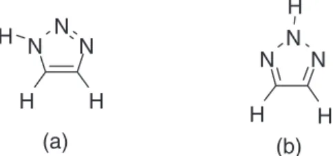
2-phenyl-2H-1,2,3-triazolyl derivatives.24 See Figure 1 for
the tautomeric forms of the parent compounds: 1H
-1,2,3-triazole and 2H-1,2,3-triazole.
The crystal structures of four of the moderately active
hydrazonyl derivatives in the glycosidase inhibition study24
have been determined, namely of (Z
)-1-phenyl-4-[((2-phenylhydrazono)methyl)]-1H-1,2,3-triazole (1a), (Z
)-
4-[(2-(2,4-dimethylphenyl)hydrazono)methyl]-2-phenyl-2H-1,2,3-triazole (2a), (E)-4-[(2-(2,4-dinitrophenyl)
hydrazono)methyl]-2-phenyl-2H-1,2,3-triazole (2b),
and (E)-N’-((2-phenyl-2H-1,2,3-triazol-4-yl)methylene)
isonicotinohydrazide dihydrate, (3·2H2O), see Scheme 1.
An antifungal activity study of related 1H
-1,2,3-triazolyl hydrazones was also published very recently.13
The crystal structure of one of the compounds, (E
)-4-[2-ClC6H4−NH−N=CH)-1-(2-ClC6H4)-1H-1,2,3-triazole (1b),
was reported and has been deposited in the Cambridge Crystallographic Data Centre (CCDC No. 976935; ref code: FONZAL), but only the molecular configuration had
been reported in the article.13 In this article, we report the
crystal structures of 1a, 2a, 2b and (3.2H2O), and make
comparisons with that of 1b.
Experimental
X-Ray crystallography
Data for compounds 1a, 2a and (3.2H
2O) were obtained
at 100(2) K, while data for compound 2b were collected
at 120(2) K. All with Mo Kα radiation by means of a
Rigaku Saturn 724+ (2 × 2 bin mode) instrument of the National Crystallography Service (NCS), University of Southampton. Data collection, data reduction and unit
cell refinement were achieved with the DENZO25 and
COLLECT26 programs. Correction for absorption was
achieved in each case by a semi-empirical method based upon the variation of equivalent reflections with the Rigaku
version of the program SADABS 2007/2.27 The program,
MERCURY28 was used in the preparation of the Figures.
SHELXL9729 and PLATON30 were used in the calculation
of molecular geometry. The structures were solved by
direct methods by SHELXT and fully refined by means of
the SHELXL using OSCAIL.31 Difference map provided
position for the N−H hydrogen atoms in all four compounds
and for the water hydrogen atoms in (3.2H
2O). All other
hydrogen atoms were placed in calculated positions. Crystal data and structure refinement details are listed in Table 1.
Results and Discussion
The compounds were prepared as previously reported24
from the corresponding aldehydes, see Scheme 1. Samples used in the structure determinations were grown by slow
evaporation of solutions in methanol for 1a and 2a, in
2-methoxyethanol for 2b, and in ethyl acetate for 3.
The cell dimensions for a sample of 2b, recrystallized
from methanol, indicated the same phase as obtained from 2-methoxyethanol. The crystals obtained from
recystallisation of 3 from ethyl acetate were of the dihydrate
(3·2H2O).
Molecular conformations
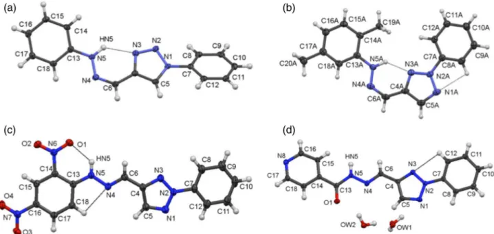
The asymmetric unit in each of 1a, 2a and 2b consists
of a single molecule, that of (3·2H2O) a molecule of 3 and
two molecules of water. Figure 2 illustrates the numbering schemes for all the molecules. Selected bond lengths and angles are listed in Table 2. Comparison of the bond lengths
in the triazolyl rings in the 1H-1,2,3-triazole, compound 1a,
and the 2H-1,2-3-triazole compounds, 2a, 2b and(3·2H2O),
indicate that the major differences are found in the
C4−C5 and C5−N1 bond lengths. The bond lengths in the
hydrazonyl linker, C13−N5−N4−C6−C4, in compounds 1a,
1b and 2a, indicate that electron delocalization occurs
within the link, as do the bond lengths in the acylhydrazonyl
linker, C14−C13(O1)−N5−N4−C6−C4, in molecule 3.
The most significant conformational result is that
compounds 1a and 2a have (Z) geometries about the
C=N bond, in contrast to the (E)-configuration in 2b
and (3·2H2O) (Figure 2); compound 1b13 also has the
(E)-configuration (Figure 3a). Generally in the absence of
special circumstances, (Z)-isomers are thermodynamically
less stable than (E)-isomers. The special circumstances
in 1a and 2a must be the formations of the classical and
strong N5−HN5···N3 intramolecular hydrogen bonds,
which enhance the stability of the (Z)-isomers. On the
other hand, the (E)-configuration in the 2,4-dinitrophenyl
derivative, 2b does permit the formation of strong classical
N4−HN5···O1 intermolecular hydrogen bonds, involving
an oxygen atom of the ortho-nitro group. Such a strong
N4−HN5···O1 intermolecular hydrogen bond in 2b must
further enhance the stability of the (E)-configuration of 1b
over that of the (Z)-isomer.For compound 1b, it is argued
that the ortho-chloro substituent prevents the formation of
a (Z)-configuration, due to the potential steric hindrance
between chlorine and adjacent atoms (see Figure 3b). In
Figure 3b are drawn the two possibilities for (Z)-(1b),
arising from the two possible positions of the chloro group in the phenyl ring. The chlorine atom would be
uncomfortably close in (i) to the N−H bond and in (ii) to
a nitrogen atom.
The arrangement about the C(O)−NH−N=CH-aryl
fragment in 3 is designated as EC(O)NH /EC=N. As reported
for many acylhydrazones, such as 3, there are two potential
configurations about the C(O)−NH bond (EC(O)NH and
ZC(O)NH), as well as the two geometric isomers about the C=N
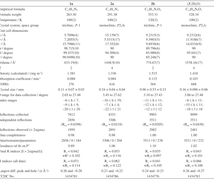
Table 1. Crystal data and structure refinement
1a 2a 2b (3.2H2O)
Empirical formula C15H13N5 C17H17N5 C15H11N7O4 C15H16N6O3
Formula weight 263.30 291.38 353.31 328.34
Temperature / K 100(2) 100(2) 120(2) 100(2)
Crystal system, space group triclinic, P-1 monoclinic, P21/n triclinic, P-1 monoclinic, P21/c
Unit cell dimensions a / Å
b / Å c / Å
α / degree
β / degree
γ / degree
5.7096(4) 7.2055(5) 15.7986(11) 98.715(10 99.437(10) 90.9490(10) 15.139(7) 5.5315(17) 17.352(8) 90 94.300(4) 90 9.2315(3) 9.5903(5) 9.6938(6) 89.796(6) 65.000(8) 85.248(7) 9.2332(6) 11.9186(7) 14.0316(9) 90 95.041(7) 90
Volume / Å3 633.19(8) 1448.9(10) 774.67(7) 1538.16(17)
Z 2 4 2 4
Density (calculated) / (mg m-3) 1.381 1.336 1.515 1.418
Absorption coefficient / mm-1 0.088 0.084 0.115 0.103
F(000) 276 616 364 688
Crystal size / mm 0.11 × 0.07 × 0.03 0.24 × 0.04 × 0.04 0.96 × 0.53 × 0.23 0.16 × 0.096 × 0.06
θ range for data collection / degree 2.65 to 27.48 3.45 to 27.62 3.10 to 27.43 3.06 to 27.48
Index ranges −6 ≤ h ≤ 7;
−9 ≤ k ≤ 9;
−20 ≤ l ≤ 20
−16 ≤ h ≤ 19;
−7 ≤ k ≤ 4;
−22 ≤ l ≤ 21
−11 ≤ h ≤ 11;
−12 ≤ k ≤ 12;
−12 ≤ l ≤ 12
−8 ≤ h ≤ 11;
−15 ≤ k ≤ 11;
−18 ≤ l ≤ 18
Reflections collected 7812 8351 9903 9099
Independent reflections 2856
(Rint = 0.0398)
3306 (Rint = 0.0218)
3511 (Rint = 0.0205)
3511 (Rint = 0.0438)
Reflections observed (> 2sigma) 1959 2891 2982 2481
Data completeness 0.98 0.98 1.00 1.00
Data/restraints/parameters 2856 / 0 / 184 3306 / 0 / 204 3511 / 0 / 238 3511 / 0 / 232
Goodness-of-fit on F2 0.89 1.06 1.10 1.02
Final R indices [I > 2sigma(I)] R1 = 0.042
wR2 = 0.102
R1 = 0.053
wR2 = 0.116
R1 = 0.035
wR2= 0.097
R1 = 0.043
wR2 = 0.101
R indices (all data) R1 = 0.071
wR2 = 0.114
R1 = 0.062
wR2 = 0.122
R1 = 0.041
wR2 = 0.105
R1 = 0.066
wR2 = 0.108
Largest diff. peak and hole / (e Å-3) 0.26 and −0.28 0.21 and −0.22 0.24 and −0.23 0.26 and −0.27
bond (EC=N and ZC=N), making four possible arrangements
in all about the C(O)−NH−N=CH-aryl fragment.32-35
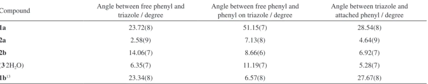
While the triazolyl ring is planar in all compounds, none of the compounds is planar overall. The deviation
from planarity is relatively small for the 2H-1,2,3-triazolyl
compounds, 2a, 2b and 3, as shown by the angles between
the aryl rings in Table 3, and very much larger for 1a and
1b (see Figure 4). The increased deviation from planarity
of the triazole and its attached phenyl ring in a H
-1,2,3-triazolyl may arise from steric repulsions between the
ortho C−H bonds in the triazole ring and the phenyl ring. Of interest, the sums of the dihedral angles between the
Table 2. Selected bond lengths (Å) and angles (degree)
1a 2a 2b (3.2H2O)
C13−N5 1.4005(18) 1.395(2) 1.3524(14) 1.3519(13)
N5−N4 1.3626(18) 1.3502(18) 1.3757(13) 1.3772(15)
N4−C6 1.2940(19) 1.295(3) 1.2812(14) 1.2769(18)
C6−C4 1.455(2) 1.446(3) 1.4551(15) 1.4494(18)
C4−C5 1.377(2) 1.403(3) 1.4031(16) 1.397(2)
C5−N1 1.3466(19) 1.323(3) 1.3245(16) 1.3245(16)
N1−N2 1.3600(17) 1.3452(17) 1.3449(13) 1.3429(16)
N2−N3 1.3155(17) 1.339(2) 1.3550(13) 1.3318(15)
N3−C4 1.375(2) 1.346(2) 1.3384(14) 1.3342(18)
C13−N5−N4 118.14(12) 119.79(14) 118.97(9) 117.58(12)
N5−N4−C6 118.39(15) 118.44(15) 115.19(10) 116.68(12)
N4−C6−C4 129.14(15) 129.27(15) 118.73(10) 118.03(13)
C18−C13−N5−N4 21.9(2) 1.3(2) 8.82(17)
C15−C14−C13−N5 8.2(2)
C18−C14−C13−N5 173.02(13)
C15−C14−C13−O1 171.29(13)
C18−C14−C13−O1 7.5(2)
C14−C13−N5−N4 159.39(14) 179.36(15) 172.05(11) 179.94(11)
C13−N5−N4−C6 177.85(13) 179.16(16) 177.03(11) 175.48(13)
N5−N4−C6−C4 1.4(2) 0.2(3) 176.56(11) 179.05(12)
N4−C6−C4−N3 1.8(3) 2.4(3) 179.51(12) 176.87(13)
N4−C6−C4−C5 178.51(15) 176.1(2) 0.7(2) 1.1(2)
Figure 2. Atom arrangements and numbering schemes for (a) 1a, (b) 2a, (c) 2b, and (d) (3.2H
2O). Ellipsoids are drawn at the 50% level. Hydrogen atoms
phenyl groups and the triazolyl ring are very similar to the single dihedral angle between the two phenyl rings in each
of 1a, 2a, 2b and 3, which indicates that the deviation from
planarity can be considered to have arisen from rotations
about the C13−N5 and C7−N2 bonds occurring in the
same sense. This is not the case in the 1H-1,2,3-triazolyl
compound, 1b. For 1b, the dihedral angles point to rotations
about the C13−N5 and C7−N2 bonds occurring in the
opposite senses, with the result that the two phenyl groups
have a small dihedral angle of ca. 6o, compared to ca. 51o
in 1a. If rotations about these C13−N5 and C7−N2 bonds
did occur in the same sense, it would place either Cl1 too close to N5, or Cl2 too close to N2.
Dihedral angles between aryl and triazolyl rings in
1-aryl-1H- and 2-aryl-2H-1,2,3-triazole compounds
have been shown to vary considerably, for example,
such angles are 0.34(17) and 87.1(2)o, respectively,
in 4-(difluoromethyl)-1H-1,2,3-triazole,36 and in one
independent molecule of 1-[5-methyl-1-(4-nitrophenyl)
methyl-1-(4-methylphenyl)-1H-1,2,3-triazol-4-yl]
ethanone.37 The aryl group substituents and crystal packing
effects have major influences on such dihedral angles. The
dihedral angles in 2a between the phenyl group and (i) the
attached ortho-nitro group, O2−N6−O1, and (ii) para-nitro
group, O3−N7−O4, are 3.93 and 13.69o, respectively.
The small angle between the phenyl and its ortho-nitro
group facilitates the formation of the N5−HN5···O1
intramolecular hydrogen bond.
Crystal structures
Compound 1a
The only classical hydrogen bond present in 1a is the
intramolecular N5−HN5···N3 hydrogen bond (Figure 2a).
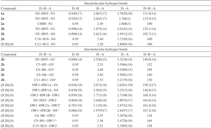
The intermolecular interactions in 1a are four C−H···π
interactions (Table 4).38
The combination of the C−H···π interactions,
C9−H9···π(phenyl-b), C12−H12···π(phenyl-b),
C15−H15···π(phenyl-a) and C18−H18···π(phenyl-a),
provides sheets of molecules in the ab plane, as shown
in Figure 5. Phenyl-a and phenyl-b are the phenyl groups
with atoms C7−C12 and C13−C18, respectively. The
triazole ring is not involved in C−H···π interactions.
The PLATON analysis31 indicates the possibility of
π(triazolyl)···π(triazolyl) stacking interactions.39 However,
Figure 3. (a) The (E)-geometric form determined for 1b in the solid state;13
(b) potential (Z)-forms of 1b. Ar = 2-chlorophenyl.
Figure 4. Molecular conformations. Hydrogen atoms have been omitted.
Table 3. Angles between the best planes through the aryl rings
Compound Angle between free phenyl and triazole / degree
Angle between free phenyl and phenyl on triazole / degree
Angle between triazole and attached phenyl / degree
1a 23.72(8) 51.15(7) 28.54(8)
2a 2.58(9) 7.13(8) 4.64(9)
2b 14.06(7) 8.66(6) 6.92(7)
(3.2H
2O) 6.35(7) 11.19(7) 5.28(7)
Table 4. Geometric parameters (Å, degree) for intra- and intermolecular interactions
Intramolecular hydrogen bonds
Compound D−H···A D−H H···A D···A D−H···A
1a N5−HN5···N3 0.949(17) 2.067(17) 2.7829(18) 131.0(14)
2a N5−HN5···N3 0.929(17) 2.042(17) 2.768(2) 133.9(15)
2a C8H8···N1 0.95 2.49 2.808(3) 100
2b N5−HN5···O1 0.898(14) 1.975(14) 2.6342(14) 129.1(14)
2b N5−HN5···N6 0.898(14) 2.621(16) 2.9512(15) 102.7(11)
2b C18−H18···N4 0.95 2.40 2.7320(16) 100
(3⋅2H2O) C12−H12···N3 0.93 2.50 2.8069(18) 100
Intermolecular hydrogen bonds
Compound D−H···A D−H H···A D···A D−H···A
2b N5−HN5···O1i 0.898(14) 2.520(15) 3.3218(14) 149.0(14)
2b C5−H5···O3ii 0.95 2.53 3.3984(18) 152
2b C6−H6···O3iii 0.95 2.60 3.0389(15) 109
2b C6−H6···O2i 0.95 2.60 3.5082(15) 160
2b C11−H11···O4iv 0.95 2.52 3.2179(18) 130
(3⋅2H2O) OW2−HW1A···O1 0.836(18) 2.072(18) 2.8281(150 150.2(17)
(3⋅2H2O) OW2−HW1A···N4 0.836(18) 2.562(19) 3.2512(16) 140.6(15)
(3⋅2H2O) OW2−HW1B···OW1 0.959(18) 1.773(18) 2.7198(16) 168.5(16)
(3⋅2H2O) N5−HN5···OW2v 0.869(18) 2.048(18) 2.8974(17) 165.6(14)
(3⋅2H2O) OW1−HW2A···OW2vi 0.787(18) 2.119(18) 2.8742(16) 161.0(18)
(3⋅2H2O) OW1−HW2B···N8vii 0.886(18) 1.975917) 2.8457(17) 167.2(16)
(3⋅2H2O) C6−H6···OW2v 0.93 2.55 3.3076(18) 138
(3⋅2H2O) C9−H9···OW1viii 0.93 2.56 3.4728(19) 169
(3⋅2H2O) C15−H15···OW2v 0.93 2.52 3.3985(18) 158
Symmetry codes: i = 1 − x, 1 − y, −1 − z; ii = 1 − x, 2 − y, −z; iii = x, y, −1 + z; iv = −1 + x, 1 + y, −1 + z; v = −x, −½ + y, ½ + z; vi = −x, 1 − y, 1 − x; vii = 1 + x, ½ − y, ½ + z; viii = 1 – x, y, z.
Y−X···π interactionsa
Compound Y−X···Cga X···Cg X
perp γ Y−X···Cg Y···Cg
1a C9−H9···Cg3i 2.68 2.67 5.81 131 3.3796(17)
1a C12−H12···Cg3ii 2.63 2.62 6.29 131 3.3403(16)
1a C15−H15···Cg2iii 2.78 2.75 8.72 131 3.4822(17)
1a C18−H18···Cg2iv 2.76 2.72 10.36 129 3.4370(16)
2a C19−H19B···Cg2v 2.81 2.68 17.59 150 3.692(2)
2b N7−O4···Cg2vi 3.7402(11) 3.37 25.73 74.54(6) 3.6125(12)
Symmetry codes: i = −x, 1 − y, 1 − z; ii = 1 − x, 2 − y, 1 − z; iii = −x, 2 − y, 1 − x; iv = 1 − x, 1 − y, 1 − z; v = x, 1 + y, z; vi = 1 − x, 2 − y, −1 − z.
π···π interactionsa
Compound Cg(I)···Cg(J) Cg···Cg α β γ CgIperp CgJperp
1a Cg1···Cg1i 4.2304(9) 0.02 34.8 34.8 3.48 3.475
2a Cg1···Cg2ii 3.726(2) 4.58(9) 27.4 22.8 3.4350(7) 3.3089(6)
2a Cg2···Cg1ii 3.726(2) 4.58(9) 22.8 27.4 3.3089(6) 3.4350(7)
2a Cg3···Cg1iii 4.065(2) 2.64(9) 33.3 30.7 3.493597) 3.3986(7)
2b Cg1···Cg1iv 3.8953(8) 0.03 32.4 32.4 3.27 3.265
2b Cg2···Cg3v 4.0523(9) 8.66 30.4 36.7 3.25 3.294
2b Cg3···Cg1v 3.8968(7) 14.05 26.6 26.2 3.50 3.484
(3⋅2H2O) Cg1···Cg2vi 3.5089(8) 5.28 12.6 7.5 3.48 3.425
(3⋅2H2O) Cg1···Cg2vii 3.9188(8) 8.00 32.3 36.0 3.17 3.314
(3⋅2H2O) Cg3···Cg2vii 3.9611(8) 14.12 44.0 23.8 3.63 3.246
Symmetry operation: i = 1 – x, 1 – y, 1 – z; ii = 1 – x, –y, 1 – z; iii = x, 1 + y, z; iv = –x, 2 – y, –1 – z; v = 1 – x, 2 – y, –1 – z; vi = 1 + x, y, z; vii = 1 + x, ½ – y, ½ + z.
aIn compounds 1a and 2b, Cg1, Cg2 and Cg3 are the centroids of the ring containing N2, C10 and C16, respectively; in compound 3, Cg1, Cg2 and Cg3
are the centroids of the ring containing N2, C16 and C10, respectively; in compound 2a, Cg1, Cg2, Cg3, Cg4, Cg5 and Cg6 are the centroids of the ring containing N2A, C10A, C16A, N2B, C10B and C16B, respectively; β is the angle between the vectors Cg···Cg and CgIperp, where CgIperp is the perpendicular
distance of CgI from the plane of ring J. Similarly, γ is the angle between the vectors Cg···Cg and CgJperp. CgJperp is the perpendicular distance of CgI from
although the perpendicular distance between parallel triazole rings is only 3.475 Å, the Cg···Cg distance is large at 4.2304(9) Å, resulting in ring offsets of 2.413 Å, which indicates that the triazole rings do not overlap.
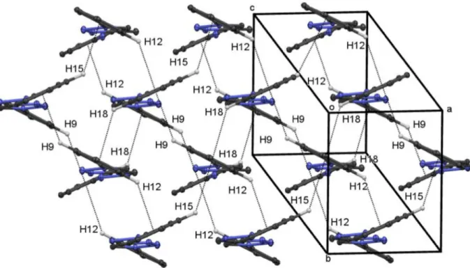
Compound 2a
The major intermolecular interactions in 2a are π···π
stacking interactions. Dimers are generated from pairs
of π(triazole)···π(phenyl-a) interactions. These dimeric
units are further linked by π(triazole)···π(phenyl-b) and
by C−H···π(phenyl-a) interactions into two molecule
wide columns, where phenyl-a and phenyl-b refer to the
phenyl group attached to the triazole and the other phenyl group, respectively (Figure 6a). These two-molecular
wide columns are free standing and so 2a has a
one-dimensional structure. As shown in Figure 6b, such columns of molecules are propagated in different directions, with
angles between the best planes of ca. 76o.
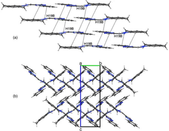
Compound 2b
As well as the classical intramolecular N−H···O
hydrogen bond, there is also a classical intermolecular
N−H···O hydrogen bond, and weaker intermolecular
C−H···O hydrogen bonds, π···π stacking and N−O···π
interactions (Table 4).40,41 In the following discussion, the
overall structure is broken down into three sub-structures.
Firstly, pairs of the classical intermolecular N5−HN5···O1
and weaker C11−H11···O4 hydrogen bonds form
centrosymmetric dimers, as shown in Figure 7a. Included
within these dimers are intramolecular N5−HN5···O1
hydrogen bonds: together, these hydrogen bonds generate a
set of R2
2(8), R22(4) and R22(8) rings. The O1···O1 distance
in the O1−NH5−O1−NH5 ring is short. Secondly, ladders
of molecules, containing R2
2(24) and R42(24) rings,42
are generated from the combination of C5−H5···O3
and C6−H6···O3 hydrogen bonds: in these ladders, the
C5−H5···O3 hydrogen bonds form the rings of the ladder
and the C6−H6···O3 form the sides (see Figure 7b). The
third sub-structure is a sheet of molecules generated from
π(triazole)···π(nitrophenyl), π(phenyl)···π(nitrophenyl)
and π(triazole)···π(triazole) stacking interactions,
N7−O4···π(phenyl) and C11−H11···O4 hydrogen bonds.
The π(triazole)···π(nitrophenyl), π(phenyl)···π(nitrophenyl)
and N7−O4···π(phenyl) interactions generate dimeric units,
which are linked into single-molecule wide columns by
the π(triazole)···π(triazole) interactions. These
single-molecule wide columns are further linked into bi-single-molecule
wide columns by the C11−H11···O4 hydrogen bonds (see
Figure 7c). Overall, a three-dimensional array is produced.
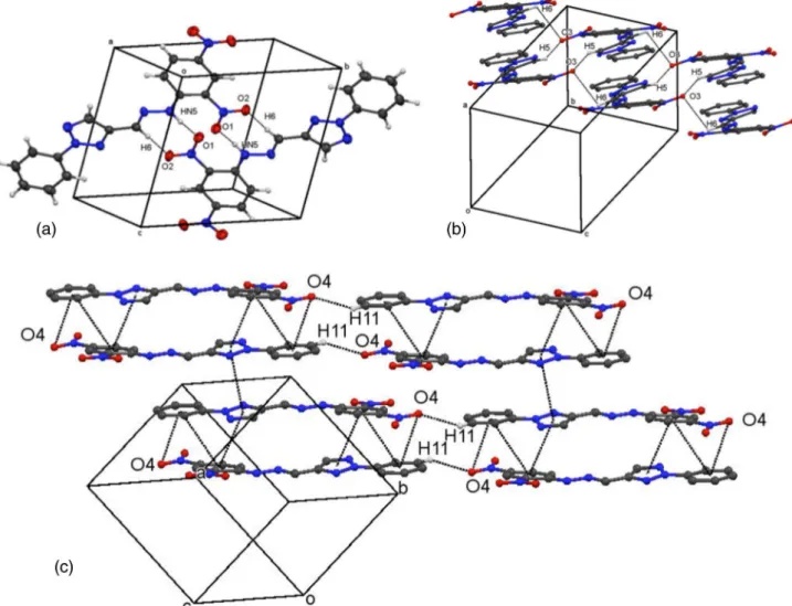
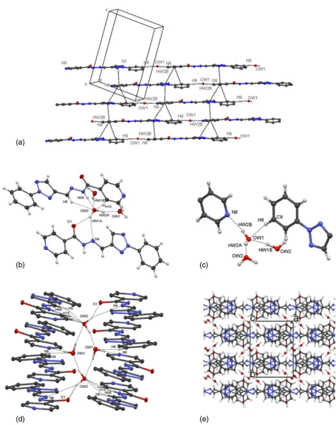
Compound (3.2H2O)
The intermolecular interactions in compound (3.2H2O)
are π···π stacking interactions and O−H···X (X = O and
N), N−H···O and C−H···O hydrogen bonds (Table 4). As
expected, the two water molecules are strongly involved in the supramolecular arrangements. A sheet containing
Figure 5. Compound 1a. Part of the sheet of molecules obtained from C9−H9···π(phenyl-a), C12−H12···π(phenyl-a), C15−H15···π(phenyl-b) and C18−
molecules of 3 and water, with oxygen atom, OW1, is
generated from combinations of π(triazolyl)···π(phenyl),
π(triazolyl)···π(pyridinyl) and π(phenyl)···π(pyridinyl)
interactions, and C9−H9···OW1 and OW1−HW2B···N8
hydrogen bonds, as illustrated in Figure 8a. Molecules
in each layer of the sheet are linked by C9−H9···OW1
and OW1−HW2B···N8 hydrogen bonds, and layers are
linked by the three different π···π interactions. The two
water molecules make very different connections to other molecules, as shown in Figures 8b and 8c. There are seven
short contacts to the water molecule, HW1A−OW2−HW2A
(Figure 8b), but there are only four short contacts to the
other water molecule, HW2A−OW1−HW2B (Figure 8c).
Such contacts to the watermolecules generate various rings of atoms. However, the most interesting ring
present in (3.2H
2O) is the R44(8) ring generated from
four water molecules, two of each HW1A−OW2−HW2A
and HW2A-OW1-HW2B, see Figure 8d. As shown in Figure 8d, the water molecules in the tetrameric rings
make short contacts with various atoms in 3. Overall, a
three-dimension array is formed, see Figure 8e.
The hydrogen bonding interactions between the
water molecules and molecule of 3 clearly stabilize the
EC(O)NH /EC=Narrangement about the C(O)−NH−N=CH-aryl
fragment in (3.2H
2O).
Conclusions
The significance of the classical intramolecular hydrogen bonds in the molecular conformations is very pronounced in this study. The formations of
(Z)-configurations about the C=N bonds in 1a and 2a
arise from the stabilizing presence of intramolecular
N5−HN5···N3 hydrogen bonds, while in 2b the presence
of intramolecular N5−HN5···O1 hydrogen bonds, in lieu
of potential N5−HN5···N3 hydrogen bonds, reinforces
the (E)-geometry. An EC(O)NH/EC=Narrangement about the
C(O)−NH−C=N fragment, and an interesting R4
4(8) ring
composed of four hydrate molecules, are features of the
crystal structure of the hydrated acylhydrazone, (3.2H
2O).
As found in this study, significant π···π interactions
are exhibited by compounds 2a and 2b, but not by the
least planar molecule, 1a. In contrast, the only important
intermolecular interactions in 1a are C−H···π interactions.
Are these differences between 1a, on one hand, and 2a and
2b, on the other, consequences of compound 1a being an
1H-1,2,3-triazole compound, while 2a and 2b are 2H
-1,2,3-triazole derivatives? To effectively answer these questions, further structures of related hydrazonyl derivatives of 1,2,3-triazoles need to be determined.
Moreover, there appears to be no obvious reason why 1a
cannot adopt a near planar configuration. Other points to be considered are the influences of steric effects or the position
of substituents. Compound 1a has no substituents in either
the two phenyl rings, while both 2a and 2b have ortho- and
para-substituents in the phenyl ring (C13-C18). The other
1H-1,2,3-triazole compound mentioned in this article, 1b,13
has ortho chloro substituents in both phenyl rings, and does
exhibit a much smaller dihedral angle between the two phenyl
rings than does 1a, but is still not planar (Table 3). As in 1a,
no π···πinteractions are exhibited by 1b, but the number of
different C−H···π intermolecular interactions is reduced to
one. The most important intermolecular interaction in 1b is
the classical intermolecular N(hydrazine)−H···N(triazole)
hydrogen bonds, with less important interactions being
C−H···Cl and N(hydrazine)−H···Cl hydrogen bonds. This
prompts the question: does the presence of substituents in
the phenyl rings reduce or even prevent C−H···π interactions
in 2a and 2b, which thus results in different sets of
intermolecular interactions being taken up? The answer awaits further study.
Supplementary Information
Full details of the crystal structure determinations in cif format are available in the online version as Supplementary
Figure 7. Compound 2b. (a) A centrosymmetric dimer generated from pairs of N5−HN5···O1 and C6−H6···O2 intermolecular hydrogen bonds: also indicated are intramolecular N5−HN5···O1 hydrogen bonds, together these hydrogen bonds generate a set of R2
2(8), R22(4) and R22(8) rings; (b) ladders of molecules,
containing R2
2(24) and R42(24) rings, are generated from the combination of C5−H5···O3 and C6−H6···O3 intermolecular hydrogen bonds; (c) part of a
Information, at http://jbcs.sbq.org.br, and have also been deposited with the Cambridge Crystallographic Data Centre
with deposition numbers, 1434781, 1434786, 1434776 and
1434783 for 1a, 2a, 2b and (3.2H2O), respectively. Copies of
Figure 8. Compound (3.2H
2O). (a) A sheet of 3 and hydrate molecules (with OW1) is generated from combinations of π(triazolyl)···π(phenyl),
π(triazolyl)···π(pyridinyl) and π(phenyl)···π(pyridinyl) interactions, and C9−H9···OW1 and OW1−HW2B···N8 hydrogen bonds; (b) and (c) short contacts to the hydrate molecules, HW1A−OW2−HW2A and HW2A−OW1−HW2B, respectively; (d) a R4
4(8) ring generated from four hydrate molecules, showing
these can be obtained free of charge on written application to CCDC, 12 Union Road, Cambridge, CB2 1EZ, UK (fax: +44 1223 336033); on request by e-mail to deposit@ccdc. cam.ac.uk or by access to http://www.ccdc.cam.ac.uk.
Acknowledgments
The use of the NCS crystallography service at Southampton and the valuable assistance of the staff there are gratefully acknowledged. JLW thanks FAPERJ and CNPq (Brazil) for support. SMSVW and JLW thank PhD John N. Low for discussions.
References
1. Dehaen, W.; Bakulev, V. A., eds., In Topics in Heterocyclic Chemistry, vol. 40; Springer: Heidelberg, 2015.
2. Rachwal, S.; Katritzky, A. R. In Comprehensive Heterocyclic Chemistry, 3rd ed.; Katritzy, A. R.; Ramsden, C. A.; Scriven, E.
F. V.; Taylor, R. J. K., eds.; Elsevier: Oxford, 2008, p. 1. 3. Bellagamba, M.; Bencivenni, L.; Gontrani, L.; Guidoni, L.;
Sadun, C.; Struct. Chem. 2013, 4, 933.
4. Ferreira, M. L. G.; Pinheiro, L. C. S.; Santos-Filho, A. O.; Peçanha, M. D. S.; Sacramento, C. Q.; Machado, V.; Ferreira, V. F.; Souza, T. M. L.; Boechat, N.; Med. Chem. Res. 2014, 23, 1501.
5. Jordão, A. K.; Afonso, P. P.; Ferreira, V. F.; de Souza, M. C.; Almeida, M. C.; Beltrame, C. O.; Paiva, D. P.; Wardell, S. M. S. V.; Wardell, J. L.; Tiekink, E. R.; Damaso, C. R.; Cunha, A. C.; Eur. J. Med. Chem. 2009, 44, 3777.
6. Himanshu; Tyagi, R.; Olsen, C. E.; Errington, W.; Parmar, V. S.; Prasad, A. K.; Bioorg. Med. Chem. 2002, 10, 963. 7. Boechat, N.; Ferreira, M. L.; Pinheiro, L. C.; Jesus, A. M.; Leite,
M. M.; Júnior, C. C.; Aguiar, A. C.; de Andrade, I. M.; Krettli, A. U.; Chem. Biol. Drug. Des. 2014, 84, 325.
8. Ferreira, M. L.; de Souza, M. V. N.; Wardell, S. M. S. V.; Wardell, J. L.; Vasconcelos, T. R. A.; Ferreira, V. F.; Lourenço, M. C. S.; J. Carb. Chem. 2010, 29, 265.
9. Jordão, A. K.; Sathler, P. C.; Ferreira, V. F.; Campos, V. R.; de Souza, M. C.; Castro, H. C.; Lannes, A.; Lourenco, A.; Rodrigues, C. R.; Bello, M. L.; Lourenco, M. C.; Carvalho, G. S.; Almeida, M. C.; Cunha, A. C.; Bioorg. Med. Chem. 2011, 19, 5605.
10. Boechat, N.; Ferreira, V. F.; Ferreira, S. B.; Ferreira, M. L. G.; da Silva, F. C.; Bastos, M. M.; Costa, M. S.; Lourenço, M. C.; Pinto, A. C.; Krettli, A. U.; Aguiar, A. C.; Teixeira, B. M.; da Silva, N. V.; Martins, P. R.; Bezerra, F. A.; Camilo, A. L.; da Silva, G. P.; Costa, C. C.; J. Med. Chem. 2011, 54, 5988. 11. Lima-Neto, R. G.; Cavalcante, N. N.; Srivastava, R. M.;
Mendonça Junior, F. J.; Wanderley, A. G.; Neves, R. P.; dos Anjos, J. V.; Molecules 2012, 17, 5882.
12. da Silva, I. F.; Martins, P. R.; da Silva, E. G.; Ferreira, S. B.; Ferreira, V. F.; da Costa, K. R.; de Vasconcellos, M. C.; Lima, E. S.; da Silva, F. C.; Med. Chem. 2013, 9, 1085.
13. Dai, Z. C.; Chen, Y. F.; Zhang, M.; Li, S. K.; Yang, T. T.; Shen, L.; Wang, J. X.; Qian, S. S.; Zhu, H. L.; Ye, Y. H.; Org. Biomol. Chem. 2015, 13, 477.
14. da Silva, F. C.; de Souza, M. C.; Frugulhetti, I. I.; Castro, H. C.; Souza, S. L.; de Souza, T. M.; Rodrigues, D. Q.; Souza, A. M.; Abreu, P. A.; Passamani, F.; Rodrigues, C. R.; Ferreira, V. F.; Eur. J. Med. Chem. 2009, 44, 373.
15. Weide, T.; Saldanha, S. A.; Minond, D.; Spicer, T. P.; Fotsing, J. R.; Spaargaren, M.; Frère, J.-M.; Bebrone, C.; Sharpless, K. B.; Hodder, P. S.; Fokin, V. V.; ACS Med. Chem. Lett. 2010, 1, 150. 16. Rogawski, M. A.; Epilepsy Res. 2006, 69, 273.
17. Jordão, A. K.; Ferreira, V. F.; Souza, T. M. L.; Faria, G. G.; Machado, V.; Abrantes, J. L.; de Souza, M. C. B. V.; Cunha, A. C.; Bioorg. Med. Chem. 2011, 19, 1860.
18. Shafi, S.; Alam, M. M.; Mulakayala, N.; Mulakayala, C.; Vanaja, G.; Kalle, A. M.; Pallu, R.; Alam, M. S.; Eur. J. Med. Chem. 2012, 49, 324.
19. Banday, A. H.; Shameem, S. A.; Ganai, B. A.; Org. Med. Chem. Lett. 2012, 2, 13.
20. Sumangala, V.; Poojary, B.; Chidananda, N.; Fernandes, J.; Kumari, N. S.; Arch. Pharm. Res. 2010, 33, 1911.
21. Senger, M. R.; Gomes, L. C.; Ferreira, S. B.; Kaiser, C. R.; Ferreira, V. F.; Silva-Jr., F. P.; ChemBioChem 2012, 13, 1584. 22. Périon, R.; Ferrières, V.; Garcia-Moreno, M. I.; Mellet, C. O.;
Duval, R.; Fernández, J. M. G.; Plusquellec, D.; Tetrahedron 2005, 61, 9118.
23. Zhou, Y.; Zhao, Y.; O’Boyle, K. M.; Murphy, P. V.; Bioorg. Med. Chem. Lett. 2008, 18, 954.
24. Gonzaga, D.; Senger, M. R.; da Silva, F. C.; Ferreira, V. F.; Silva-Jr., F. P.; Eur. J. Med. Chem. 2014, 74, 461.
25. Otwinowski, Z.; Minor, W.; Methods in Enzymology, Vol. 276, Macromolecular Crystallography, Part A; Carter, C. W.; Sweet, R. M., eds.; Academic Press: New York, 1997, p. 307-326. 26. Hooft, R. W. W.; Nonius, B. V.; COLLECT, Data Collection
Software;Delft: The Netherlands, 1998.
27. Sheldrick, G. M.; SADABS Version 2007/2; Bruker AXS Inc.: Madison, Wisconsin, 2007.
28. MERCURY 3.3.1 Cambridge Crystallographic Data Centre, UK.
29. Sheldrick, G. M.; Acta Crystallogr. 2008, A64, 112. 30. Spek, A. L.: J. Appl. Crystallogr. 2003, 36, 7.
31. McArdle, P.; Oscail - Windows Software for Crystallography and Molecular Modelling; National University of Ireland: Galway, 2016.
33. Lopes, A. B.; Miguez, E.; Kümmerle, A. E.; Rumjanek, V. M.; Fraga, C. A.; Barreiro, E. J.; Molecules 2013, 18, 11683. 34. da Silva, Y. K.; Reyes, C. T.; Rivera, G.; Alves, M. A.; Barreiro,
E. J.; Moreira, M. S.; Lima, L. M.; Molecules 2014, 19, 8456. 35. Palla, G.; Predieri, G.; Domiano, P.; Vignali, C.; Turner, C. W.;
Tetrahedron 1986, 42, 3649.
36. Costa, M. S.; Boechat, N.; Ferreira, V. F.; Wardell, S. M. S. V.; Skakle, J. M. S.; Acta Crystallogr. Sect. E 2006, E62, o1925. 37. Vinutha, N.; Madan Kumar, S.; Nithinchandra; Balakrishna, K.;
Lokanath, N. K.; Revannasiddaiah, D.; Acta Crystallogr. Sect. E 2013, E69, o1724.
38. Desiraju, G. R.; Angew.Chem., Int. Ed. 2007, 46, 8342.
39. Tiekink, E. R. T.; Zukerman-Schpector, J., eds., In The Importance of π-Interactions in Crystal Engineering: Frontiers
in Crystal Engineering, 2nd ed.; Wiley: Singapore, 2012.
40. Tiekink, E. R. T. In Supramolecular Chemistry: from Molecules to Nanomaterials; Steed, J. W.; Gale, P. A., eds.; John Wiley & Sons Ltd: Chichester, UK, 2012, p. 2791.
41. Huang, L.; Massa, L.; Karle, J.; Proc. Natl. Acad. Sci. 2008, 105, 13720.
42. Bernstein, J.; Davis, R. E.; Shimoni, L.; Chang, N. L.; Angew. Chem. Int. Ed. 1995, 34, 1555.
Submitted: November 24, 2015