during Anaerobic Respiration of
Shewanella
piezotolerans
WP3
Xin-Wei Yang1, Ying He1, Jun Xu1, Xiang Xiao1, Feng-Ping Wang1,2*
1State Key Laboratory of Microbial Metabolism and School of Life Sciences and Biotechnology, State Key Laboratory of Ocean Engineering, Shanghai Jiao Tong University, Shanghai, PR China,2Key Laboratory of Systems Biomedicine, Ministry of Education, Shanghai Jiao Tong University, Shanghai, PR China
Abstract
Ferric uptake regulator (Fur) is a global regulator that controls bacterial iron homeostasis. In this study, afurdeletion mutant of the deep-sea bacteriumShewanella piezotoleransWP3 was constructed. Physiological studies revealed that the growth rate of this mutant under aerobic conditions was only slightly lower than that of wild type (WT), but severe growth defects were observed under anaerobic conditions when different electron acceptors (EAs) were provided. Comparative transcriptomic analysis demonstrated that Fur is involved not only in classical iron homeostasis but also in anaerobic respiration. Fur exerted pleiotropic effects on the regulation of anaerobic respiration by controlling anaerobic electron transport, the heme biosynthesis system, and the cytochromecmaturation system. Biochemical assays demonstrated that levels ofc-type cytochromes were lower in thefur mutant, consistent with the transcriptional profiling. Transcriptomic analysis and electrophoretic mobility shift assays revealed a primary regulation network for Fur in WP3. These results suggest that Fur may act as a sensor for anoxic conditions to trigger and influence the anaerobic respiratory system.
Citation:Yang X-W, He Y, Xu J, Xiao X, Wang F-P (2013) The Regulatory Role of Ferric Uptake Regulator (Fur) during Anaerobic Respiration ofShewanella piezotoleransWP3. PLoS ONE 8(10): e75588. doi:10.1371/journal.pone.0075588
Editor:Ligia M. Saraiva, Instituto de Tecnologia Quimica e Biologica, Portugal
ReceivedApril 8, 2013;AcceptedAugust 16, 2013;PublishedOctober 4, 2013
Copyright:ß2013 Yang et al. This is an open-access article distributed under the terms of the Creative Commons Attribution License, which permits unrestricted use, distribution, and reproduction in any medium, provided the original author and source are credited.
Funding:This work was supported by National Science Foundation of China (Grant No. 31290232, 41076078), National Basic Research Program of China (Grant No.2011CB808800), China Ocean Mineral Resources R & D Association (Grant No.DY125-15-T-04), ‘‘ShuGuang’’ Project supported by Shanghai Municipal Education Commission and Shanghai Education Development Foundation. The funders had no role in study design, data collection and analysis, decision to publish, or preparation of the manuscript.
Competing Interests:The authors have declared that no competing interests exist.
* E-mail: [email protected]
Introduction
Iron is one of the most important micronutrients for bacterial growth and an essential cofactor for several proteins that participate in major cellular processes [1]. Due to the scarcity of available iron under aerobic conditions, as well as the toxicity of free iron at elevated concentrations via the Fenton reaction [2], bacteria employ a number of strategies by which to regulate intracellular iron concentrations, such as the synthesis and export of chelators [3], reduction by ferric reductases [4], and the expression of oxidative stress genes [5].
In most bacterial species, iron homeostasis is controlled by the ferric uptake regulator (Fur). Generally, Fur can act as both a positive and negative regulator of transcription. Fur senses excess intracellular Fe2+
and binds to the promoter regions of genes that participate in cellular processes, thereby directly obstructing or activating the transcription of these genes [6–8]. Even in its iron-free (apo) form, Fur can act as a transcriptional repressor [9]. Most indirect Fur regulation occurs at the posttranscriptional level in the presence of iron through the repression of a non-coding regulatory RNA (ryhB), to allow its target genes to be expressed [10–12]. In addition to its major role in the regulation of gene expression in the iron homeostasis system, Fur also functions as a pleiotropic transcriptional regulator and is involved in the control of diverse cellular processes, such as acid tolerance, redox-stress responses, flagellar chemotaxis, and virulence factor production [13–16].
Recent studies on the effects of iron concentration or fur
inactivation have provided some evidence for the regulation of anaerobic respiration by Fur/iron in different bacterial species. In
Salmonella enterica,nitrate respiration is controlled by Fur through the regulators NarP and NarL [17]. InShewanella oneidensisMR-1, a
furmutation results in the reduced expression of genes encoding proteins that are involved in electron transport and cytochrome systems, such as cymA (tetraheme cytochrome c), omcA/B
(decaheme cytochromec), and ccmH/E(cytochromec biogenesis protein), under anaerobic conditions [18]. In Bacillus subtilis, several cytochrome systems (e.g.,cydABCD)have been reported to be repressed by iron limitation [19], and inPasteurella multocida,the expression of genes that are involved in energy metabolism and electron transport (e.g., fumarate reductase, dimethyl sulfoxide reductase, and NapF) is decreased in response to iron restriction [20]. Moreover, FurA can act as a heme sensor protein [21] and directly control the tetrapyrrole biosynthesis pathway, which is involved in many metabolic processes, including anaerobic respiration, inAnabaena sp. PCC 7120 [22]. These studies have indicated a close relationship between iron regulation (primarily by Fur) and anaerobic respiration.
bioremediation of metal and organic contaminants [24]. The majority of isolated Shewanella species are capable of iron respiration, in which iron plays essential roles as a both a protein cofactor and an EA [25]. Furthermore, the electron transfer chain in anaerobic respiration inShewanellais predominantly composed of cytochromecproteins, which contain heme groups that use iron as a cofactor [25,26], indicating a relationship between iron regulation by Fur and anaerobic respiration inShewanella.
The Fur protein is well conserved in theShewanellagenus [27]. In the modelShewanellaspeciesShewanella oneidensisMR-1 (hereafter abbreviated as MR-1), which was originally isolated from Oneida Lake [28], Fur has been suggested to coordinate the regulation of energy metabolism. This conclusion was reached because muta-tions infurin MR-1 affected the transcription of several genes that are involved in the electron transport system, energy metabolism, and regulation [15,18,29]. However, only a small number of physiological studies have compared the WT and fur mutant of MR-1, and these studies did not reveal any substantial differences in the growth or utilization of different EAs under anaerobic conditions [15,18,29]. Consequently, the role of Fur in the anaerobic respiration ofShewanellaremains elusive.
Here, the role of Fur in anaerobic respiration was investigated in Shewanella piezotolerans WP3 (hereafter abbreviated as WP3), which was isolated from deep-sea sediments of the west Pacific [23,30]. Deep-sea sediments contain high levels of authigenic ferric oxides [31] and low levels of oxygen. WP3 can use various external EAs under anaerobic conditions [32], and it is able to reduce hydrous ferric oxide to produce superparamagnetic magnetite particles with an average grain size of 4–6 nm [33]. The WP3 genome includes 55 putative cytochrome c genes, explaining the versatile respiratory capabilities of this strain [23]. To investigate the role of Fur in anaerobic respiration of the deep-sea bacterium WP3, a comparative transcriptomic analysis of WT WP3 and its fur mutant under anaerobic conditions was performed. In addition, physiological studies, cytochrome c
content measurements, and DNA binding experiments were performed to verify the role of Fur in anaerobic respiration. Fur is shown to have important roles in regulating anaerobic respiration in WP3. This work calls for more attention on elucidating the general roles and molecular mechanisms of Fur regulation in deep-sea bacteria.
Materials and Methods
Bacterial Strains, Culture Conditions, and Physiological Studies
All bacterial strains and plasmids used in the present study are listed in Table S1. Cultures ofEscherichia coli(E. coli) were grown aerobically in Luria–Bertani medium at 37uC. The Shewanella
strains were cultured at 20uC under aerobic and anaerobic conditions. For aerobic cultivation, a modified 2216E culture (5 g tryptone, 1 g yeast extract, 34 g sodium chloride, and 50 mg FePO4 per liter) was used; for anaerobic cultivation, an oligotrophic medium (0.1 g tryptone, 0.2 g yeast extract, 34 g sodium chloride, 4.8 g HEPES, and 3.4 ml sodium lactate per liter) was dispensed into serum bottles gassed with O2-free nitrogen. After the media were autoclaved, the EAs were added at the required concentrations (2 mM nitrate; 20 mM dimethyl sulfoxide (DMSO); 20 mM fumarate, and 15 mM hydrous ferric oxide (HFO)) [34]. Chloramphenicol (25mg ml21for E. coli; 12.5mg ml21for WP3) was added to the medium when required. Siderophores were detected under anaerobic conditions (Coy anaerobic glove box) on solid culture medium via the application of chrome azurol-S (CAS)-based techniques. CAS screening plates
were prepared using a previously described procedure [35,36]. The HFO solution was prepared according to a previously described procedure [37]. The Fe2+
concentration was determined by measuring the absorbance at 562 nm on a SHIMADZU UV-2550 spectrophotometer (SHIMADZU CO., Kyoto, Japan) following the ferrozine method [33] after extraction with 1 N HCl. The OD600 was measured with a SHIMADZU UV-2550 spectrophotometer to obtain a growth curve. All of the physio-logical studies were performed in triplicate, and the average values and standard deviations were calculated.
Generation of Mutant and Complementation Strains
The genes fur (Ferric uptake regulator), ccmC (cytochrome c
biogenesis protein), andfccA(flavocytochromec) were deleted in-frame using a fusion PCR method with the pRE112 plasmid, as previously described [38]. Chromosomal mutants were selected by resistance to chloramphenicol and sucrose, and deletions were confirmed using PCR sequencing.
For complementation, we used the Shewanella–E. coli shuttle plasmid vector pSW2, which was developed from the WP3 filamentous phage SW1 (unpublished data). The completefurgene was ligated into the phage-based vector pSW2 to generate the pSW2-Fur plasmid. The plasmid was introduced into WM3064 by calcium chloride transformation and then mobilized into thefur
mutant by mating. Complementation of thefur locus in the fur
mutant strain was confirmed using PCR. The primers that were used to generate the PCR products are listed in Table S2.
RNA Isolation and RNA Sequencing
Total RNA from WP3 WT and furmutant strain at mid-log phase under anaerobic conditions using 20 mM fumarate as the EA were extracted in triplicate using Trizol reagent, respectively. The triplicate samples were mixed for RNA sequencing. Ribosomal RNA was removed using the RiboMinusTM Tran-scriptome Isolation Kit (Invitrogen, Carlsbad, CA, USA). RNA sequencing was performed on the Illumina HiSeq 2000 (Illumina, San Diego, CA, USA) at the Beijing Genomics Institute (BGI, China), following the manufacturer’s instructions. The accession code of our RNA-Seq dataset is GSE47773.
RNA-seq Data Analysis and RT-PCR Validation
The raw sequencing data were trimmed of linker sequences, and a set of unique sequences was created by combining all of the reads with identical sequences. Unique sequences were mapped to the WP3 genome with SOAPaligner (soap2) [39]. The uniquely mapped reads were collected and analyzed with the DEG-seq package [40] to identify differentially expressed genes (estimation of gene expression based on RPKM values). The results of this analysis yield P- and Q-values for each gene to denote the difference in its expression between libraries [40]. In order to validate the data generated by RNA-seq, the expression levels of 8 randomly selected genes (swp0429, swp1175, swp1055, swp3209, swp3979, swp3980, swp3981 and swp4950) were quantified using RT-PCR. The RT-PCR log2 ratio values were plotted against the RNA-seq data log2 values.
Fur Box Analysis and Logo Graph
the RegPredict web server. A graphical representation of the matrix through a Logo graph was obtained with Weblogo software, which is available at http://weblogo.berkeley.edu.
RT-PCR Analysis
RT-PCR was performed using 7500 System SDS software and 20ml reaction mixtures containing 10ml SYBR Green-I Universal PCR Master Mix (Applied Biosystems, Warrington, UK), 0.5mM of each primer, and 1ml cDNA template. The primer pairs for the selected genes were designed using Primer Express software (Applied Biosystems, Foster City, CA, USA) (Table S2). In this method, pepN, which exhibits stable expression under various conditions, was used as the reference gene. The gene transcription levels of the targets were normalized topepNin both the WT and
furmutant WP3 strains under anaerobic conditions [23]. RT-PCR assays were performed in triplicate for each sample. The mean value and standard deviation of the relative RNA expression levels were calculated.
Cytochromec Content Measurement
The WP3 WT andfurmutant strain were incubated at 20uC under aerobic and anaerobic conditions using 20 mM fumarate as the EA. The cells were harvested at mid-log phase by centrifu-gation and resuspended in the phosphate buffered saline (PBS). After the cells were lysed with an ultrasonic cell disruptor, the soluble protein was measured by Bradford protein assay. For equal part (50mg), reduce the heme iron from Fe3+
to Fe2+
by adding a few grains of sodium dithionite to the sample, cover, and mix by slowly inverting until a color change upon reduction of the sample was observed. The cytochrome c content was evaluated in a spectrophotometer (Amersham Ultrospec 3100, GE Healthcare, USA), recording from 400 to 600 nm using the untreated protein as blank.
Expression and Purification of the Fur Protein
The entirefur ORF (a 429-bp DNA fragment) containing an EcoRI site (59-end) and an XholI site (39-end) was PCR-amplified and then cloned into the EcoRI/XholI sites of the plasmid pET28a which carries an N-terminal His-tag. The resulting fur
recombinant expression plasmid, pET28a-fur, was transformed intoE. coliBL21 (DE3) cells. The cell cultures were incubated at 37uC in LB medium until an OD600of 1.0 was reached. Protein expression was then induced by adding 0.1 mM IPTG (final concentration), and the cells were subsequently grown at 37uC for 4 h. The cells were then harvested by centrifugation and resuspended in 20 ml PBS. After the cells were lysed with an ultrasonic cell disruptor, the cell lysate was purified using a nickel-nitrilotriacetic acid (Ni-NTA) agarose column as directed by the manufacturer (GE Healthcare). Recombinant Fur was eluted with elution buffer containing 500 mM imidazole.
Purified Fur from the elution buffer was concentrated in the phosphate buffered saline using Amicon Ultra-15 Centrifugal Filter Unit with Ultracel-10 membrane, according to the manufacturer’s protocol (Millipore Corporation, Bedford, MA). The concentration of the protein was determined by the Bradford assay.
Electrophoretic Mobility Shift Assay (EMSA)
Double-stranded DNA probes were generated by PCR with DIG-labeled dNTPs using the primers listed in Table S2 and purified with the Cycle Pure Kit (Omega). The binding reaction was performed with ,0.2 pmol DIG-labeled probes and ,200 pmol purified Fur protein in 20ml binding buffer
contain-ing 40 mM KCl, 12.5 mM Tris (pH 7.5), 125mM MnCl2, 1.25 mM MgCl2, 5% glycerol, 0.5 mM DTT, 0.01% BSA and 50mg/ml Salmon Sperm DNA. Specific competitors (2 pmol and 20 pmol unlabeled probes) were added when necessary. The reaction mixtures were incubated at 20uC for 30 min and then loaded onto 6% non-denaturing polyacrylamide gels. Following non-denaturing polyacrylamide gel electrophoresis, gel was transferred onto positively charged nylon membrane (Amersham, GE Healthcare, USA), and UV-cross linked. The membrane was then subjected to detection by chemiluminescent EMSA kit (Pierce, Thermo Scientific, USA) following the manufacturer’s protocol.
Results and Discussion
Generation and Physiological Evaluation of the WP3fur
Mutant
A fur deletion mutant was constructed in WT WP3. When grown aerobically at 20uC on 2216E agar medium, thefurmutant formed smaller colonies than the WT strain. Similarly, the mutant exhibited a lower growth rate than the WT strain when cultivated in liquid 2216E medium (Figure 1), indicating thatfurinactivation resulted in a slight growth deficiency under aerobic conditions. The colonies of thefurmutant also appeared paler in color than WT WP3 colonies, potentially indicating the presence of lower heme levels because the pink pigmentation of WT WP3 cells has been attributed to high heme content [26]. To further investigate the behavior of thefurmutant, an iron chelator (2, 29-dipyridyl) was added to the liquid 2216E medium to mimic iron depletion. In the presence of 60mM iron chelator, both strains displayed clear growth inhibition. Notably, the fur mutant displayed a much shorter lag phase than the WT strain and achieved a higher cell density at stationary phase (Figure 1), suggesting that the fur
mutant had a higher tolerance to the stress of iron depletion, consistent with the findings in MR-1 [15]. Anaerobic incubations on CAS screening plates revealed that thefurmutant produced a larger yellow halo around the colony periphery than the WT strain, indicating that thefurmutant possessed an enhanced ability to produce a diffusible, Fe(III)-chelating compound that outcom-peted the WT strain for Fe(III) (Figure S1). This enhancement also explains the better growth of thefurmutant under iron-depleted conditions compared to the WT strain.
To elucidate the roles of Fur related to the anaerobic respiration in WP3 cells, the fur mutant WP3Dfur was cultivated in oligotrophic medium with fumarate, nitrate, DMSO, or HFO as the sole EA. The initial growth rate of thefurmutant on fumarate was lower than that of the WT strain; however, the growth rates of the strains were nearly identical at stationary phase (Figure 2A). In the presence of nitrate, thefurmutant exhibited a pronounced lag phase in growth, with a much lower growth rate and lower cell densities compared to the WT strain (Figure 2B). The most significant growth deficiency was observed for the mutant grown in the presence of DMSO (Figure 2C), and the initial reduction in HFO was decreased severely (Figure 2D). To confirm the casual relationship between the disruption of the fur gene and the differences in the growth of thefurmutant and the WT strain, a complementation assay was conducted by cloning and introducing thefurgene back into the mutant strain. As shown in Figure 2, the recovery of the fur gene in the mutant restored its respiratory ability when any of the EAs were provided. These results confirmed the role of Fur in the regulation of respiration, particularly in the anaerobic respiration of WP3 cells.
Previous physiological studies of MR-1 demonstrated that the
anaerobically on different EAs such as MnO2, Fe(OH)3, Fe(III) citrate, nitrate, nitrite, DMSO, TMAO, fumarate, thiosulfate, sulfite, and AQDS [29]. Recently, the growth rate of the fur
mutant was also tested inSalmonella entericaserovar Typhimurium,
Dichelobacter nodosus, and Desulfovibrio vulgaris; in these strains, the absence offurdid not cause any notable changes in growth under anaerobic conditions [42–44]. Here, a series of physiological experiments confirmed the roles of Fur in the regulation of anaerobic respiration in WP3. Iron is a cofactor of heme, an important component of cytochromes for electron delivery during the anaerobic respiration of Shewanella [45]. Fur is a major regulator in iron homeostasis, and it is thus likely that Fur is utilized in the regulation of the anaerobic respiration system. However, the mechanism by which Fur exerts its influence in anaerobic respiration remains to be elucidated.
All sequencedShewanellagenomes include a large number ofc -type cytochrome genes; for example, 55 c-type cytochrome-encoding genes were detected in WP3 [23], and the products of these genes are believed to transfer electrons to EAs [46]. To assess the impact of Fur on the cellular levels of c-type cytochromes, reduced-minus-oxidized difference spectra were obtained (Figure 3). The absorption maxima peak of c-type cytochromes occurred at<550 nm. The results revealed that (1) the amount of
c-type cytochromes in each strain was higher under anaerobic conditions than aerobic conditions and (2) the fur mutant contained greatly reduced levels ofc-type cytochromes compared to the WT strain, particularly under anaerobic conditions. These results suggest that the Fur protein might regulate anaerobic respiration by affecting the levels ofc-type cytochromes.
Genes Regulated by Fur
To examine the global impact of Fur in anaerobic respiration, comparative transcriptomic analysis was performed for the WP3Dfurand WT WP3 strains. The data generated using
RNA-seq were validated by quantitative PCR, and a high correlation (r2= 0.839, n = 8) was observed between the two transcriptional datasets.
In total, 1160 genes (approximately 23% of the WP3 genome) exhibited differential expression under anaerobic conditions (at least 2-fold difference), and the COG annotations of these genes are displayed in Table 1. The absence of fur resulted in the increased expression of 988 genes and the decreased expression of 172 genes, indicating that Fur acts primarily as a repressor in the global regulation in WP3.
Among the genes with defined functions (Table 1), the following two groups were highly enriched with differentially expressed products: (1) genes involved in translation, ribosomal structure and biogenesis (2) genes related to energy production and conversion. Interestingly, the expression of genes encoding polar flagellum (swimming) and phages were also induced significantly in thefur
mutant, indicating the role of Fur in motility and phage formation. Furthermore, 101 genes in 53 operons were identified having putative Fur binding sites in their corresponding upstream promoter regions (Table S3). The putative element with a 10-1-10 inverted repeat in WP3 (Figure S2) showed high sequence identity to the consensus sequence of MR-1 [18].
(1) Genes with functions in iron homeostasis. The largest gene module identified was composed nearly exclusively of an iron acquisition system, in agreement with the crucial role of Fur in iron homeostasis (Table 2). This result is also consistent with the results of the CAS-based analysis, which indicated increased iron absorption in the fur mutant under anaerobic conditions. Four homologous TonB systems have been annotated in the WP3 genome (swp3077–3080, swp3204–3207, swp3979–3981, and swp4948–4954). In gram-negative bacteria, TonB systems utilize the proton motive force across the cytoplasmic membrane to transduce the energy for delivering iron-siderophore complexes into the periplasmic space [47]. In thefurmutant WP3Dfur, the
Figure 1. Growth curves of WT WP3 and thefur mutant in liquid 2216E with or without 60mM iron chelator under aerobic conditions.(&) WT WP3, (
N
)furmutant, (m) WT WP3 with 60mM iron chelator, and (.)furmutant with 60mM iron chelator. The data represent averages of triplicate cultures.TonB1 (swp3979–3981) and TonB2 (swp4948–4954) transporting systems were significantly induced, while there were much smaller or no detectable changes in the mRNA levels of the other systems (TonB3 and TonB4 transport systems). Notably, Fur Box motifs were identified upstream of the two TonB operons but not upstream of the other two operons (Table S3). The differences in the expression and gene regulation of the four TonB systems suggest that they may possess different functions in facilitating the uptake of various iron sources. Similar findings inVibriospp. and MR-1 have been reported [18,48].
Interestingly, the iron storage proteins (ferritin and bacterio-ferritin) that displayed increased expression in the MR-1 fur
mutant [18] displayed repressed expression patterns in the WP3fur
mutant constructed in the present study (Table 2). Iron storage systems can sequester excess iron, decrease iron toxicity, and decrease the production of reactive oxygen species (ROS) via the Fenton reaction [49]. In E. coli, Fur indirectly regulates intracellular iron storage by repressing the expression of a the small RNA RyhB in the presence of iron [50]. In this study, Fur Box motifs were identified upstream of genes implicated in iron storage (Table S3), suggesting that these genes are directly
regulated by Fur in WP3. This result is in accordance with the direct positive regulation reported inV. cholera,Neisseria meningitides, andE. coli[6–8].
(2) Genes encoding secondary regulators. The indirect expression pattern suggested that Fur may act with other regulators to coordinate anaerobic respiration. Several secondary regulators or regulatory proteins were observed to be regulated by the Fur protein. Among these regulators, ArcA is a global regulator that controls hundreds of genes involved in aerobic/ anaerobic respiration in a few gram-negative bacterial species [51– 54]. ThearcAmutant of MR-1 exhibits impaired aerobic growth and defective utilization of DMSO in the absence of O2[53,55], and ArcA was previously shown to be required for the regulation of cytochromecproteins [54]. The change inarcAexpression (, 4-fold increase inDfur) in WP3 suggests that Fur regulates anaerobic respiration indirectly through ArcA. The Crp/Fnr family transcriptional regulator swp3806, which is an ortholog of theV. cholerae cAMP-binding protein Crp (66% identity), was up-regulated (,16-fold increase) in the furmutant, with a Fur Box located upstream of the Crp-like regulator gene. Crp was previously reported to be a major regulator of anaerobic
Figure 2. Growth curves of the WT WP3,furmutant, andfurcomplement strains with different electron acceptors under anaerobic conditions.(a–c) Growth on 20 mM fumarate, 2 mM nitrate, and 20 mM DMSO as the electron acceptor, respectively. (d) Time course of Fe2+ concentration with 15 mM HFO as an electron acceptor. The following abbreviations are used for all of the panels: (&) WT WP3, (
N
)furmutant, (m)respiration; in MR-1, crp mutants are defective in using several EAs [56,57]. It is very likely that Fur regulates anaerobic respiration in WP3 by interacting with the Crp-like protein directly, as there is a Fur Box in its promoter region. The TetR family transcriptional regulator SO1415 was characterized as a transcriptional factor that is involved in anaerobic energy metabolism in MR-1 [58]; its homolog in WP3, swp4152, displayed a ,2-fold decrease in expression in the Dfur mutant, suggesting that it may be a novel transcriptional factor in anaerobic respiration. A relationship was also observed between Fur and histone-like nucleoid structuring protein (H-NS) (swp3473,,3-fold increase inDfur). InS. typhimurium,Fur regulates HilA expression and virulence by negatively regulating H-NS [59]. InE. coli, Fur-mediated activation offtnAtranscription is due to Fur binding to theftnApromoter region, resulting in competition for H-NS binding [7]. In WP3, Fur likely exerts its influence on anaerobic respiration at least partially through interaction with secondary regulators and regulatory proteins.
(3) Genes with functions in the anaerobic electron transport system. Eleven genes that are involved in anaerobic electron transport were significantly repressed in the fur mutant (Table 3); this finding may explain the deficient growth of thefur
mutant under anaerobic conditions with a variety of EAs. The tetraheme c cytochrome CymA, a key protein that controls respiration in the presence of a variety of EAs, such as metals, DMSO, nitrate, and nitrite [60,61], was repressed in the fur
mutant. AcymA gene deletion mutant of WP3 (constructed in a previous study) displayed growth deficiencies when a variety of EAs were tested, including fumarate, HFO, DMSO, nitrate, and
nitrite [34]. However, mutation ofcymAonly partially influenced fumarate respiration, indicating that other proteins are involved in receiving electrons from the menaquinone pool under fumarate-respiring conditions. No Fur-binding box motif was identified upstream of thecymAgene, suggesting potential indirect regulation by Fur.
In addition to cymA, a variety of genes encoding c-type cytochromes were also regulated by Fur in WP3 (Table 3), including a periplasmic protein (MtrA), a cell-surface decaheme cytochrome c (MtrC/OmcA), and an integral outer-membrane protein (MtrB), which are all essential for metal reduction [46]. The repression of theomcA-mtrABC operon may have prevented the fur mutant from reducing HFO during the initial phase of growth. A Fur Box motif was identified within the upstream sequence in the putative promoter region of omcA, suggesting positive regulation through the direct binding of Fur.
There are two functional periplasmic dissimilatory (NAP) nitrate-reducing systems in WP3 (NAP-a and NAP-b), and deletion of either system has little effect on the ability of the cells to respire nitrate [34]. In this study, both of these NAP systems were regulated by Fur; the genes in the NAP-asystem (swp2272– 2275) were up-regulated in thefurmutant, while those of the NAP-b system (swp4456–4458) were down-regulated. Because our experiment was conducted at 20uC, the NAP-bsystem dominated the nitrate reduction. Moreover, a conserved Fur Box was identified upstream of napD (swp4456, NAP-b system). In conjunction with the growth deficiencies under nitrate-respiring conditions, these results indicate that Fur is involved in nitrate respiration. A similar involvement of Fur in nitrate/nitrite respiration was observed inSalmonella enterica[17].
The transcriptomic data revealed that a variety of putative fumarate reductase genes are differentially regulated by Fur. Among these genes, flavocytochromec (swp4352) displayed the highest identity (63%) with the periplasmic fumarate reductase FccA in MR-1. FccA is the sole fumarate reductase given that the MR-1 FccA deletion mutant is unable to reduce fumarate [62]. Similarly, thefccAdeletion mutant of WP3 did not grow under fumarate-respiring conditions. However, the expression of the flavocytochromec was only slightly changed in WP3, indicating that other factors are involved in the clear physiological change at mid-log phase under fumarate-respiring conditions.
Overall, the results clearly demonstrate that Fur plays an important role in controlling the expression of genes that are involved in anaerobic electron transport. The identification of a conserved Fur Box motif in the promoter regions of the genes mentioned above suggests that these genes may be regulated through direct binding of Fur.
(4) Genes with functions in heme biosynthesis and transport. Heme is an iron-containing cofactor that is involved in redox reactions within cells [63]. Most heme-containingc-type cytochromes, such as CymA, NrfA, and MtrA, facilitate electron transport during anaerobic respiration [34,46]. Heme can be obtained from external sources or produced by a dedicated biosynthetic pathway [63]. The expression of genes that are involved in heme biosynthesis, including hemA (swp3892), hemB
(swp0440),hemC(swp0402), andhemK(swp0051 and swp4046), was significantly decreased in thefurmutant (Figure S3). The glutamyl tRNA reductase gene (hemA) is the first committed step in heme biosynthesis [64]. Meanwhile, putative genes for heme transport, such as the ABC-type heme transport system (hmuUTV, swp3982-swp3984) and the TonB-dependent heme/hemoglobin receptor (hugA, swp3978), were largely up-regulated (.20-fold, Table 2) in thefurmutant. The deficiency in heme biosynthesis was also reflected by the paler color of mutant cells. These data
Figure 3. Spectrum analysis of the cytochromeccontent of WT andfurmutant strains under aerobic and anaerobic conditions.
The reduced-minus-oxidized difference spectra of equal amounts of total protein from the WT andfurmutant strains treated with sodium dithionite were recorded. The absorbance levels of the corresponding untreated strains were set as a control. Line 1 Absorbance of the aerobically grown WT strain. Line 2 Absorbance of the aerobically grownfurmutant strain.Line 3Absorbance of the anaerobically grown WT strain.Line 4Absorbance of the anaerobically grownfurmutant strain.
demonstrate that both the heme biosynthesis and electron transport systems are regulated by Fur, influencing the cytochrome
ccontent and anaerobic respiration of cells.
(5) Genes with functions in the cytochromecmaturation system. The c-type cytochromes are ubiquitous hemoproteins that function primarily as electron carriers between enzymes involved in cellular energy transduction processes, such as photosynthesis and/or respiration [65]. In addition to its role in the maturation ofc-type cytochromes, the cytochromec matura-tion (CCM) system regulates cytochrome c content in bacterial cells [66]. The significant decrease in the c-type cytochrome content in the WP3furmutant indicates a relationship between the CCM system and the Fur regulation system. The complex CCM system is composed of ten components (CcmABCDEFGH, DsbA, and DsbD) and functions in transporting heme from the cytoplasm to the periplasm and to maturated apo-cytochromec[66]. All of the CCM system genes were observed to be repressed in the fur
mutant. To validate the transcriptomic results, RT-PCR assays were conducted to detect changes in expression levels (Figure S4). Among the CCM system genes, theccmCgene displayed the most significant decrease in expression, by approximately 75% in thefur
mutant compared to WT WP3. AccmCgene deletion mutant was constructed to evaluate the potential role of the system in anaerobic respiration. TheccmCmutant cells exhibited a whitish color, similar to that of thefurmutant, and were unable to respire anaerobically when fumarate, Fe(III) and DMSO were provided
as EAs (data not shown). These data suggest that repression of the CCM system may cause a significant loss in cytochromeccontent in thefurmutant under anaerobic conditions.
Experimental Validation of the Predicted Fur Box by EMSA
A Fur Box motif was identified upstream of several genes/ operons involved in anaerobic respiration, such asomcA,napD, and the Crp-like regulator gene (Table S3). In MR-1, omcA also possesses a potential Fur-binding site in its upstream region [18]. TheomcA,napD, and Crp-like regulator genes were all repressed in thefurmutant under fumarate-respiring conditions. To evaluate the functionality of the predicted Fur-binding sites, the regulatory regions of these three genes were subjected to PCR amplification and a gel mobility shift assay with the WP3-purified Fur protein. The WP3 Fur protein was purified as a recombinant His-tagged protein expressed in E. coli, and its activity was confirmed by binding to a known Fur Box in the TonB receptor promoter region. Non-Fur Box DNA fragment (swp1869 promoter) with a size similar to that of each of the investigated fur-binding regions was used as a negative controls (Figure S5).
All three tested fragments were shifted in the presence of the purified Fur protein (Figure 4). The binding of Fur to the target promoters were not influenced by addition of the nonspecific competitor salmon sperm DNA, but were outcompeted by adding excess unlabeled probes (Figure 4). These results demonstrated the
Table 1.Number of differentially expressed genes inDfur.
Differentially Expressed Genes inDfur
Cluster of Orthologous Groups
Number of ‘‘Fur Repressed’’a Genes
Number of ‘‘Fur Activated’’b
Genes Total
No COG Assigned 174 57 231
Energy production and conversion (C) 76 14 90
Cell cycle control, cell division, and chromosome partitioning (D) 7 1 8
Amino acid transport and metabolism (E) 71 6 77
Nucleotide transport and metabolism (F) 27 1 28
Carbohydrate transport and metabolism (G) 20 1 21
Coenzyme transport and metabolism (H) 48 7 55
Lipid transport and metabolism (I) 33 4 37
Translation, ribosomal structure, and biogenesis (J) 99 5 104
Transcription (K) 39 27 66
Replication, recombination, and repair 43 1 44
Cell wall/membrane/envelope biogenesis (M) 60 4 64
Cell motility (N) 25 3 28
Posttranslational modification, protein turnover, and chaperones (O) 40 5 45
Inorganic ion transport and metabolism (P) 54 9 63
Secondary metabolites biosynthesis, transport, and catabolism (Q) 14 0 14
General function prediction only (R) 72 12 84
Function unknown (S) 62 10 72
Signal transduction mechanisms (T) 34 13 47
Intracellular trafficking, secretion, and vesicular transport (U) 49 3 52
Defense mechanisms (V) 17 3 20
Total 1064 186 1250
Categorized According to Cluster of Orthologous Groups (COGs).
aGenes with increased expression in the absence of
fur.
bGenes with decreased expression in the absence offur.
in vitrospecific binding of Fur to all three of the DNA fragments with predicted Fur-binding sites. The interaction between Fur and the Crp-like regulator confirmed the hypothesis that Fur indirectly regulates anaerobic respiration through secondary regulators. Furthermore, the transcriptomic data showed that the napD
operon, which is involved in nitrate reduction, and theomcAgene, which is involved in iron reduction, were both down-regulated in the fur mutant. Together with the confirmed Fur Box in the promoter regions, expression patterns of these two genes had indicated a Fur-dependent activation of each under anaerobic conditions.
Fur Regulation Model in WP3
Based on our results, we have proposed a model for Fur-related regulation of iron homeostasis and anaerobic respiration in WP3 (Figure 5). According to the model, Fur acts primarily as a negative regulator in the iron uptake system, where gene expression is regulated by the direct binding of Fur and Fur Box sequences. Alternatively, Fur could function as a positive regulator in the iron storage system. Fur regulates anaerobic respiration through various mechanisms. First, Fur may regulate anaerobic respiration indirectly by regulating the expression of secondary regulators such as the ArcA regulator, a transcriptional factor that is involved in aerobic/anaerobic respiration [53,56,67]; the TetR family transcriptional regulator [27]; the H-NS protein, which is involved in iron storage and virulence; and the Crp-like regulator. A conserved Fur Box was identified in the promoter region of the Crp-like regulator (as shown by EMSA), suggesting that Fur directly controls the expression of the Crp-like regulator gene.
Notably, some genes in WP3 contain both Fur Box and Crp Box in their promoter regions. For example, the gene cluster involved in ferrous iron transport (swp3271-swp3270), gene cluster encod-ing fumarate reductase (swp0428-swp0431), and gene cluster involved in iron reduction (swp3272-swp3275). It is thus possible that dual regulatory role occurred between the Crp and Fur transcriptional regulators. The detail regulatory interplay between Fur and CRP warrants further investigation. Second, as reported for other bacteria [10,50], non-coding small RNAs are involved in the regulation of iron-containing proteins, such as Fe-S proteins, that provide iron for central metabolic processes. Using MR-1ryhB
as a seed against the WP3 genome revealed a region of strong conservation, suggesting that this WP3 sequence may be aryhB -like gene (5199865–5200032). The RNA chaperone Hfq was also identified in the WP3 genome (swp0789). Further studies are required to determine the role of Hfq in Fur regulation of anaerobic respiration in WP3. Third, Fur could directly regulate genes that are involved in anaerobic respiration, such asnapDand
omcA, and positively regulate genes that are closely related to anaerobic respiration, such as those involved in the heme biosynthesis system and the CCM system. Because only a small number of genes that are involved in anaerobic respiration possess upstream Fur Box motifs, indirect regulation likely plays a predominant role in the Fur regulation system.
In the deep-sea iron-reducing bacterium WP3, the anaerobic iron respiration pathway produces iron in ferrous form (Fe2+),
which can be taken up easily by WP3 itself [33]. Under these conditions, WP3 has a stable iron source, and iron can be used as a stable environmental signal molecule. Oxygen has the highest
Table 2.Fur-responsive modules for iron acquisition and storage systems with fumarate as the EA.
Functional category ORF Gene product
WT/Dfur
log2 ratio p-value q-value
Ferredoxin swp0771 Bacterioferritin-associated ferredoxin (Bfd) 24.92 0 0
Ferrous iron transport system swp3270 Ferrous iron transport protein B (FeoB) 22.10 6.5E-131 2.4E-129 swp3271 Ferrous iron transport protein A (FeoA) 21.93 7.87E-37 9.78E-36
TonB-dependent receptor swp0083 TonB-dependent siderophore receptor 23.84 1.62E-18 1.14E-17
swp3978 TonB-dependent Heme/hemoglobin receptor (HmuA)
26.45 0 0
swp5150 TonB-dependent siderophore receptor 24.56 4.4E-277 3.6E-275
Energy-transducing TonB system swp3979 TonB, C-terminal 28.35 7.91E-37 9.79E-36
swp3980 ExbB proton channel 25.44 1.19E-44 1.78E-43
swp3981 Biopolymer transport protein ExbD 25.13 3.61E-25 3.13E-24
swp4950 Biopolymer transport protein ExbD 25.34 1.04E-93 2.95E-92
swp4952 Biopolymer transport protein 25.31 1.31E-60 2.52E-59
swp4953 ExbB proton channel 25.30 9.34E-61 1.81E-59
Siderophore biosynthesis system swp0084 Siderophore biosynthesis protein 26.34 3.01E-12 1.49E-11
ABC transporter system swp3982 ABC hemin transporter (HmuB) 25.27 1.32E-64 2.72E-63
swp3983 ABC hemin transporter (HmuC) 26.07 1.46E-32 1.64E-31
swp3984 ABC hemin transporter (HmuD) 26.18 8.24E-69 1.79E-67
swp4105 ABC iron (III) transporter (FbpA) 23.73 8.08E-17 5.16E-16
swp4106 ABC iron (III) transporter (FbpB) 23.16 2.5E-10 1.1E-09
swp4107 ABC iron (III) transporter (FbpC) 24.08 0 0
Iron storage system swp0167 Ferritin and Dps 1.21 2.54E-07 8.82E-07
swp1175 Bacterioferritin 4.00 9.82E-52 1.65E-50
swp1176 Bacterioferritin 2.31 8.25E-18 5.59E-17
redox potential of the EAs [68] and is consumed at a low level in deep-sea sediments. In such cases, sensitive sensors would be
produced by microorganisms to detect oxygen concentrations. Oxygenation of ferrous iron occurs immediately in the presence of O2inside of the cell, and the Fur-Fe
2+complex, which contains
the remaining free Fe2+, is able to bind to the Fur-box. Therefore,
the Fur protein appears to act as a sensor for anoxic conditions by responding to environmental redox changes and regulating various metabolic pathways, including the anaerobic respiration system. Previously, two different pathways (Fnr and ArcA) were known to control anaerobic metabolism and were associated with either the cellular oxidation or reduction (redox) status [69,70]. The ability of Fur to function as a sensor of anoxic conditions in association with free Fe2+ suggests that the Fur sensor is more
sensitive to and responds more rapidly to changes in the cellular redox status than to the cellular oxidation status, which is characterized by the [4Fe-4S] cluster or the oxidation state of membrane-bound quinines. InSalmonella enterica, Fur is involved in the control of nitrate/nitrite respiration by sensing the cellular redox status [17]. In summary, Fur could play important roles, such as an iron sensor, in response to environmental redox changes and in the regulation of various metabolic pathways, including anaerobic respiration.
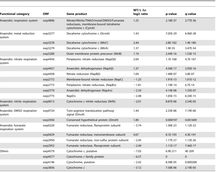
Table 3.Fur-responsive modules for anaerobic electron transport with fumarate as the EA.
Functional category ORF Gene product
WT/Dfur
log2 ratio p-value q-value
Anaerobic respiration system swp4806 Nitrate/Nitrite/TMAO/metal/DMSO/Fumarate reductases, membrane-bound tetraheme cytochromec(CymA)
1.25 2.18E-37 2.77E-36
Anaerobic metal reduction system
swp3277 Decaheme cytochromec(OcmA) 1.43 7.05E-29 6.96E-28
swp3278 Decaheme cytochromec(MtrC) 2.44 2.8E-182 1.4E-180
swp3279 Decaheme cytochromec(MtrA) 1.37 1.9E-55 3.47E-54
swp3280 Outer membrane protein precursor (MtrB) 1.19 2.44E-16 1.53E-15
Anaerobic nitrate respiration system
swp4456 Periplasmic nitrate reductase (NapDb) 2.04 1.1E-168 4.7E-167
swp4457 Anaerobic dehydrogenases (NapAb) 1.37 4.56E-17 2.95E-16
swp4458 Nitrate reductase (NapBb) 1.69 1.48E-07 3.8E-07
swp2772 Membrane-bound nitrate reductase (NapC) 21.25 1.91E-13 1.01E-12 swp2773 Periplasmic nitrate reductase, (NapBa) 21.81 1.19E-14 6.7E-14
swp2774 Anaerobic dehydrogenase (NapAa) 22.34 4.14E-08 1.55E-07
swp2775 NapDa 22.48 1.05E-15 6.34E-15
Anaerobic nitrite respiration system
swp0613 Cytochromecnitrite reductase (NrfA) 22.01 8.87E-06 2.54E-05 Anaerobic DMSO respiration
system
swp0724 Twin-arginine translocation pathway signal (DmsA)
1.44 2.33E-06 7.19E-06
swp3456 Conserved hypothetical protein (DmsH) 1.80 0.000747 0.001609
Anaerobic fumarate respiration system
swp0220 Fumarate reductase, flavoprotein subunit 23.74 1.36E-23 1.12E-22 swp0429 Fumarate reductase, transmembrane subunit 4.67 8.1E-193 4.3E-191
swp2950 Fumarate reductase, iron-sulfur protein subunit 22.45 1.17E-27 1.12E-26 swp2952 Fumarate reductase, flavoprotein subunit 22.49 1.11E-17 7.46E-17
Others swp4579 Cytochromec, putative 27.03 6.9E-211 4E-209
swp4577 Cytochromecfamily protein 26.57 0 0
swp4146 Cytochrome, putative 22.42 8.38E-05 0.000208
swp3856 Cytochromec 23.12 7.58E-06 2.19E-05
doi:10.1371/journal.pone.0075588.t003
Figure 4. Fur binding to selected promoters (omcA, Crp-like regulator gene and napD) by EMSA. The binding assays were performed in the presence of 200 pmol Fur (lanes 2–4, lanes 6–8, lanes 10–12) and 0.2 pmol DIG-labeled (lanes 1–12) promoter DNA. Non-specific competitor DNA (50mg/ml Salmon Sperm DNA) was used in all these binding reactions to control for the presence of unspecific binding. Specific competitors (2 pmol and 20 pmol unlabeled probes) were added respectively in lane 3, 4; lane 7, 8; lane 11, 12 to verify the specificity of a band resulting from protein-binding to the labeled probe.
Supporting Information
Figure S1 Anaerobic incubation of the WT WP3 andfur
mutant strains on a CAS screening plate.
(TIF)
Figure S2 The identification of a predicted consensus of the Fur-binding motif in WP3 using the web-based tool RegPredict (http://regpredict.lbl.gov). A sequence logo repre-sentation of a palindromic-motif model was derived based on those sites located upstream of the genes listed in Table S3. The error bars indicate the standard deviations of the sequence conservation. (TIF)
Figure S3 The heme biosynthesis pathway in WP3.The pathway begins from L-Glutamate and proceeds through the formation of porphobilinogen, hydroxymethylbilane, and uropor-phyrinogen III to coproporuropor-phyrinogen III, aided by five distinct enzymes (HemA-HemE). Next, HemF catalyzes the conversion of coproporphyrinogen III to protoporphyrinogen IX, and HemK catalyzes the subsequent formation of protoporphyrin IX. Lastly, heme is formed by HemH. The genes exhibiting attenuated expression in thefurmutant are highlighted in grey. Adapted from the KEGG database (http://www.genome.jp/kegg).
(TIF)
Figure S4 Gene transcription levels of the 10 (CcmABC-DEFGH, DsbA and DsbD) components involved in the
cytochromecmaturation system in the WT WP3 andfur
mutant strains under anaerobic conditions using fuma-rate as an EA.The ATP-hydrolyzing CcmA subunits (swp2043) and their membrane integral partners CcmB (swp2042), CcmC (swp2041), and CcmD (swp2040) load heme onto the heme
chaperone CcmE (swp2039). Meanwhile, apocytochrome c (apocyt c) translocates through the secretion system (signal sequence cleavage) and is oxidized by DsbA (swp2175). The electron transport complex (DsbD, swp 4520, and CcmG, swp2047) then reduces the disulfide bond of apocyt c. Lastly, the CcmF (swp2046) and CcmH (swp2048) complex ligate heme to apocyt c, and holocytochrome c is produced. The transcription level of WT WP3 was set as 1. The WP3pepNgene was used to normalize the RNA concentration of each sample. The data shown represent 3 independent experiments, and the error bars indicate standard deviations.
(TIF)
Figure S5 Fur binding to target promoters of napD,
omcAand the Crp-like regulator gene.Fur binding to the
TonB receptor promoter and swp_1869 promoter (not predicted to be bound by Fur) were used as the positive and negative control, respectively. The DNA probe was pre-incubated with the purified Fur protein at the indicated molar ratios. The amount of DNA is 1 pmol, and 0, 4, 8, 16 pmol purified His tag fusion Fur were used in the DNA binding assays. The probes remained unbound in the absence of Fur binding, and reduced mobility was observed with increasing Fur concentration for all three of the Fur targets. (TIF)
Table S1 Bacterial strains and plasmids used in the present study.
(PDF)
Table S2 Primers used in this study. (PDF)
Table S3 Genes containing a putative Fur binding site in WP3. (XLSX)
Figure 5. A model of the Fur regulatory system in WP3.In this model, Fur acts as both a direct and indirect regulator of iron homeostasis and anaerobic respiration. As a direct regulator, the Fur protein generally binds to a Fur Box region to down- or up-regulate the expression of genes. As an indirect regulator, the Fur protein represses an antisense non-coding regulatory RNA or controls secondary regulators, such as Crp, ArcA, the H-NS protein, and the TetR family regulator. These proteins are also capable of regulating genes in the anaerobic respiration system as well as those that encode iron using proteins. The genes regulated by Fur are involved in iron homeostasis, the anaerobic electron transport system, the heme biosynthesis and transport systems, and the CCM system. Direct regulation is depicted with a solid arrow, and indirect regulation is depicted with a dotted arrow.
Acknowledgments
We thank Huahua Jian and Ping Sun for assistance with Cytochromec
content measurement and data analysis.
Author Contributions
Conceived and designed the experiments: XY JX XX FW. Performed the experiments: XY. Analyzed the data: XY YH FW. Contributed reagents/ materials/analysis tools: XX FW JX. Wrote the paper: XY YH FW.
References
1. Andrews SC, Robinson AK, Rodriguez-Quinones F (2003) Bacterial iron homeostasis. FEMS Microbiol Rev 27: 215–237.
2. Touati D (2000) Iron and oxidative stress in bacteria. Arch Biochem Biophys 373: 1–6.
3. Wandersman C, Delepelaire P (2004) Bacterial iron sources: from siderophores to hemophores. Annual Review of Microbiology 58: 611–647.
4. Schroder I, Johnson E, de Vries S (2003) Microbial ferric iron reductases. FEMS Microbiol Rev 27: 427–447.
5. Cornelis P, Wei Q, Andrews SC, Vinckx T (2011) Iron homeostasis and management of oxidative stress response in bacteria. Metallomics 3: 540–549. 6. Delany I, Rappuoli R, Scarlato V (2004) Fur functions as an activator and as a
repressor of putative virulence genes inNeisseria meningitidis. Mol Microbiol 52: 1081–1090.
7. Nandal A, Huggins CC, Woodhall MR, McHugh J, Rodriguez-Quinones F, et al. (2010) Induction of the ferritin gene (ftnA) ofEscherichia coliby Fe(2+)-Fur is mediated by reversal of H-NS silencing and is RyhB independent. Mol Microbiol 75: 637–657.
8. Craig SA, Carpenter CD, Mey AR, Wyckoff EE, Payne SM (2011) Positive Regulation of theVibrio choleraePorin OmpT by Iron and Fur. J Bacteriol 193: 6505–6511.
9. Miles S, Carpenter BM, Gancz H, Merrell DS (2010)Helicobacter pyloriapo-Fur regulation appears unconserved across species. J Microbiol 48: 378–386. 10. Gaballa A, Antelmann H, Aguilar C, Khakh SK, Song KB, et al. (2008) The
Bacillus subtilisiron-sparing response is mediated by a Fur-regulated small RNA and three small, basic proteins. Proc Natl Acad Sci U S A 105: 11927–11932. 11. Masse E, Salvail H, Desnoyers G, Arguin M (2007) Small RNAs controlling iron
metabolism. Curr Opin Microbiol 10: 140–145.
12. Masse E, Vanderpool CK, Gottesman S (2005) Effect of RyhB small RNA on global iron use inEscherichia coli. J Bacteriol 187: 6962–6971.
13. Escolar L, Perez-Martin J, de Lorenzo V (1999) Opening the iron box: transcriptional metalloregulation by the Fur protein. J Bacteriol 181: 6223–6229. 14. Ratledge C, Dover LG (2000) Iron metabolism in pathogenic bacteria. Annual
Review of Microbiology 54: 881–941.
15. Yang Y, Harris DP, Luo F, Wu L, Parsons AB, et al. (2008) Characterization of theShewanella oneidensisFur gene: roles in iron and acid tolerance response. BMC Genomics 9 Suppl 1: S11.
16. Ellermeier JR, Slauch JM (2008) Fur regulates expression of theSalmonella
pathogenicity island 1 type III secretion system through HilD. J Bacteriol 190: 476–486.
17. Teixido L, Cortes P, Bigas A, Alvarez G, Barbe J, et al. (2010) Control by Fur of the nitrate respiration regulators NarP and NarL in Salmonella enterica. Int Microbiol 13: 33–39.
18. Wan XF, Verberkmoes NC, McCue LA, Stanek D, Connelly H, et al. (2004) Transcriptomic and proteomic characterization of the Fur modulon in the metal-reducing bacteriumShewanella oneidensis. J Bacteriol 186: 8385–8400. 19. Baichoo N, Wang T, Ye R, Helmann JD (2002) Global analysis of theBacillus
subtilisFur regulon and the iron starvation stimulon. Mol Microbiol 45: 1613– 1629.
20. Paustian ML, May BJ, Kapur V (2001)Pasteurella multocidagene expression in response to iron limitation. Infect Immun 69: 4109–4115.
21. Pellicer S, Gonzalez A, Peleato ML, Martinez JI, Fillat MF, et al. (2012) Site-directed mutagenesis and spectral studies suggest a putative role of FurA from
Anabaenasp. PCC 7120 as a heme sensor protein. FEBS J 279: 2231–2246. 22. Gonzalez A, Bes MT, Valladares A, Peleato ML, Fillat MF (2012) FurA is the
master regulator of iron homeostasis and modulates the expression of tetrapyrrole biosynthesis genes inAnabaenasp. PCC 7120. Environ Microbiol 14: 3175–3187.
23. Wang F, Wang J, Jian H, Zhang B, Li S, et al. (2008) Environmental adaptation: genomic analysis of the piezotolerant and psychrotolerant deep-sea iron reducing bacteriumShewanella piezotoleransWP3. PLoS One 3: e1937. 24. Venkateswaran K, Moser DP, Dollhopf ME, Lies DP, Saffarini DA, et al. (1999)
Polyphasic taxonomy of the genusShewanella and description of Shewanella oneidensissp. nov. Int J Syst Bacteriol 49 Pt 2: 705–724.
25. Fredrickson JK, Romine MF, Beliaev AS, Auchtung JM, Driscoll ME, et al. (2008) Towards environmental systems biology ofShewanella. Nature Reviews Microbiology 6: 592–603.
26. Meyer TE, Tsapin AI, Vandenberghe I, de Smet L, Frishman D, et al. (2004) Identification of 42 possible cytochrome c genes in the Shewanella oneidensis
genome and characterization of six soluble cytochromes. OMICS 8: 57–77. 27. Rodionov DA, Novichkov PS, Stavrovskaya ED, Rodionova IA, Li X, et al.
(2011) Comparative genomic reconstruction of transcriptional networks controlling central metabolism in theShewanella genus. BMC Genomics 12 Suppl 1: S3.
28. Myers CR, Nealson KH (1988) Bacterial manganese reduction and growth with manganese oxide as the sole electron acceptor. Science 240: 1319–1321.
29. Thompson DK, Beliaev AS, Giometti CS, Tollaksen SL, Khare T, et al. (2002) Transcriptional and proteomic analysis of a ferric uptake regulator (fur) mutant of Shewanella oneidensis: possible involvement of fur in energy metabolism, transcriptional regulation, and oxidative stress. Appl Environ Microbiol 68: 881– 892.
30. Wang F, Wang P, Chen M, Xiao X (2004) Isolation of extremophiles with the detection and retrieval ofShewanellastrains in deep-sea sediments from the west Pacific. Extremophiles 8: 165–168.
31. Chen S-Y, Ambe S, Takematsu N, Ambe F (1996) The Chemical States of Iron in Marine Sediments by Means of Mo¨ssbauer Spectroscopy in Combination with Chemical Leachings. J Oceanography 52: 705–715.
32. Xiao X, Wang P, Zeng X, Bartlett DH, Wang F (2007)Shewanella psychrophilasp. nov. and Shewanella piezotoleranssp. nov., isolated from west Pacific deep-sea sediment. Int J Syst Evol Microbiol 57: 60–65.
33. Wu W, Li B, Hu J, Li J, Wang F, et al. (2011) Iron reduction and magnetite biomineralization mediated by a deep-sea iron reducing bacteriumShewanella piezotoleransWP3. J Geophys Res 116: G04034.
34. Chen Y, Wang F, Xu J, Mehmood MA, Xiao X (2010) Physiological and evolutionary studies of NAP systems inShewanella piezotoleransWP3. ISME J 5: 843–855.
35. Schwyn B, Neilands JB (1987) Universal chemical assay for the detection and determination of siderophores. Anal Biochem 160: 47–56.
36. Fennessey CM, Jones ME, Taillefert M, DiChristina TJ (2010) Siderophores are not involved in Fe(III) solubilization during anaerobic Fe(III) respiration by
Shewanella oneidensisMR-1. Appl Environ Microbiol 76: 2425–2432.
37. Lovley DR, Holmes DE, Nevin KP (2004) Dissimilatory Fe(III) and Mn(IV) reduction. Adv Microb Physiol 49: 219–286.
38. Edwards RA, Keller LH, Schifferli DM (1998) Improved allelic exchange vectors and their use to analyze 987P fimbria gene expression. Gene 207: 149–157. 39. Li R, Li Y, Kristiansen K, Wang J (2008) SOAP: short oligonucleotide
alignment program. Bioinformatics 24: 713–714.
40. Wang LK, Feng ZX, Wang X, Wang XW, Zhang XG (2010) DEGseq: an R package for identifying differentially expressed genes from RNA-seq data. Bioinformatics 26: 136–138.
41. Novichkov PS, Rodionov DA, Stavrovskaya ED, Novichkova ES, Kazakov AE, et al. (2010) RegPredict: an integrated system for regulon inference in prokaryotes by comparative genomics approach. Nucleic Acids Res 38: W299–307.
42. Bender KS, Yen HC, Hemme CL, Yang Z, He Z, et al. (2007) Analysis of a ferric uptake regulator (Fur) mutant ofDesulfovibrio vulgarisHildenborough. Appl Environ Microbiol 73: 5389–5400.
43. Troxell B, Fink RC, Porwollik S, McClelland M, Hassan HM (2011) The Fur regulon in anaerobically grownSalmonella enterica sv.Typhimurium: identification of new Fur targets. BMC Microbiol 11: 236.
44. Parker D, Kennan RM, Myers GS, Paulsen IT, Rood JI (2005) Identification of aDichelobacter nodosusferric uptake regulator and determination of its regulatory targets. J Bacteriol 187: 366–375.
45. Gao H, Barua S, Liang Y, Wu L, Dong Y, et al. (2010) Impacts ofShewanella oneidensis c-type cytochromes on aerobic and anaerobic respiration. Microb Biotechnol 3: 455–466.
46. Shi L, Squier TC, Zachara JM, Fredrickson JK (2007) Respiration of metal (hydr)oxides by Shewanella and Geobacter: a key role for multihaem c-type cytochromes. Mol Microbiol 65: 12–20.
47. Postle K, Kadner RJ (2003) Touch and go: tying TonB to transport. Mol Microbiol 49: 869–882.
48. Kuehl CJ, Crosa JH (2010) The TonB energy transduction systems inVibrio
species. Future Microbiol 5: 1403–1412.
49. Beliaev AS, Klingeman DM, Klappenbach JA, Wu L, Romine MF, et al. (2005) Global transcriptome analysis ofShewanella oneidensisMR-1 exposed to different terminal electron acceptors. J Bacteriol 187: 7138–7145.
50. Masse E, Gottesman S (2002) A small RNA regulates the expression of genes involved in iron metabolism inEscherichia coli. Proc Natl Acad Sci U S A 99: 4620–4625.
51. Iuchi S, Lin EC (1988)arcA(dye), a global regulatory gene inEscherichia coli
mediating repression of enzymes in aerobic pathways. Proc Natl Acad Sci U S A 85: 1888–1892.
52. Evans MR, Fink RC, Vazquez-Torres A, Porwollik S, Jones-Carson J, et al. (2011) Analysis of the ArcA regulon in anaerobically grownSalmonella enterica sv.
Typhimurium. BMC Microbiol 11: 58.
53. Gao H, Wang X, Yang ZK, Palzkill T, Zhou J (2008) Probing regulon of ArcA inShewanella oneidensisMR-1 by integrated genomic analyses. BMC Genomics 9: 42.
55. Gralnick JA, Brown CT, Newman DK (2005) Anaerobic regulation by an atypical Arc system inShewanella oneidensis. Mol Microbiol 56: 1347–1357. 56. Gao H, Wang X, Yang ZK, Chen J, Liang Y, et al. (2010) Physiological roles of
ArcA, Crp, and EtrA and their interactive control on aerobic and anaerobic respiration inShewanella oneidensis. PLoS One 5: e15295.
57. Saffarini DA, Schultz R, Beliaev A (2003) Involvement of cyclic AMP (cAMP) and cAMP receptor protein in anaerobic respiration ofShewanella oneidensis. J Bacteriol 185: 3668–3671.
58. Yang Y, Harris DP, Luo F, Xiong W, Joachimiak M, et al. (2009) Snapshot of iron response inShewanella oneidensis by gene network reconstruction. BMC Genomics 10: 131.
59. Troxell B, Sikes ML, Fink RC, Vazquez-Torres A, Jones-Carson J, et al. (2011) Fur negatively regulates hns and is required for the expression of HilA and virulence inSalmonella enterica sv.Typhimurium. J Bacteriol 193: 497–505. 60. Myers JM, Myers CR (2000) Role of the tetraheme cytochrome CymA in
anaerobic electron transport in cells ofShewanella putrefaciensMR-1 with normal levels of menaquinone. J Bacteriol 182: 67–75.
61. Murphy JN, Saltikov CW (2007) The cymA gene, encoding a tetrahemec-type cytochrome, is required for arsenate respiration inShewanellaspecies. J Bacteriol 189: 2283–2290.
62. Maier TM, Myers JM, Myers CR (2003) Identification of the gene encoding the sole physiological fumarate reductase in Shewanella oneidensis MR-1. J Basic Microbiol 43: 312–327.
63. Cavallaro G, Decaria L, Rosato A (2008) Genome-based analysis of heme biosynthesis and uptake in prokaryotic systems. J Proteome Res 7: 4946–4954. 64. Wang LY, Brown L, Elliott M, Elliott T (1997) Regulation of heme biosynthesis inSalmonella typhimurium: activity of glutamyl-tRNA reductase (HemA) is greatly elevated during heme limitation by a mechanism which increases abundance of the protein. J Bacteriol 179: 2907–2914.
65. Sanders C, Turkarslan S, Lee DW, Daldal F (2010) Cytochromecbiogenesis: the Ccm system. Trends Microbiol 18: 266–274.
66. Sanders C, Turkarslan S, Lee DW, Onder O, Kranz RG, et al. (2008) The cytochrome c maturation components CcmF, CcmH, and CcmI form a membrane-integral multisubunit heme ligation complex. J Biol Chem 283: 29715–29722.
67. Shroff NP, Charania MA, Saffarini DA (2010) ArcB1, a Homolog ofEscherichia coliArcB, Regulates Dimethyl Sulfoxide Reduction inShewanella oneidensisMR-1. J Bacteriol 192: 3227–3230.
68. Orcutt BN, Sylvan JB, Knab NJ, Edwards KJ (2011) Microbial ecology of the dark ocean above, at, and below the seafloor. Microbiol Mol Biol Rev 75: 361– 422.
69. Spiro S, Guest JR (1990) FNR and its role in oxygen-regulated gene expression inEscherichia coli. FEMS Microbiol Rev 6: 399–428.