Major Article
Corresponding author:Dr. Ervina Rosmarwati.
e-mail: [email protected]
Received 8 February 2017
Accepted 10 August 2017
Strong renal expression of heat shock protein 70,
high mobility group box 1, inducible nitric oxide synthase,
and nitrotyrosine in mice model of severe malaria
Loeki Enggar Fitri
[1], Ervina Rosmarwati
[2], Yesita Rizky
[2], Niniek Budiarti
[3],
Nur Samsu
[4]and Karyono Mintaroem
[5][1]. Department of Parasitology, Faculty of Medicine, Universitas Brawijaya, Malang, Indonesia. [2]. Master Program in Biomedical Sciences, Faculty of Medicine, Universitas Brawijaya, Malang, Indonesia. [3]. Tropical Medicine Division, Internal Medicine Department, Faculty of Medicine, Universitas Brawijaya, dr. Saiful Anwar Public Hospital, Malang, Indonesia. [4]. Renal and Hypertension Division, Internal Medicine Department, Faculty of Medicine,
Universitas Brawijaya, dr Saiful Anwar Public Hospital, Malang, Indonesia. [5]. Department of Pathology Anatomy, Faculty of Medicine, Universitas Brawijaya, Malang, Indonesia.
Abstract
Introduction: Renal damage is a consequence of severe malaria, and is generally caused by sequestration of Plasmodium falciparum-infected erythrocytes in the renal microcirculation, which leads to obstruction, hypoxia, and ischemia. This triggers high mobility group box 1 (HMGB1) to send a danger signal through toll-like receptors 2 and 4. This signal up-regulates inducible nitric oxide (iNOS) and nitrotyrosine to re-perfuse the tissue, and also increases heat shock protein 70 (HSP70) expression. As no study has examined the involvement of intracellular secondary molecules in this setting, the present study compared the renal expressions of HSP70, HMGB1, iNOS, and nitrotyrosine between mice suffered from severe malaria and normal mice.
Methods: C57BL/6 mice were divided into an infected group (intraperitoneal injection of 106 P. berghei ANKA) and a non-infected group. Renal damage was evaluated using hematoxylin eosin staining, and immunohistochemistry was used to evaluate the expressions of HSP70, HMGB1, iNOS, and nitrotyrosine. Results: Signiicant inter-group differences were observed in the
renal expressions of HSP70, HMGB1, and iNOS (p=0.000, Mann-Whitney test), as well as nitrotyrosine (p=0.000, independent t test). The expressions of HSP70 and HMGB1 were strongly correlated (p=0.000, R=1.000). No correlations were observed between iNOS and HMGB, HMGB1 and nitrotyrosine, HSP70 and nitrotyrosine, or iNOS and nitrotyrosine. Conclusions: It appears that HMGB1, HSP70, iNOS, and nitrotyrosine play roles in the renal damage that is observed in mice with severe malaria. Only HSP70 expression is strongly correlated with the expression of HMGB1.
Keywords: HMGB1. HSP70. iNOS. Nitrotyrosine. Severe malaria.
INTRODUCTION
According to the World Health Organization, approximately 3.2 billion people were at-risk of malaria in 2015, with 214 million new cases in 106 countries. Among the new cases, approximately 88% were located in Africa, followed by Southeast Asia (10%) and the East Mediterranean (2%). There were approximately 438,000 deaths related to malaria in 2015, with 90% occurring in Africa, 7% occurring in Southeast Asia, and 2% occurring in East ekMediterranean1. As malaria is endemic in Indonesia, approximately 70% of the Indonesian population is at-risk and approximately 38,000 deaths are caused by malaria every year, with most occurring in east Indonesia (80%)2.
The most common cause of severe malaria is Plasmodium falciparum infection3, and acute kidney injury (AKI) is a common manifestation of severe malaria4. Infected erythrocytes express P. falciparum erythrocyte membrane protein 1 (PfEMP1) and P. falciparum histidine-rich protein (PfHRP), which play roles in knob formation. In addition, the infected erythrocytes easily adhere to endothelial cells using PfEMP1, which binds to endothelial adhesive factors, such as thrombospondin, intercellular adhesion molecule 1 (ICAM), vascular cell adhesion molecule (VCAM), and endothelial leukocyte adhesion molecule 1 (ELAM-1)5. In cases of severe malaria, the parasite also produces glycophosphatidylinositol (GPI), which is a toxin that induces a renal cytoadherence
process that leads to pro-inlammatory cytokine production in
the circulatory obstruction, hypoxia, and ischemia. It is also possible that the damage is mediated by immune complexes that are deposited in the glomerulus. Alternatively, the damage could be related to a hemodynamic disturbance that is caused by dehydration and hypovolemia, as well as alteration of the renal microcirculation8. Prolonged hypoxia generates alternate oxidative phosphorylation product coupling in the mitochondria and depletion of adenosine triphosphate (ATP) stores as adenosine is metabolized into inosine and hypoxanthine. The depletion of ATP subsequently leads to cellular edema, increased osmotic pressure, and cellular decompartementilization9,10, which induces high mobility group box 1 (HMGB1) to generate a danger signal through toll-like receptors 2 and 411. This signal
strongly promotes the production of inlammatory cytokines,
which increase renal stress and stimulate resident macrophages that can destroy renal tissue12.
During stressful and hypoxic conditions, the body activates inducible nitric oxide (iNOS) to catalyze the production of nitric oxide (NO) and peroxynitrite, which act as vasodilators and help re-perfuse the hypoxic tissue13. Peroxynitrite production can be measured using nitrotyrosine levels14. The body also increases the production of heat shock protein 70 (HSP70), which is a chaperone protein with many injury-related functions15, such
as repairing unfolded or mis-folded protein, anti-inlammatory
effects, stimulating regulatory T-cells, and stabilizing the cytoskeleton by repairing its organelle and architecture. In addition, HSP70 prevents further renal damage and restores renal function16. The present study aimed to compare the expressions of HSP70, HMGB1, iNOS, and nitrotyrosine in the renal tissue of mice that were and were not infected with P. berghei ANKA. This information will be useful in examining these molecules’ involvement in renal injury that is caused by severe malaria.
METHODS
Animals
The study’s protocol was approved by the ethical committee of our Faculty of Medicine (Universitas Brawijaya, Malang; 410/EC/KEPK/07/2014, date: July 10, 2014), and the study was performed between April 2014 and September 2015 in Parasitology Laboratory and Biomedical Laboratory Faculty of Medicine, Universitas Brawijaya, Malang. Eighteen female C57BL/6 mice (weight: 20-25g, age: 12-16 weeks) were purchased from the Eijkman Institute Jakarta. The mice were then divided into a non infected group (9 mice) and a group of 9 mice that were infected with 106 P. berghei ANKA in 0.2mL of blood via an intra-peritoneal injection.
Evaluating parasitemia
Parasitemia was evaluated daily until day 14 using thin
blood smears. A previous study revealed that pro-inlammatory mediators are increased at day 7 and that pro-inlammatory
hypercellularity peaks on days 14-1517. Infected erythrocytes were counted in a population of 1,000 erythrocytes using
microscopy (magniication: ×1,000), and the counted number
was converted into a percentage (%).
Hematoxylin and eosin staining
On day 14, the mice were anesthetized using chloroform and
then sacriiced before the kidneys were collected and preserved
in 10% formalin. Histopathology using hematoxylin and eosin staining were performed at the Pathology Anatomy Laboratory of Dr. Soetomo Hospital, Surabaya. The kidney specimens were dehydrated for 30 min at each step of an alcohol gradient: 30%, 50%, 70%, 85%, 95%, and twice at 100%. Clearing was performed twice using xylol for 1h. The specimens were
subsequently immersed twice in liquid parafin (1h at 42-46°C), blocked using solid parafin in a parafin cast, and incubated at 46-52°C for 1 day. Deparafinization was performed for 5 min
at each step of an alcohol gradient: 30%, 50%, 70%, 85%, 95%, and twice at 100%. The specimens were subsequently rinsed twice using H2O for 5 min, stained using hematoxylin and eosin, and then mounted using 5% gelatin.
Immunohistochemistry
The immunohistochemistry was performed in our Biomedical Laboratory (Faculty of Medicine, Universitas Brawijaya). The slides were incubated at 60oC for 1h, and then sequentially
treated using the following solutions: xylol (2× 10 min), absolute ethanol (2× 10 min), 90% ethanol (1× 5 min), 80% ethanol (1× 5 min), 70% ethanol (1× 5 min), and sterile distilled water (3× 5 min). Antigen retrieval was performed by heating the
slides in a water bath at 95ºC for 20 min, cooling the slides to room temperature for 20 min, and then soaking the slides in a citric buffer chamber (pH 6.0). The slides were subsequently
washed using phosphate-buffered saline [(PBS); 3× 2 min],
treated using 3% H2O2 in methanol, incubated for 15 min, and
then washed using PBS (3× 2 min). Blocking was performed
using a background sniper solution (15 min at room temperature),
and then the slides were washed using PBS (3× 2 min).
The antibodies that targeted NOS2 (N-20, sc-651), nitrotyrosine (39B6, sc-32757), and HMG-1 (J2E1, sc: 135809) were manufactured by Santa Cruz Biotechnology. The antibodies to HSP70 (2A4) were manufactured by Stress Marq Biosciences. The slides were incubated with the primary antibodies, PBS buffer, and 2% BSA overnight at 4oC. On the
next day, the slides were rinsed using PBS (3× 2 min), incubated
with secondary antibodies for 30 min at room temperature, and
then rinsed using PBS (3× 2 min). The slides were subsequently
incubated with horseradish peroxidase-conjugated streptavidin for 20 min at room temperature, and then rinsed using PBS
(3× 2 min) and then distilled water. Next, the slides were
incubated with diaminobenzidine (DAB) and DAB buffer (1:50) for 3-10 min at room temperature, and then washed using PBS
(3× 2 min) and distilled water (3× 2 min). Finally, the slides were
rinsed using a Mayer and tap water solution (1:10), incubated for 5-10 min at room temperature, rinsed using tap water, dried,
and observed under a microscope (magniication: ×1,000).
Histopathological examination
Renal damage can be histopathologically observed in the medulla and cortex as glomerulus necrosis, inflammatory
sclerosis. The expressions of HSP70, HMGB1, iNOS, and nitrotyrosine were measured by counting dark brown cells in
20 ields of view at ×1,000 magniication (quantitative scoring:
3+ to 4+). The evaluations were performed using a light microscope (Nikon Eclipse Ci), a calibrated camera (Optilab Plus 12 Megapixel), and Raster 3 software.
Statistical analysis
Normality was evaluated using the Kolmogorov-Smirnov test, and sample homogeneity was evaluated using the Levene test. Protein expressions were compared between the infected and non-infected groups using the Mann-Whitney and independent t tests. Spearman and Pearson correlation testing was used to evaluate correlations between the protein expressions. All data were analyzed using International Business Machines Statistical Package for the Social Sciences (IBM SPSS) software (version 22.0; IBM Corp., Chicago, IL), and differences were considered
statistically signiicant at a p-value of <0.05.
RESULTS
Figure 1 shows the degree of parasitemia over time and the parasites’ morphology. The infected group exhibited histopathological evidence of glomerular damage, including
glomerular necrosis, inlammatory cell iniltration, mesangial
proliferation, deposition of hemozoin pigment, and glomerular sclerosis (data not shown).
Immunohistochemical indings
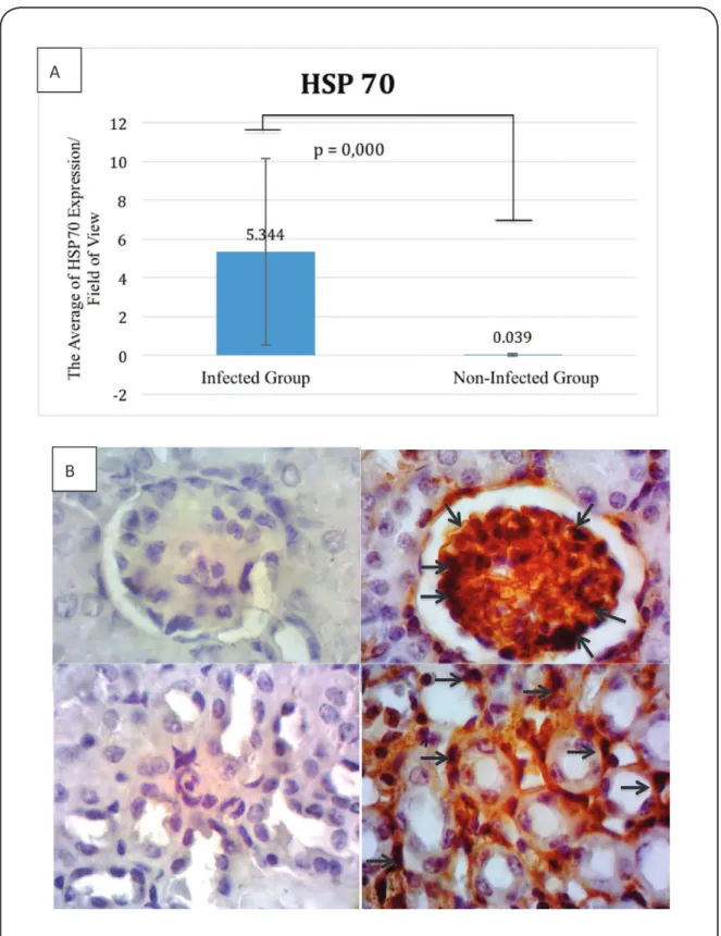
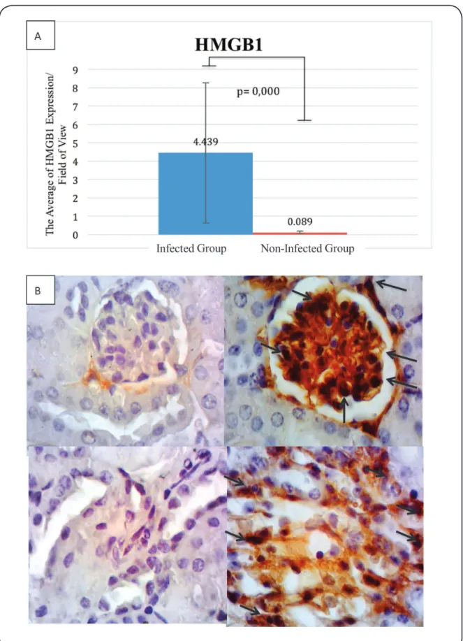
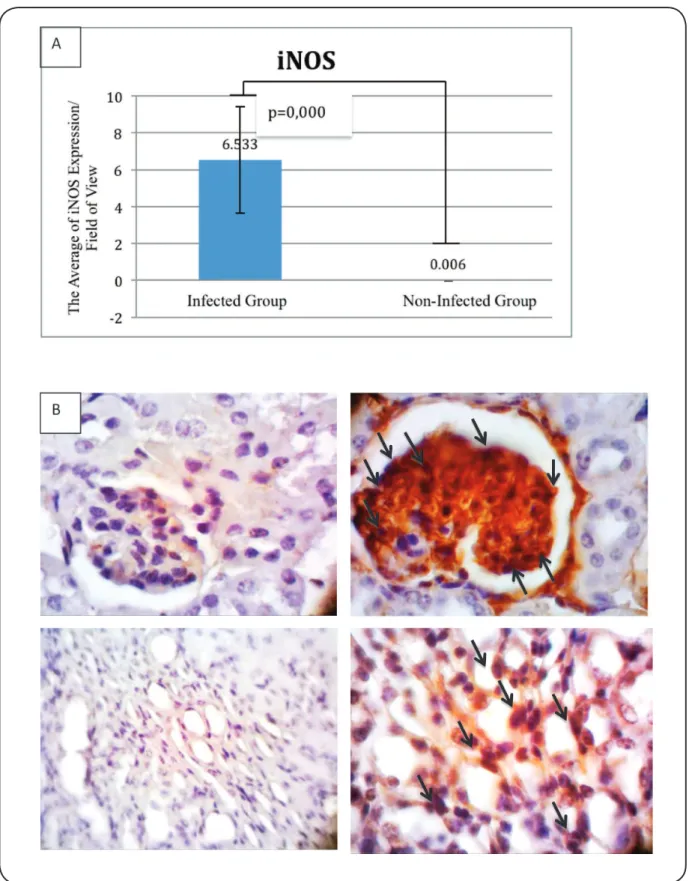
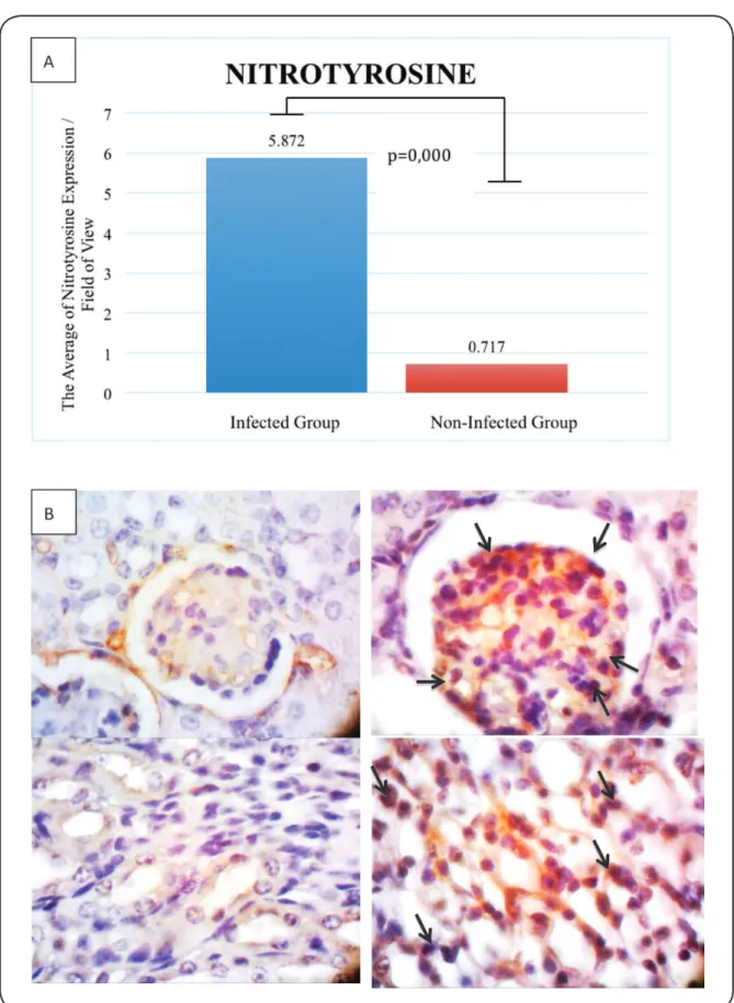
Expressions of HMGB1, HSP70, iNOS, and nitrotyrosine were predominantly observed in the cytoplasm of glomerular and tubular cells (Figure 2, Figure 3, Figure 4 and Figure 5). When the infected and non-infected groups were compared, the Mann-Whitney test revealed significant differences in the expressions of HSP70 (5.344 vs. 0.039; p=0.000), HMGB1 (4.439 vs. 0.089; p=0.000), and iNOS (6.533 vs. 0.006; p=0.000). In addition, the independent t test revealed a
signiicant difference in the expression of nitrotyrosine (5.872
vs. 0.717; p=0.000).
The Spearman correlation test results revealed a signiicant
correlation between the expressions of HSP70 and HMGB1 (p=0.000, R=1.000). However, there was no correlation between iNOS and degree of parasitemia (p=0.501, R=-0.259), nitrotyrosine and degree of parasitemia (p=0.189, R=0.482), HSP70 and degree of parasitemia (p=0.212, R=-0.461), HMGB1 and degree of parasitemia (p=0.212, R=0.461), iNOS and HMGB1 (p=0.244, R=-0.433), iNOS and HSP70 (p=0.244, R=-0.433), HMGB1 and nitrotyrosine (p=0.406, R=0.317), HSP70 and nitrotyrosine (p=0.406, R=0.317), or iNOS and nitrotyrosine (p=0.154, R=0.517).
DISCUSSION
The infected group exhibited a significant increase in parasitemia during day 5-10, which may have been related to parasite proliferation, and a subsequent decline starting on
day 12, which could relect the effects of the inlammatory process and immune response. However, the pro-inlammatory
hypercellularity failed to eliminate the parasite, and the
parasitemia subsequently increased signiicantly until day 14.
During this period, we detected histopathological evidence of glomerular necrosis, infiltration of polymorphonuclear and mononuclear cells, mesangial proliferation, hemozoin
deposition, and tubular edema. These indings are similar to the histopathological proile of acute renal failure caused by malaria,
which manifests as acute tubular necrosis, interstitial nephritis, and glomerulonephritis18. Furthermore, glomerulonephritis can be characterized by glomerular cell proliferation (e.g., mesangial, podocyte, and endothelial cells) as well as
changes in the basal membranes, inlammatory cell iniltration,
deposition of hemozoin, presence of infected erythrocytes in the glomerulus capillaries, necrosis, and deposition of immune complexes19. The glomerular cell proliferation may be related to the secondary immune response to cytokines that are released by the host in response to the infection20.
The present study revealed a signiicant difference in the
infected and non-infected groups’ expressions of HSP70, which is consistent with the theory that HSP70 expression is increased in cases of malaria. Interestingly, hemolysis is involved in the
pathogenesis of malaria, as Deuel et al. observed signiicant
increases in the genetic regulation of HSP70 and HMOX1 expression in proximal tubular cells after heme exposure21,22. In this context, the release of hemoglobin (Hb) causes endothelial dysfunction, oxidative vascular toxicity, and renal failure through several mechanisms23. First, Hb can rapidly convert into a heterodimer that can exit the blood vessels and enter various organs, including the kidneys. Second, Hb interacts with various non-oxygen ligands, such as NO and peroxides, which can lead to oxidative tissue damage. Third, ferritin Hb (Fe3+) is a product of the hemoglobin auto-oxidation reaction or the reaction of Hb with endogenous oxidants, which can release free heme
and lead to lipid peroxidation and subsequently inlammation.
Furthermore, high concentrations of heme can trigger unfolded protein receptors24.
During normal physiological conditions, the amount of free hemoglobin is controlled by plasma protein and haptoglobin,
although only glomerular iltration can remove free hemoglobin
from the circulation during pathological conditions. This can lead to short-term exposure to high concentrations of free hemoglobin, which can subsequently lead to AKI25. The body uses HSP70 over-expression as a protective mechanism15, which can help protect the kidneys from ischemic injury16.
The present study also revealed a signiicant infection-related
difference in the expression of HMGB1, which can be passively released by ischemic and necrotic cells, and actively released by macrophages. Furthermore, the Plasmodium genome encodes two types of HMGB (HMGB1 and HMGB2), and the release of these proteins from red blood cells is suspected to contribute to the pathogenesis of AKI. For example, Wu et al. reported that HMGB1 expression was increased in AKI, and reportedly causes kidney damage through activation of the innate immune system26. In this context, HMGB1 triggers cellular signaling by binding to TLR4, which activates NFkB through a
FIGURE 2 - A: HSP70 expression (positive cell) per ield of view. B: The expression of HSP70 in the non-infected group (left, ×1,000) and
infected group (right, ×1,000). HSP70 was expressed by glomerular cells and tubular cells (dark brown cells, black arrow) in the infected
FIGURE 3 - A: HMGB1 expression (positive cells) per ield of view. B: The expression of HMGB1 in the non-infected group (left, ×1,000)
and infected group (right, ×1,000). HMGB1 was expressed by glomerular cells and tubular cells (dark brown cells, black arrows) in the
FIGURE 4 -A: iNOS expression (positive cells) per ield of view. B: The expression of iNOS in the non-infected group (left, ×1,000) and infected
group (right, ×1,000). iNOS was expressed by glomerular cells and tubular cells (dark brown cells, black arrows) in the infected group. iNOS:
FIGURE 5 - A: Nitrotyrosine expression (positive cells) per ield of view. B: The expression of nitrotyrosine in the non-infected group
(left, ×1,000) and infected group (right, ×1,000). Nitrotyrosine was expressed by glomerular cells and tubular cells (dark brown cells, black
by releasing pro-inlammatory cytokines and chemokines27. Moreover, HMGB1 directly induces prothrombin expression
on endothelial cells, which can contribute to the inlammatory
response and exacerbate any renal impairment12.
Spearman testing revealed a very strong correlation between the expressions of HSP70 and HMGB1 in the infected group, which suggests that these two variables were practically equal in the sample rank results. However, it is very common to have perfect correlation in a small sample size, as the Spearman test uses a rank system to evaluate correlation. During the
inlammatory process, HMGB1 expression is associated with
tissue damage (i.e., a damage-associated molecular protein) and
is inluenced by abiotic stressors, such as heat28. In addition, Mezayen et al. found that triggering expressed on myeloid cells-1 (TREM-1) is an HMGB-1 mediator that induces cytokine production, which suggests that HMGB-1 is released as an
endogenous danger signal that induces the pro-inlammatory
response of macrophages and leads to exacerbated kidney damage29. However, HSP70 expression increases in the presence of pathogenic stressors, such as heat, ischemia, and elevate
levels of pro-inlammatory cytokines that are induced by the
expression HMGB1. Thus, HSP70 can protect the kidneys from severe renal damage16 by interfering with macrophages’
pro-inlammatory response and the mis-folding of proteins30. In addition, expressions of HSP70 and HMGB1 are induced sequentially, although the mechanisms of their regulation and the relationship between these molecules remain unknown28.
The infected and non-infected group had significantly different expressions of iNOS, and Elias et al. also observed similar results in BALB/C mice that were infected with P. berghei. This difference may be related to the cytoadherence of erythrocytes that are infected with P. berghei, which leads to interactions between the parasite antigens and endothelial receptors17. For example, infected erythrocytes can bind to ICAM-1, which is upregulated when endothelial cells are exposed to oxidative stress and parasites, and plays an important role in the microvascular dysfunction that can lead to increased renal expression of iNOS31,32. Thus, Elias et al. concluded
that oxidative stress and pro-inlammatory cytokines play an
important role in the pathogenesis of acute renal failure that is caused by P. berghei infection17.
Although we detected a significant difference in the expression of nitrotyrosine, there is limited information regarding the expression of nitrotyrosine in models of Plasmodium infection. However, nitrotyrosine is a marker that is used to detect peroxynitrite (ONOO/ONOOH), which is mainly formed by the reaction of superoxide radicals with NO. Furthermore, reactive oxygen species (ROS) are produced during parasite vacuole digestion in infected erythrocytes, which can induce the rapid conversion of oxyhemoglobin into methemoglobin, as well as the production of H2O214,32.
We did not detect correlations between the expressions of iNOS and nitrotyrosine, or between their expressions and the degree of parasitemia (p>0.05). Elias et al. have suggested that parasitemia can be detected on day 3 and increases until day 14 or the last day of the study period17. However, no signiicant
change was observed in iNOS expression during days 3-12, although a general positive trend was observed during the study period. Thus, it appears that the maximum expression of iNOS and other markers occurs at approximately 3 days after Plasmodium infection. Although no exponential increases were observed in the expression values, it is also possible that the expressions will continue to increase, unlike the degree of parasitemia29. Furthermore, the degree of parasitemia is a weak predictor of malaria severity and mortality, as it is not related
to the levels of inlammation and organ damage. In contrast,
infected erythrocyte sequestration is a strong predictor of malaria severity and vital organ damage (e.g., the brain in cases of cerebral malaria). Moreover, erythrocyte sequestration and malaria severity are associated with microvascular congestion by infected and uninfected erythrocytes33.Another study also found that there was no association between the degree of peripheral parasitemia and the incidence of acute renal failure34.
Previous studies have indicated that NO and nitrotyrosine expression are related, although NO does not appear to be directly related to the expression of HSP70 or HMGB1. In this context, the direct interaction between ROS and NO produces nitrotyrosine and peroxynitrite, and the reaction of peroxynitrite with tyrosine can also generate nitrotyrosine. In addition, ROS can activate tyrosine residues to produce tyrosyl radicals that oxidize NO to produce nitrotyrosine. As nitrotyrosine can be formed by the reaction of tyrosine with other reactive molecules, the absence of a correlation between iNOS and nitrotyrosine is likely related to the multi-pathway production of peroxynitrite. However, the present study only considered two products of these pathways, and the dominant pathway remains unclear35.
In conclusion, the expressions of HMGB1, HSP70, iNOS, and nitrotyrosine were elevated in mice model of AKI that is caused by severe malaria. However, only HSP70 expression was strongly correlated with the expression of HMGB1, with the expression of HMGB1 preceded the increased expression
of HSP70. The design of the present study was not suficient to determine the causality of this relationship, and the indings
are also limited by the lack of a test for renal function and AKI severity. Moreover, there were no correlations between the degree of parasitemia and the expressions of HSP70, HMGB1, iNOS, or nitrotyrosine.
Acknowledgements
We thank the staff of the Parasitology Laboratory (Faculty of Medicine, Universitas Brawijaya) for housing the animals, and the staff of the Biomedical Laboratory (Faculty of Medicine, Universitas Brawijaya) for their invaluable assistance with the immunohistochemistry.
Conlicts of interest
The authors declare that they have no conlicts of interest .
Financial support
REFERENCES
1. World Health Organization. Fact Sheet on the World Malaria Report 2015. Geneva: WHO; 2015 [updated 2016 April 19; cited 2016 October 9]. Available from: http://www.who.int/malaria/media/ world-malaria-report-2015/en/
2. Kemenkes RI. Epidemiologi malaria di Indonesia. 1st edition. Jakarta: Ditjen P2PL; 2011. 13p.
3. World Health Organization. Severe Malaria. Trop Med Int Health. 2014;19(Suppl 1):7-131.
4. Koopmans LC, Wolfswinkel ME, Hesselink DA, Hoorn EJ, Koelewijn R, Hellemond JJ, et al. Acute kidney injury in imported Plasmodium falciparum malaria. Malar J. 2015;14:523.
5. Autino B, Corbett Y, Castelli F, Taramelli D. 2012. Review Articles: Pathogenesis of malaria in tissues and blood. Mediterr J Hematol Infect Dis. 2012;4(1):e2012061.
6. Ho M, White NJ. Invited Reviews: Molecular mechanism of cytoadherence in malaria. Amer J Physiol. 1999;276(6 Pt 1):C1231-42.
7. Mayor A, Haiz A, Bassat Q, Rovira-Vallbona E, Sanz S, Machevo S, et al. Association of Severe Malaria Outcomes with Platelet-Mediated Clumping and Adhesion to a Novel Host Receptor. PLoS One. 2011;6(4):e19422.
8. Das BS. Renal failure in malaria. J Vector Borne Dis. 2008;45(2): 83-97.
9. Chuanyiu L, Jackson RM. Reactive species mechanisms of cellular hypoxia- reoxygenation injury. Am J Physiol Cell Physiol. 2002;282(2):C227-41.
10. Percário S, Moreira DR, Gomes BA, Ferreira ME, Gonçalves AC, Laurindo PS, et al. Oxidative stress in malaria. Int J Mol Sci. 2012;13(12):16346-72.
11. Liu F, Zhang Y, Peng Z, Gao H, Xu L, Chen M. High expression of high mobility group box 1 (hmgb1) predicts poor prognosis for hepatocellular carcinoma after curative hepatectomy. J Transl Med. 2012;10:135.
12. Li J, Gong Q, Zhong S, Wang L, Guo H, Xiang Y, et al. Neutralization of the extracellular HMGB1 released by ischaemic damaged renal cells protects against renal ischemia–reperfusion injury. Nephrol Dial Transplant. 2010;26(2):469-78.
13. Chua CL, Brown G, Hamilton JA, Regerson S, Boeuf P. Monocyte and macrophages in malaria: protection or pathology. Trends in Parasitol. 2013;29(1):26-34.
14. Nathan C, Ding A. SnapShot: Reactive Oxygen Intermediates (ROI). Cell. 2010;140(6):951-951.e2.
15. Aufricht C. Heat-shock protein 70: molecular supertool? Paediatr Nephrol. 2005;20(6):707-13.
16. O’Neill S, Harrison EA, Ross JA, Wigmore SJ, Hughes J. Heat-shock proteins and acute ischaemic kidney injury. Nephron Exp Nephrol. 2014;126(4):167-74.
17. Elias RM, Correa-Costa M, Barreto CR, Silva RC, Hayashida CY,
Castoldi A, et al. Oxidative stress and modiication of renal vascular
permeability are associated with acute kidney injury during P. berghei ANKA Infection. PLoS One. 2012;7(8):e44004.
18. Klopleisch R. Multiparametric and semiquantitative scoring systems for the evaluation of mouse model histopathology - a systematic review. BMC Vet Res. 2013;9:123.
19. Nguansangiam S, Day NP, Hien TT, Mai NTH, Chaisri U, Riganti M, et al. A quantitative ultrastructural study of renal pathology in fatal Plasmodium falciparum malaria. Trop Med Int Health. 2007;12(9):1037-50.
20. Wichapoon B, Punsawad C, Chaisri U, Viriyavejakul P. Glomerular changes and alterations of zonula occludens-1 in the kidneys of Plasmodium falciparum malaria patients. Malaria J. 2014;13:176.
21. Deuel JW, Vallelian F, Schaer CA, Puglia M, Buehler PW, Schaer
DJ. Different target speciicities of haptoglobin and hemopexin deine a sequential protection system against vascular hemoglobin
toxicity. Free Radic Biol Med. 2015;89:931-43.
22. Deuel JW, Schaer CA, Boretti FS, Opitz L, Garcia-Rubio I, Baek JH, et al. Hemoglobinuria-related acute kidney injury is driven by intrarenal oxidative reactions triggering a heme toxicity response. Cell Death Dis. 2016;7(1):e2064.
23. Schaer DJ, Buehler PW, Alayash A, Belcher JD, Vercellotti GM. Hemolysis and free hemoglobin revisited: exploring hemoglobin and hemin scavengers as a novel class of therapeutic proteins. Blood. 2013;121(8):1276-84.
24. Vallelian F, Deuel JW, Opitz L, Schaer CA, Puglia M, Lönn M, et al. Proteasome inhibition and oxidative reactions disrupt cellular homeostasis during heme stress. Cell Death Differ. 2015;22(4):597-611.
25. Schaer DJ, Buehler PW, Cell-free hemoglobin and its scavenger proteins: new disease models leading the way to targeted therapies. Cold Spring Harb Perspect Med. 2013;3(6) :pii a013433.
26. Wu H, Ma J, Wang P, Corpuz TM, Panchapakesan U, Wyburn KR, et al. HMGB1 contributes to kidney ischemia reperfusion injury. J Am Soc Nephrol. 2010;21(11): 1878-90.
27. Briquet SB, Gissot C, Tissandié M, Sevilla E, Franetich E, Thierry JF, et al. High-mobility-group box nuclear factors of Plasmodium falciparum. Eukaryot Cell. 2006;5(4):672-82.
28. Norouzitallab P, Baruah K, Muthappa D, Bossier P. Non-lethal heat shock induces HSP70 and HMGB1 protein production sequentially to protect Artemia franciscana against Vibrio campbellii. Fish
Shellish Immunol. 2015;42(2):395-9.
29. Mezayen RE, El Gazzar M, Seeds MC, McCall CE, Dreskin SC, Nicolls MR. Endogenous signals released from necrotic cells
augment inlammatory responses to bacterial endotoxin. Immunol
Lett. 2007;111(1):36-44.
30. Kampinga HH, Hageman J, Vos MJ, Kubota H, Tanguay RM, Bruford EA, et al. Guidelines for the nomenclature of the human heat-shock proteins. Cell Stress Chaperones. 2009;14(1):105-11.
31. Chakravorty SJ, Craig A. The role of ICAM-1 in Plasmodium falciparum cytoadherence. Eur J Cell Biol. 2005;84(1):15-27.
32. Nathan C. 1992. Nitric oxide as a secretory product of mammalian cells. FASEB J. 1992;6(12):3051-64.
33. Ponsford MJ, Medana IM, Prapansilp P, Hien TT, Lee SJ, Dondorp AM, et al. Sequestration and Microvascular Congestion Are Associated with Coma in Human Cerebral Malaria. J Infect Dis. 2012;205(4):663-71.
34. Dondorp AM, Pongponratn E, White N.J. Reduced microcirculatory
low in severe falciparum malaria: pathophysiology and
electron-microscopic pathology. Acta Trop. 2004;89(3):309-17.
35. Zhou XJ, Vaziri ND, Zhang J, Wang HW, Wang XQ. Association of
renal injury of nitric oxide deiciency in aged SHR: prevention by