348
Corresponding author: Dra. Roberta Vieira de Morais Bronzoni.
e-mail: [email protected]
Received 20 January 2016
Accepted 14 March 2016
Serological evidence of hantavirus infection in an
urban area in Mato Grosso State, Brazil
Carla Julia da Silva Pessoa Vieira
[1], David José Ferreira da Silva
[1], Eriana Serpa Barreto
[1],
Carlos Eduardo Hassegawa Siqueira
[2], Vivaldo Gomes da Costa
[3], Fábio José Lourenço
[1],
Marcos Lázaro Moreli
[3]and Roberta Vieira de Morais Bronzoni
[1][1]. Instituto de Ciências da Saúde, Universidade Federal de Mato Grosso, Sinop, Mato Grosso, Brasil. [2]. Laboratório Municipal de Análises Clínicas, Secretaria Municipal de Saúde de Sinop, Sinop, Mato Grosso, Brasil.
[3]. Instituto de Ciências da Saúde, Universidade Federal de Goiás, Jataí, Goiás, Brasil.
Abstract
Introduction: In Brazil, Mato Grosso (MT) has the highest number of hantavirus cardiopulmonary syndrome cases. Our
study aimed to identify anti-hantavirus antibodies in the sera of patients from Sinop, MT, presenting with acute febrile illness.
Methods: A retrospective analysis of data for 198 sera samples assessed using enzyme-linked immunosorbent assay (ELISA)
was conducted. Results: Immunoglobulins G (IgGs) against the hantavirus nucleoprotein were found in 13.6% of the tested sera. No sample had immunoglobulin M (IgM) antibodies to hantavirus. Seropositivity occurred mainly in female residents in urban areas who worked around the household. Conclusions: Our indings suggest circulation of hantavirus in Sinop.
Keywords: Zoonosis. Serosurvey study. Hantavirus.
Hantaviruses (genus Hantavirus, family Bunyaviridae) are emerging zoonotic viruses of great importance for public health. In South America, the rodents of the Sigmodontinae subfamily are reservoirs of these viruses. Some hantaviruses infect humans, causing either hemorrhagic fever with renal syndrome (HFRS) and/or hantavirus cardiopulmonary syndrome (HCPS)(1).
The irst cases of HCPS diagnosed in Brazil occurred in
1993. Since then, more than 1,920 cases of hantavirus have been
notiied in all Brazilian regions, with a case fatality rate of up to
39.5%. Mato Grosso (MT) is the leading state for HCPS cases
and accounts for 16.1% of the total notiied cases in Brazil. In
a previous study conducted in the municipality of Sinop, Mato Grosso State, we carried out an epidemiological surveillance for arboviruses in 198 sera samples from patients presenting with acute febrile illness and clinical-epidemiological diagnoses of dengue. The results showed 46 samples positive for arboviruses. However, it was intriguing that more than 70% of the patients did not have dengue or other arbovirus infections that were tested for(2). To address the possibility of hantavirus infection,
we investigated the presence of antibodies against the hantavirus nucleoprotein in these febrile patients.
Sinop is a municipality located 503km from the capital Cuiabá, in a geographical transition zone between a savannah
and rainforest. The city was founded in 1974 and currently has an estimated population of approximately 130,000 inhabitants with 17.1% of them living in rural areas. The economic activities include an intensive agriculture based on the grain production, animal husbandry, logging, and human services. All of the sera analyzed in this study were part of the serum bank of the Laboratory of Immunology and Molecular Biology of Universidade Federal de Mato Grosso (UFMT), Campus of Sinop, which have been previously described in detail(2). At the time of blood
collection, the patients illed out a questionnaire that contained
their epidemiological data, sex, age, occupation, address and region of birth, as well as information about any contact with rural or forested areas, and contact with rodents. These data were collected to analyze the risk factors associated with infection. The sera were tested with enzyme-linked immunosorbent assay (ELISA) for the presence of immunoglobulin M (IgM) and immunoglobulins G (IgG) antibodies to hantavirus, by using the
recombinant nucleocapsid (N) protein of the Araraquara virus
(ARAV), according to the description of Figueiredo et al.(3).
Samples with an optical density equal to or greater than the
average of the negative controls plus three standard deviations at a dilution of 1:400 were considered positives. The positive samples were titrated using serial dilutions (1:200 to 1:3,200) and were tested by using ELISA in order to determine serum
antibody titers to hantavirus. The Chi-square test was used to
compare the hantavirus antibody status according to sex and
age at a signiicance level of 0.05. Odds ratios corresponding to 95% conidence intervals were calculated for proportions.
Rev Soc Bras Med Trop 49(3):348-350, May-June, 2016 doi: 10.1590/0037-8682-0026-2016
349 Vieira CJSP et al. - Evidence of hantavirus infection in an urban area
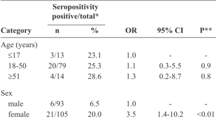
TABLE 1 - IgG seropositivity to hantavirus according to age and sex.
Seropositivity positive/total*
Category n % OR 95% CI P**
Age (years)
≤17 3/13 23.1 1.0 - -18-50 20/79 25.3 1.1 0.3-5.5 0.9
≥51 4/14 28.6 1.3 0.2-8.7 0.8
Sex
male 6/93 6.5 1.0 - -female 21/105 20.0 3.5 1.4-10.2 <0.01
IgG: immunoglobulin G; OR: odds ratio; 95% CI: 95% conidence interval. *The variables were analyzed by using only the available data. **p-values were determined via two-tailed chi-square or binomial test for two proportions.
TABLE 2 - Epidemiological data of hantavirus seropositive participants.
Positivity/participants*
Epidemiological data n %
Occupation
housewife 9/19 47.4 student 3/19 15.8 retired 1/19 5.3 rural worker 1/19 5.3 other occupations** 5/19 26.3 Home
rural 1/27 3.7
urban 26/27 96.3 Region of birth
Central West 6/16 37.5
North 1/16 6.3
South 4/16 25.0
Southeast 5/16 31.3 Contact with rodents
no 1/16 6.3
yes 15/16 93.8
*The variables were analyzed independently by using only the available data.
**Other occupations: receptionist, illing station attendant, woodworker,
contractor, and social worker.
Because of the missing information, the other epidemiological data (occupation, home, the region of birth, and contact with rodents) were analyzed only among the seropositive participants. The statistical analysis was carried out using the R v2.15.1(4).
Out of the 198 tested samples, 27 (13.6%) had IgG antibodies to hantavirus, at titers of 400 to 3,200. No samples had IgM antibodies to hantavirus. The study participants were between 5 and 64 years
old. Furthermore, no signiicant difference in the seropositivity
to hantavirus was observed among various age ranges (Table 1). A signiicant difference in the seropositivity was observed
between men (6.5%) and women (20%, p<0.01) (Table 1). Most of the seropositive participants were housewives (47.4%) and students (15.8%), and just one was a rural worker (5.3%) (Table 2). There was a higher incidence of infection in the seropositive participants living in urban areas (96.3%) than in those living in rural areas (3.7%). These participants are native to different regions of Brazil, including Central West (37.5%), Southeast (31.3%), South (25%), and North (6.3%) (Table 2). Fifteen (93.8%) participants reported a previous contact with rodents.
Our results showed a higher rate of seropositivity to hantavirus (13.6%) when compared with other serological studies conducted in all regions of Brazil by using the same methodology (0.6% to 4.7%) (5) (6) (7) (8) (9) (10). In this study, we
used ELISAs that were based on the recombinant N protein of ARAV, despite not having this virus described in MT yet. We observed high antibodies titers in the positive samples (1:400 to 1:3,200). In fact, the N protein induces a strong humoral immune response based on the epitopes that cross-react with members of the Hantavirus genus(3).
A high seropositivity rate was observed in three studies carried out in rural areas in MT (9.43%, 13%, and 51.1%)(11) (12) (13).
These studies reported an association between the infection and
agricultural activities. In our study, seropositivity was signiicantly associated with women living in urban areas. This inding is unique because previous studies observed a higher proportion
of male patients among the HCPS cases, who were probably infected with the hantavirus while performing general farm duties(1). The urbanization that occurred in the city could explain
why most seropositive patients were from an urban area. Over the last four decades, extensive native forest areas in Sinop have been deforested for agricultural purposes, especially for soybean and corn production, as well as for urbanization. As a result, there was a disordered growth of human settlements on the outskirts of the city, with the most susceptible people living close to the forests and in rural areas. The majority of the individuals that were analyzed have reported living in these new neighborhoods (data not shown) and having a previous contact with rodents. Therefore, it is possible that they had contact with the natural reservoirs of the hantavirus. In fact, deforestation promotes rodent proliferation, especially those prone to anthropogenic change, such as the rodents belonging to Sigmodontinae, thus driving them to invade human dwellings(14) (15).Humans are typically infected via contaminated
aerosolized secretions (feces, urine, and saliva) of the reservoir animals. Considering that 47.4% of the seropositive participants were housewives, primarily responsible for household tasks, it is possible that the practice of activities that allow the dispersion of
aerosols or dust, such as cleaning in the domestic environment, has been the main form of contamination for these women. These
activities were reported as risk factors for acquiring the disease(16).
350
Rev Soc Bras Med Trop 49(3):348-350, May-June, 2016
asymptomatic hantavirus infections than clinical. The severity of hantavirus infections can be associated with the virus type involved as well as with the different roles of the pathogenesis(15).
The Castelo dos Sonhos virus and Laguna Negra virus are associated with HCPS in both Pará and MT(13). There are no
data regarding the strains circulating in Sinop. However, it is possible that less virulent strains of hantavirus have been causing
the disease. Further studies are required to identify hantavirus
species and rodent reservoirs in Sinop.
An additional explanation for the seropositivity found in our study could be related to migration. Over the past few decades, Sinop has received a large number of migrants from other parts of the country. Most of the participants, who tested positive in this serosurvey, have migrated from the South (25%) and Southeast (31.3%) regions of Brazil. These two regions together
account for more than two-thirds of the HCPS notiications from
the country. Therefore, the participants may have been infected with the virus while they were living in those regions. This was also observed in a previous study carried out in MT(12).
Although the seroprevalence of hantavirus is high, to date, there have been no hantavirus cases reported in Sinop. Over the
past decade, the Sinop Municipal Secretary of Health notiied 26
allochthonous cases of HCPS. These cases occurred in patients living in the surrounding municipalities. It is possible that most of the infections in Sinop, especially those manifesting as mild forms of the disease, might remain undiagnosed. In dengue-endemic areas, such as Sinop, fever due to other arboviruses or even hantaviruses can be misdiagnosed as dengue based only on clinical-epidemiological data(2) (8). Taking this into account, it is
important to screen for hantavirus in acute febrile illness patients
using rapid and sensitive diagnostic methods. Our indings suggest
hantavirus circulation in Sinop and emphasize the need to expand the current diagnostic criteria and surveillance programs to improve the understanding of hantavirus dissemination in the region.
ETHICAL CONSIDERATIONS
The study was approved by the Ethical Committee for Human Research of Júlio Müller University Hospital - UFMT (266.568/2013).
Acknowledgments
The authors gratefully acknowledge Raquel de Souza Ferreira from the Sinop Municipal Department of Health.
Conlict of interest
The authors declare that there is no conlict of interest.
Financial Support
Conselho Nacional de Desenvolvimento Cientíico e Tecnológico (CNPq) research grant nº 478876/2010-6.
REFERENCES
1. Figueiredo LT, Souza WM, Ferrés M, Enria DA. Hantaviruses and cardiopulmonary syndrome in South America. Virus Res 2014; 187:43-54.
2. Vieira CJ, Silva DJ, Barreto ES, Siqueira CE, Colombo TE, Ozanic K, et al. Detection of Mayaro virus infections during a dengue outbreak in Mato Grosso, Brazil. Acta Trop 2015; 147:12-16. 3. Figueiredo LTM, Moreli ML, Borges AA, Figueiredo GG, Badra
SJ, Bisordi I, et al. Evaluation of an enzyme-linked immunosorbent assay based on Araraquara virus recombinant nucleocapsid protein. Am J Trop Med Hyg 2009; 81:273-276.
4. R Core Team. R: A language and environment for statistical computing. R Foundation for Statistical Computing, Vienna, Austria. URL http://www.R-project.org/
5. Souza WM, Machado AM, Figueiredo LTM, Boff E. Serosurvey of hantavirus infection in humans in the border region between Brazil and Argentina. Rev Soc Bras Med Trop 2011; 44:131-135.
6. Pereira GW, Teixeira AM, Souza MS, Braga AD, Santos Júnior GS, Figueiredo GG, et al. Prevalence of serum antibodies to hantavirus in a rural population from the Southern State of Santa Catarina, Brazil. Rev Soc Bras Med Trop 2012; 45:117-119.
7. Badra SJ, Maia FGM, Figueiredo GG, Santos Júnior GS, Campos GM, Figueiredo LTM, et al. A retrospective serological survey of hantavirus infections in the county of Cássia dos Coqueiros, State of São Paulo, Brazil. Rev Soc Bras Med Trop 2012; 45:468-470. 8. Lima DM, Sabino-Santos Junior G, Oliveira ACA, Fontes RM,
Colares JKB, Araújo FMC, et al. Hantavirus infection in suspected dengue cases from State of Ceará, Brazil. Rev Soc Bras Med Trop 2011; 44:795-796.
9. Moreli ML, Costa VG, Pariz FR. A seroepidemiological survey of hantavirus in Ilheus county. Am J Virol 2012; 1:18-23.
10. Gimaque JBL, Bastos MS, Braga WSM, Oliveira CMC, Castilho MC, Figueiredo RMP, et al. Serological evidence of hantavirus infection in a rural and urban regions in the State of Amazonas, Brazil. Mem Inst Oswaldo Cruz 2012; 107:135-137.
11. Medeiros DBA, Rosa EST, Marques AAR, Simith DB, Carneiro AR, Chiang JO, et al. Circulation of hantaviruses in the inluence area of the Cuiabá-Santarém Highway. Mem Inst Oswaldo Cruz 2010; 105:665-671.
12. Santos IO, Figueiredo GG, Figueiredo LTM, Azevedo MRA, Novo NF, Vaz CAC. Serologic survey of hantavirus in a rural population from the northern State of Mato Grosso, Brazil. Rev Soc Bras Med Trop 2013; 46:30-33.
13. Terças AC, Atanaka dos Santos M, Pignatti MG, Espinosa MM, Via AV, Menegatti JA. Hantavirus pulmonary syndrome outbreak, Brazil, December 2009-January 2010. Emerg Infect Dis 2013; 19:1824-1827. 14. Pinto Junior VL, Hamidad AM, Albuquerque Filho DO, Santos
VM. Twenty years of hantavirus pulmonary syndrome in Brazil: a review of epidemiological and clinical aspects. J Infect Dev Ctries 2014; 8:137-142.