Cop
yright
© ABE&M t
odos os dir
eit
os r
eser
vados
.
1 Divisão de Endocrinologia,
Hospital Agamenon Magalhães, Sistema Único de Saúde (SUS), Ministério da Saúde, Universidade de Pernambuco (UPE), Centro de Osteoporose de Pernambuco, Recife, PE, Brasil
2 Centro de Pesquisa Médica,
Fundação Oswaldo Cruz (Fiocruz), Ministério da Saúde, Recife, PE, Brasil
Correspondence to:
Francisco Bandeira
Divisão de Endocrinologia, Hospital Agamenon Magalhães, UPE Estrada do Arraial, 2723 52021-380 − Recife, PE, Brasil [email protected]
Received on Feb/4/2009 Accepted on Oct/8/2009
Vitamin D deiciency and its
relationship with bone mineral
density among postmenopausal
women living in the tropics
Deiciência de vitamina D e sua relação com a densidade mineral óssea em mulheres na pós-menopausa residentes nos trópicos
Francisco Bandeira1, Luiz Griz1, Eduardo Freese2,
Daniela Castro Lima1, Ana Carolina Thé1, Erik Trovão Diniz1,
Thyciara Fontenele Marques1, Cynthia Salgado Lucena1
ABSTRACT
Objective: To determine vitamin D (25OHD) status and its relationship with bone mineral den-sity (BMD) in 93 postmenopausal women. Subjects and methods: Patients were distributed in two groups: Group 1 – 51 to 65 years (n = 45) and Group 2 – 66 to 84 years (n = 48); 25OHD and PTH serum were analyzed and a DXA scan of the lumbar spine (LS) and femoral neck (FN) were taken. Results: Mean ± SD of serum 25OHD levels were 80.6 ± 43.3 nmol/L (Group 1) and 63.7 ± 27.6 nmol/L (Group 2); 24% had 25OHD levels < 25 nmol/L and 43.7% < 50 nmol/L. The prevalence of vitamin D deiciency at the 62.5 nmol/L cutoff increased signiicantly with age. Patients with hypovitaminosis D had a lower BMD at the FN (0.738 ± 0.102 vs. 0.793 ± 0.115 g/ cm, p = 0.03) and had been postmenopausal for longer (21.0 ± 8.4 vs. 16.2 ± 8.4 years, p = 0.01).
Conclusion: We found a high prevalence of hypovitaminosis D in postmenopausal women. Age, years elapsed since menopause and low BMD in the FN were associated with deiciency.
Arq Bras Endocrinol Metab. 2010;54(2):227-32
Keywords
Vitamin D; osteoporosis; bone mineral density; PTH; hyperparathyroidism
RESUMO
Objetivo: Determinar o peril da vitamina D (25OHD) e sua relação com a densidade mineral óssea (DMO) em 93 mulheres na pós-menopausa. Sujeitos e métodos: As pacientes foram dis-tribuídas em dois grupos: grupo 1 – 51 a 65 anos (45 pacientes) e 2 – 66 a 84 anos (48 pacientes); foram analisados 25OHD e PTH séricos e realizou-se exame de DXA em coluna lombar (LS) e colo do fêmur (FN). Resultados: As médias ± desvio-padrão (DP) dos níveis de 25OHD foram 80,6 ± 43,3 nmol/L (grupo 1) e 63,7 ± 27,6 nmol/L (grupo 2); 24% tinham 25OHD < 25 nmol/L e 43,7%, < 50 nmol/L. A prevalência de deiciência de 25OHD, considerando 62,5 nmol/L como ponto de corte, aumentou signiicativamente com a idade. Pacientes com hipovitaminose D tinham uma menor DMO no FN (0,738 ± 0,102 vs. 0,793 ± 0,115 g/cm2, p = 0,03) e maior tempo
de pós-menopausa (21,0 ± 8,4 vs. 16,2 ± 8,4 anos, p = 0,01). Conclusão: Encontrou-se alta preva-lência de hipovitaminose D em mulheres na pós-menopausa. Idade, anos desde a menopausa e baixa DMO no FN estavam associados à deiciência. Arq Bras Endocrinol Metab. 2010;54(2):227-32
Descritores
Cop
yright
© ABE&M t
odos os dir
eit
os r
eser
vados
.
INTRODUCTION
I
n countries close to the Equator the ultraviolet ra-diation of the sun penetrates the ozone layer of the Earth’s stratosphere suficiently to permit the produc-tion of vitamin D by the skin throughout the year. It should be emphasized, however, that the very process of aging leads to a diminution in the skin’s ability to produce vitamin D because of the diminution in the amount of 7-dehydrocholesterol. A 70-year-old indivi-dual exposed to the same amount of ultraviolet sunrays manages to produce only 20% of the amount produced by a young person (1).Although severe vitamin D deiciency leading to rickets or osteomalacia is rare in Brazil, there is accu-mulating evidence of the frequent occurrence of sub-clinical vitamin D deiciency in several other countries, especially in the elderly. As a result, we ind secondary hyperparathyroidism, increased bone remodeling, a de-crease in bone mineral density (BMD), particularly in the proximal femur, and an increased risk of osteopo-rotic fractures, when compared with individuals consid-ered to have a suficient supply of vitamin D (2).
Some initial data suggested that at least 50 nmol/L would be needed to satisfactorily meet metabolic re-quirements, especially in elderly persons, since below this there would be a rise in serum parathyroid hormone (PTH) and increased bone remodeling (3). When these individuals were given a vitamin D supplement raising the serum 25-hydroxyvitamin D (25OHD) to values above 5 nmol/L, PTH levels fell by approximately 40%, and bone mass increased.
There is clearly no consensus regarding ideal serum levels of 25OHD, and there are data in the literature indicating 50, 62.5, 75, and even 92.5 nmol/L as the minimum level required (4-7). Levels below 37.5 nmol/L are regarded as representing a signiicant dei-ciency. In patients visiting an osteoporosis clinic, PTH levels clearly increase when the serum levels of 25OHD fall below 62.5 nmol/L and there is a signiicant in-crease in bone remodeling and loss of bone mass with levels even lower than 75 nmol/L (8).
In Brazil, a mainly tropical country, a proper amount of vitamin D in the population is to be expected. In two studies conducted in elderly patients living in the south-east of Brazil (latitude 20-30°S), a high prevalence of vitamin D deiciency was demonstrated (9-12). Thus the aim of this study was to evaluate the prevalence of vitamin D deiciency and its relationship with BMD in
postmenopausal women visiting an endocrine outpa-tient clinic in Recife, which is situated at 10 degrees of latitude south of the Equator.
SUBJECTS AND METHODS
This was a cross-sectional study, which involved ninety-three postmenopausal women evaluated on an outpa-tient basis at Agamenon Magalhães Hospital, Recife. The following inclusion criterion was applied: post-menopausal women visting our endocrine outpatient clinic for routine medical evaluation. The exclusion cri-teria adopted were as follows: patients on drugs, such as glucocorticoids and anticonvulsants, known to lead to bone loss; patients on hormone replacement therapy; and those with diseases that affect bone metabolism, contributing to osteoporosis, such as multiple my-eloma, diseases of the kidney or liver, malabsorption diseases, Paget’s disease, primary hyperparathyroidism and uncontrolled hypo- or hyperthyroidism.
Menopause was deined as the absence of menstrua-tion for at least 2 years in women over 45 years. For determining the diagnosis and extent of osteoporosis WHO criteria were used.
Patients were divided into two groups according to age: Group 1 – 51 to 65 years (n = 45) and Group 2 – 66 to 84 years (n = 48).
The study protocol was approved by the Ethics Committee of Agamenon Magalhães Hospital, Univer-sity of Pernambuco.
Vitamin D and PTH measurements
25-hydroxyvitamin D was measured in the serum by ra-dioimmunoassay (DiaSorin Inc., Stillwater, USA). The sensitivity of the assay, deined as the lowest value dif-ferent from zero, was 3.75 nmol/L and the interassay coeficient of variation was 5%. In our laboratory the ref-erence values for healthy premenopausal women range from 25.7 to 148 nmol/L, with a median of 57 nmol/L. PTH was measured by automated electrochemolumines-cence (Elecsys – Roche Diagnostics GmbH, Mannheim, Germany), with a detection limit of 1.2 pg/mL.
Determination of bone mineral density
tro-Cop
yright
© ABE&M t
odos os dir
eit
os r
eser
vados
.
chanter. The results are expressed in g/cm2, T-score.
We used a local database (reference population aged 20 to 29 years) to calculate the T-score. The mean (± SD) of normal values for women was 1.085 g/cm2 (
± 0.1) at the lumbar spine, 0.913 g/cm2 (
± 0.12) at the femo-ral neck and 0.316 g/cm2 (± 0.07) at the distal radius
and for men it was 1.188 g/cm2 (
± 0.12), 0.966 g/cm2
(± 0.13) and 0.332 g/cm2 (
± 0.08) at the lumbar spine, femoral neck and distal radius, respectively. The in vivo
precision error of the equipment employed in the study expressed in percentage coeficient of variation (%CV = SD + mean BMD of repeated measurements) was 0.9% for the lumbar spine on the anteroposterior view and 1.2% for the femoral neck.
Statistical analysis
The prevalences of vitamin D deiciency in different age groups were compared using the Mantel-Haenszel chi-square test. Comparison of BMD values in patients with and without vitamin D deiciency was made by analysis of variance (ANOVA) and the Wilcoxon test for two samples. P values < 0.05 were considered sta-tistically signiicant. The Pearson correlations between age and 25OHD and between PTH and 25OHD were also obtained.
RESULTS
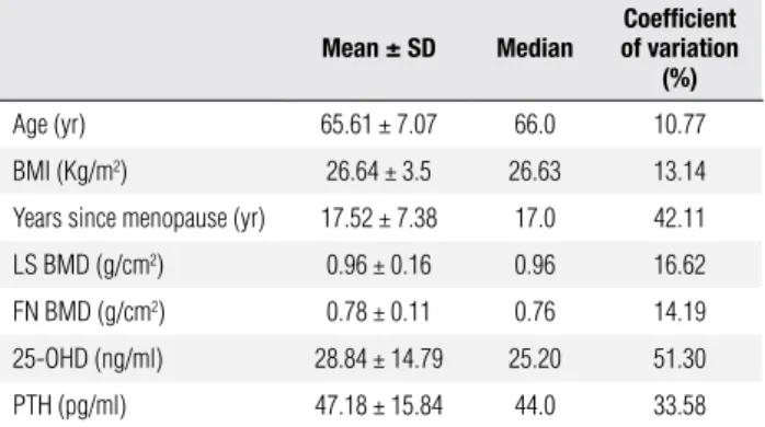
The mean ± SD age was 65.6 ± 7.07 years. Mean serum 25OHD was 71.9 ± 36.9 nmol/L (Table 1); for groups 1 and 2, these values were, respectively, 80.6 ± 43.3 nmol/L and 63.7 ± 27.6 nmol/L.
Table 1. Mean values of studied variables for 93 postmenopausal women
Mean ± SD Median
Coeficient of variation
(%)
Age (yr) 65.61 ± 7.07 66.0 10.77
BMI (Kg/m2) 26.64 ± 3.5 26.63 13.14
Years since menopause (yr) 17.52 ± 7.38 17.0 42.11
LS BMD (g/cm2) 0.96 ± 0.16 0.96 16.62
FN BMD (g/cm2) 0.78 ± 0.11 0.76 14.19
25-OHD (ng/ml) 28.84 ± 14.79 25.20 51.30
PTH (pg/ml) 47.18 ± 15.84 44.0 33.58
SD = standard deviation; BMI = body mass index; LS BMD = bone mineral density at lumbar spine; FN BMD = bone mineral density at femoral neck.
(95% CI = 45.2-66.4) above 62.5 nmol/L. Eight per cent had signiicant vitamin D deiciency (serum 25 OHD levels lower than 37.5 nmol/L).
With regard to age group, the prevalence of vitamin D deiciency was 30% in patients from 50 to 59 years old, 32.5% in patients from 60 to 69 years old, 54.5% in pa-tients from 70 to 79 years old, and 83% in papa-tients over the age of 80 (Figure 1).
Twenty-four percent (95% CI = 45.2-66.4) of the patients had levels below 50 nmol/L, 19.7% (95% CI = 12.2-29.4) between 50 and 62.5 nmol/L, and 56%
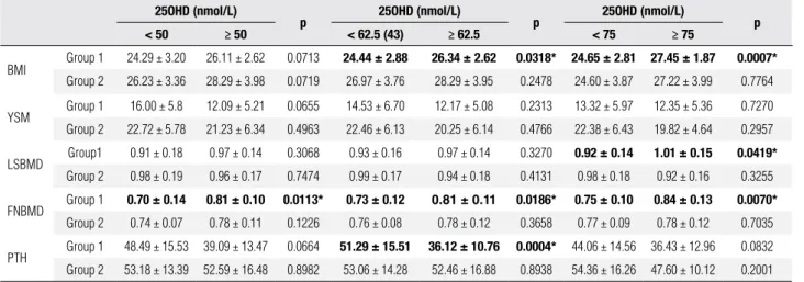
In Group 1 patients with serum 25OHD levels below 50 nmol/L we observed lower values for bone mineral density at the femoral neck (FNBMD) than in those whose 25OHD levels were above 50 nmol/L (0.70 ± 0.14 g/cm2vs. 0.81 ± 0.10 g/cm2; p = 0.01).
The difference remained signiicant when cutoffs of 62.5 nmol/L (0.73 ± 0.12 g/cm2vs. 0.81 ± 0.11 g/
cm2; p = 0.01) and 75 nmol/L (0.75 ± 0.10 g/cm2vs.
0.84 ± 0.13 g/cm2; p = 0.007) were used. Regarding
bone mineral density at the lumbar spine (LSBMD), using a cutoff of 75 nmol/L, we also observed signii-cant differences in the younger group 1 patients (0.92 ± 0.14 g/cm2vs 1.01 ± 0.15 g/cm2; p = 0.04) (Table 2).
A signiicant negative correlation was detected be-tween 25OHD and age (r = - 0.2310; p < 0.05) and between PTH and 25OHD (r = - 0.2899; p < 0.05).
Regarding the 62.5 nmol/L cutoff for determing whether a patient has vitamin D deiciency or in-suficiency, we found statistically signiicant differences in comparison with the patients with vitamin D levels greater than 62.5 nmol/L. Patients with vitamin D de-iciency were older (68.7 ± 8.8 vs 64.7 ± 7.1 years, p =
0.02), had been postmenopausal for longer (21.0 ± 8.4
vs 16.2 ± 8.4 years, p = 0.01), and had a lower bone density at the femoral neck (0.738 ± 0.102 vs 0.793 ±
0.115 g/cm, p = 0.03) (Table 3). Mean serum PTH was 52.9 ± 14.5 pg/mL in patients with 25OHD levels lower than 62.5 nmol/L and 39.7 ± 7.8 pg/mL in pa-tients with levels equal to or greater than 62.5 nmol/L, p = 0.002 (Figure 2).
Figure 1. Prevalence of vitamin D deficiency / insufficiency by age group.
0 10 20 30 40 50 60 70 80 90
50-59 60-69 70-79 80-89
Vitamin D deficiency (%)
Cop
yright
© ABE&M t
odos os dir
eit
os r
eser
vados
.
Table 3. Characteristics of the study patients according to serum levels of 25-hydroxivitamin D at the cut point of 62.5 nmol/L
Characteristics (mean ± SD)
25OHD < 62.5 nmol/L
25OHD
≥ 62.5 nmol/L p
Age (years) 68.7 ± 8.8 64.7 ± 7.1 0.02
BMI (kg/m2) 26.7 ± 4.3 26.3 ± 4.8 0.6
Years since menopause 21.0 ± 8.4 16.2 ± 8.4 0.01
LSBMD (g/cm2) 0.945 ± 0.183 0.957 ± 0.149 0.8
FNBMD (g/cm2) 0.738 ± 0.102 0.793 ± 0.115 0.03
PTH (pg/ml) 52.95 ± 14.5 39.7 ± 10.8 0.002
SD = standard deviation; BMI = body mass index; LSBMD = bone mineral density at lumbar spine; FNBMD = bone mineral density at femoral neck, PTH = parathyroid hormone.
Table 2. Differences in BMI, years since menopause, LSBMD, FNBMD and PTH among postmenopausal women according to age and various serum 25OHD cut points
25OHD (nmol/L)
p 25OHD (nmol/L) p 25OHD (nmol/L) p
< 50 ≥ 50 < 62.5 (43) ≥ 62.5 < 75 ≥ 75
BMI Group 1 24.29 ± 3.20 26.11 ± 2.62 0.0713 24.44 ± 2.88 26.34 ± 2.62 0.0318* 24.65 ± 2.81 27.45 ± 1.87 0.0007* Group 2 26.23 ± 3.36 28.29 ± 3.98 0.0719 26.97 ± 3.76 28.29 ± 3.95 0.2478 24.60 ± 3.87 27.22 ± 3.99 0.7764
YSM Group 1 16.00 ± 5.8 12.09 ± 5.21 0.0655 14.53 ± 6.70 12.17 ± 5.08 0.2313 13.32 ± 5.97 12.35 ± 5.36 0.7270 Group 2 22.72 ± 5.78 21.23 ± 6.34 0.4963 22.46 ± 6.13 20.25 ± 6.14 0.4766 22.38 ± 6.43 19.82 ± 4.64 0.2957
LSBMD Group1 0.91 ± 0.18 0.97 ± 0.14 0.3068 0.93 ± 0.16 0.97 ± 0.14 0.3270 0.92 ± 0.14 1.01 ± 0.15 0.0419* Group 2 0.98 ± 0.19 0.96 ± 0.17 0.7474 0.99 ± 0.17 0.94 ± 0.18 0.4131 0.98 ± 0.18 0.92 ± 0.16 0.3255
FNBMD Group 1 0.70 ± 0.14 0.81 ± 0.10 0.0113* 0.73 ± 0.12
0.81 ± 0.11 0.0186* 0.75 ± 0.10 0.84 ± 0.13 0.0070*
Group 2 0.74 ± 0.07 0.78 ± 0.11 0.1226 0.76 ± 0.08 0.78 ± 0.12 0.3658 0.77 ± 0.09 0.78 ± 0.12 0.7035
PTH Group 1 48.49 ± 15.53 39.09 ± 13.47 0.0664 51.29 ± 15.51 36.12 ± 10.76 0.0004* 44.06 ± 14.56 36.43 ± 12.96 0.0832 Group 2 53.18 ± 13.39 52.59 ± 16.48 0.8982 53.06 ± 14.28 52.46 ± 16.88 0.8938 54.36 ± 16.26 47.60 ± 10.12 0.2001
Group 1 = women 51-65 years; Group 2 = women 66-84 years; 25OHD = 25-hidroxi-vitamin D; BMI = body mass index; YSM = years since menopause; LSBMD = lumbar spine bone mineral density; FNBMD = femoral neck bone mineral density; PTH = parathyroid hormone.
* p < 0.05.
DISCUSSION
This was the irst study of vitamin D deiciency in post-menopausal women conducted in northeast Brazil. The other Brazilian studies were carried out in places more distant from the Equator, namely São Paulo, Minas Gerais and Rio Grande do Sul.
In a study of 250 elderly individuals from São Pau-lo (23ºS) with a mean age of 79 years, mean serum 25OHD was 19.8 ng/mL, and overall 57% of them showed values below 50 nmol/L (9). A study con-ducted in the same city with 177 inpatients (mean age 76.6 years) and 243 outpatients (mean age 79.1 years) showed a prevalence of hypovitaminosis D (25OHD < 50 nmol/L) of 71.2% and 43.8%, respectively (10). This prevalence is higher than that found in our study even when considering only the outpatients. Mean-while, two other studies (13,14) also conducted in São Paulo with young and elderly patients found mean 25OHD levels of 78.5 nmol/L and 77.4 nmol/L, re-spectively, only slightly above the mean found in this study (71.98 ± 36.91 nmol/L).
In Rio Grande do Sul (latitude 30°S) Premaor and cols. (11) evaluating 81 elderly inpatients found a preva-lence of hypovitaminosis D (25OHD < 50 nmol/L) of 77.8% (11), while Scalco and cols. (12) studying 102 in-stitutionalized elderly with mean age of 77.8 years dem-onstrated a vitamin D deiciency in 85.7% of them (12). In Minas Gerais (latitude 19°S) a study conducted by Silva and cols. (15) showed a vitamin D deiciency (25OHD < 80 nmol/L) in 42.4% of 180 patients with a mean age of 58.8 years, predominantly women (15).
Vitamin D has also been evaluated in other Latin American countries. In Buenos Aires (latitude 34°S), Argentina, 27% of women between 40 and 90 years old had serum vitamin D < 50 nmol/L in summer, which is similar to what was found in southeast Brazil in winter. Figure 2. Serum PTH according to 25-hydroxy-vitamin D (25OHD) levels.
0 10 20 30 40 50 60
< 62.5 ≥ 62.5
PTH (pg/mL)
Cop
yright
© ABE&M t
odos os dir
eit
os r
eser
vados
.
On the other hand, the prevalence increased to 71% in the winter months (16). Furthermore, only 2 of 40 postmenopausal women evaluated in Chile had hypo-vitaminosis D. However, a 25OHD level < 37 nmol/L was considered (17).
Our data showed a prevalence similar to that seen in the USA, but greater than that of Canada and the Scan-dinavian countries (4), reinforcing the idea that the abundant presence of sunlight may not prevent vitamin D deiciency in postmenopausal women. Moreover, the Brazilian diet is very deicient in vitamin D, the main source of which is fatty ish. In Canada and the Scandi-navian countries 25OHD levels are signiicantly higher than those of the patients in the present study. In those countries, despite the lower amount of sunlight there is a broader range of natural food sources and there is also fortiication of milk with vitamin D.
It is also important to bear in mind that in countries with an arid or semi-arid climate, with a very low amount of rainfall and therefore sunny weather throughout the year, vitamin D deiciency attains one of the highest prevalence rates on the whole planet (18,19). Moreover, the populations of such countries have speciic dietary habits and wear clothing that may explain these results. Even though the city of Recife has a humid tropical cli-mate (latitude 10°S), these data from arid and semi-arid regions also serve to strengthen the notion that, at least in postmenopausal women, the abundance of sunlight may not prevent vitamin D deiciency.
Although we have not performed any sun exposure evaluations, a recent study from Honolulu, Hawaii (lat-itude 21°N) demonstrated a high prevalence of vitamin D deiciency despite abundant sun exposure in a popu-lation of 93 adults over 18 years of age (20).
Our data also demonstrated that the mean of se-rum 25OHD levels was similar to that found in our postmenopausal patients who had primary asymptom-atic hyperparathyroidism (21) and was also no differ-ent from the levels reported in the North American patients in the MORE study (4).
As vitamin D deiciency may be asymptomatic, al-beit predisposing to a greater loss of bone and conse-quent increased risk of fractures, it is important that each region should attempt to establish the lowest limit of normality for serum 25OHD, deined as the level at which mean serum PTH levels begin to rise, char-acterizing secondary hyperparathyroidism (22). We found signiicant differences in the serum PTH lev-els up to the 62.5 nmol/L cutoff for serum 25OHD.
A Brazilian study, using the cutoff of 50 nmol/L, found a prevalence of secondary hyperparathyroidism of 54% in outpatients, showing a negative correlation between 25OHD and PTH (10). In another Brazilian study, in which a 25OHD cutoff of 80 nmol/L was used, the in-crease in PTH was signiicant and an inverse correlation between both measurements was also observed (15).
The report by Binkley and cols. (23) highlighted the importance of validation of circulating 25OHD assays in the user’s laboratory regardless of the manufacturer’s claims, as we did for our assay. They compared the re-sults of serum 25OHD measurement from samples of postmenopausal women referred to different laborato-ries. The DiaSorin RIA which we use in our laboratory demonstrated excellent results when compared with the HPLC standard method and has been very effec-tive in detecting endogenous 25OHD2 and 25OHD3 in human serum (23,24).
In conclusion, we found a high prevalence of vi-tamin D deiciency among healthy postmenopausal women who were seen for routine medical evaluation irrespective of age group. Patients with 25OHD lev-els lower than 62.5 nmol/L were signiicantly older, longer past the menopause, and had a signiicantly much lower BMD at the femoral neck and higher PTH levels.
Disclosure: no potential conlict of interest relevant to this article was reported.
REFERENCES
1. Holick M, Matsuoka LY, Wortsman J. Age, vitamin D, and solar ultraviolet radiation. Lancet. 1989;4:1104-5.
2. Sahota O, Masud T, San P. Vitamin D insuficiency increases bone turnover at the hip in patients with established vertebral osteopo-rosis. Clin Endocrinol (Oxf). 1999;51:217-21.
3. Rosen CJ, Morrison A, Zhou H. Elderly women in northern New England exhibit seasonal changes in bone mineral density and calciotropic hormones. Bone Miner. 1994; 25:83-92.
4. Lips P, Duong T, Oleksik A, Black DM, Cummings S, Cox D, et al. The Multiple Outcomes of Raloxifene Evaluation Study Group. A global study of vitamin D status and parathyroid function in postmenopausal women with osteoporosis: baseline data from the multiple outcomes of raloxifene evaluation clinical trial. J Clin Endocrionol Metab. 2001;86:1212-18.
5. Haden ST, Fuleihan GE, Agell JE, Cotran NM. Calcidiol and PTH levels in women attending an osteoporosis program. Calcif Tis-sue Int. 1999;64:275-79.
6. Chapuy MC, Preziosi P, Maaner P, Delmas P. Prevalence of vita-min D insuficiency in an adult normal population. Osteoporos Int. 1997;7:439-43.
Cop
yright
© ABE&M t
odos os dir
eit
os r
eser
vados
.
8. Wagman RB, Marcus M. Beyond bone mineral density – navi-gating the laboratory assessment of patients with osteoporosis. J Clin Endocrinol Metab. 2002;87:4429-30.
9. Saraiva GL, Cendoroglo MS, Ramos LR, Araújo LM, Vieira JG, Ku-nii I, et al. Inluence of ultraviolet radiation on the production of 25 hydroxyvitamin D in the elderly population in the city of São Paulo (23º 34’S) Brazil. Osteoporos Int. 2005;16:1649-54. 10. Saraiva GL, Cendoroglo MS, Ramos LR, Araújo LMQ, Vieira JGH,
Maeda SS, et al. Prevalência da deiciência, insuiciência de vita-mina D e hiperparatiroidismo secundário em idosos institucio-nalizados e moradores na comunidade da cidade de São Paulo, Brasil. Arq Bras Endocrinol Metab. 2007;51(3):437-42.
11. Premaor MO, Alves GV, Crossetti LB, Furlanetto TW. Hyperpara-thyroidism secondary to hypovitaminosis D in hypoalbuminemic is less intense than in normoalbuminemic patients: a prevalence study in medical inpatients in southern Brazil. Endocrine. 2004;24(1):47-53. 12. Scalco R, Premaor MO, Fröehlich PE, Furlanetto TW. High preva-lence of hypovitaminosis D and secondary hyperparathyroidism in the elderly living in nonproit homes in South Brazil. Endocri-ne. 2008;33(1):95-100.
13. Maeda SSK, Lazaretti-Castro M. Inluência sazonal sobre as con-centrações de 25-hidroxivitamina D em população idosa ativa na cidade de São Paulo. Arq Bras Endocrinol Metab. 2003;48:503. 14. Maeda SSK, Hayashi L, Pereira RL, Lazaretti-Castro M. Inluência
dos aspectos ocupacionais e da sazonalidade nas concentrações de 25-hidroxivitamina D em população jovem saudável da cidade de São Paulo. Arq Bras Endocrinol Metab. 2003;48:501.
15. Silva BC, Camargos BM, Fujii JB, Dias EP, Soares MM. Prevalên-cia de deiciênPrevalên-cia e insuiciênPrevalên-cia de vitamina D e sua correlação
com PTH, marcadores de remodelação óssea e densidade mine-ral óssea, em pacientes ambulatoriais. Arq Bras Endocrinol Me-tabol. 2008;52(3):482-8.
16. Fradinger EE, Zanchetta JR. Vitamin D status in women living in Buenos Aires. Medicina (B Aires). 1999;59:449-52.
17. Rodríguez Portales JA. Hipovitaminosis D en mujeres postme-nopáusicas con masa ósea baja en la región metropolitana. Rev Med Chile. 2001;129:849-52.
18. Alagol F, Shihadeh Y, Boztepe H, Tanakol R. Sunlight exposure and vitamin D deiciency in Turkish women. J Endocrinol Invest. 2000;23:173-77.
19. Ganage-Yared MH, Chemali R, Yaacoub N, Asmar A. Hypovitami-nosis D in a sunny country: relation to lifestyle and bone markers. J Bone Miner Res. 2000;15:1856-62.
20. Binkley N, Novotny R, Krueger D, Kawahara T, Daida YG, Lens-meyer G, et al. Low Vitamin D Status despite Abundant Sun Ex-posure. J Clin Endocrinol Metab. 2007;92:2130-35.
21. Bandeira F, Caldas G, Freese E, Griz L, Faria M, Bandeira C. Rela-tionship between serum vitamin D status and clinical manifesta-tions of primary hyperparathyroidism. Endocr Pract. 2002;8:266-70. 22. Thomas MK, Lloyd-Jones DM, Thadhani RI, Demay MB. Hypovita-minosis D in medical inpatients. N Engl J Med. 1998;338:777-83. 23. Binkley N, Krueger D, Cowgill CS, Plum L, Lake E, Hansen KE,
et al. Assay variation confounds the diagnosis of hypovita-minosis D: a call for standardization. J Clin Endocrinol Metab. 2004;89:3152-57.