Cop
yright
© ABE&M t
odos os dir
eit
os r
eser
vados
.
1 Unidade de Doenças Metabólicas Ósseas, Divisão de Endocrinologia, Departamento de Medicina, College of Physicians and Surgeons, Columbia University, Nova York, Estados Unidos
Correspondence to: John P. Bilezikian Division of Endocrinology Department of Medicine College of Physicians and Surgeons
630 W. 168th Street 10032 − New York, NY, United States
Received on Nov/20/2009 Accepted on Feb/28/2010
Hypoparathyroidism: clinical
features, skeletal microstructure and
parathyroid hormone replacement
Hipoparatiroidismo: aspectos clínicos, microestrutura esquelética e reposição do paratormônio
Mishaela R. Rubin1, John P. Bilezikian1
ABSTRACT
Objective: Hypoparathyroidism is a disorder in which parathyroid hormone is deicient in the circulation due most often to immunological destruction of the parathyroids or to their surgical removal. The objective of this work was to deine the abnormalities in skeletal microstructure as well as to establish the potential eficacy of PTH(1-84) replacement in this disorder. Subjects and methods: Standard histomorphometric and µCT analyses were performed on iliac crest bone biopsies obtained from patients with hypoparathyroidism. Participants were treated with PTH(1-84) for two years. Results: Bone density was increased and skeletal features relected the low turnover state with greater BV/TV, Tb. Wi and Ct. Wi as well as suppressed MS and BFR/BS as compared to controls. With PTH(1-84), bone turnover and bone mineral density increased in the lumbar spine. Requirements for calcium and vitamin D fell while serum and urinary calcium concentrations did not change. Conclusion: Abnormal microstructure of the skeleton in hypo-parathyroidism relects the absence of PTH. Replacement therapy with PTH has the potential to correct these abnormalities as well as to reduce the requirements for calcium and vitamin D. Arq Bras Endocrinol Metab. 2010;54(2):220-6
Keywords
Calcium; hypoparathyroidism; parathyroid hormone; vitamin D; bone density
RESUMO
Objetivo: O hipoparatiroidismo é uma doença em que há diminuição dos níveis circulantes do paratormônio, em geral, causada por destruição autoimune ou exerese cirúrgica. O objetivo deste estudo foi descrever as anormalidades microestrutrurais esqueléticas, como também o potencial terapêutico do uso do PTH(1-84). Sujeitos e métodos: Histomorfometria padrão e análise de micro-CT foram realizadas em biópsias de crista ilíaca de indivíduos com hipoparati-roidismo. Os participantes foram tratados com PTH(1-84) por dois anos. Resultados: A densida-de óssea aumentou e os achados esqueléticos reletiram o estado densida-de baixa remodensida-delação óssea com maior BV/TV, Tb Wi e CT Wi, como também supressão de MS e BFR/BS quando comparado com o grupo controle. Com o uso de PTH(1-84), a remodelação óssea aumentou e a densidade óssea aumentou na coluna lombar. As necessidades de cálcio e vitamina D diminuíram e a calciúria não mudou. Conclusão: A microestrutura esquelética anormal no hipoparatiroidismo relete a ausência do PTH. A terapia de reposição com PTH tem o potencial de reverter essas anormalidades, como também reduzir as necessidades de cálcio e vitamina D. Arq Bras Endocrinol Metab. 2010;54(2):220-6
Descritores
Cop
yright
© ABE&M t
odos os dir
eit
os r
eser
vados
.
H
ypoparathyroidism is a disorder of parathyroidhormone (PTH) deiciency caused by two main etiologies (1-4). Autoimmune destruction of the para-thyroid glands can occur as an isolated endocrine dei-ciency syndrome or in connection with failure of other endocrine glands. The other common etiology of hy-poparathyroidism is after neck surgery in which all pa-rathyroid tissue is removed, either in the context of sur-gery for primary hyperparathyroidism or after extensive neck surgery for thyroid cancer. The hypoparathyroi-dism that occurs after neck surgery does not always pre-sent itself soon, post-operatively, but rather can become apparent months to years thereafter. Although these two causes of hypoparathyroidism constitute the vast majo-rity of patients with this disorder, it can also occur as a congenital disorder in which the parathyroids and other
derivatives of the 3rd and 4th pharyngeal pouches do not
develop (DiGeorge syndrome). Very rarely, intracellular processing defects or activating mutations of the calcium sensing receptor can lead to hypoparathyroidism (5-7).
CLINICAL FEATURES
The clinical features of hypoparathyroidism, when symptomatic, can include any of the following: circum-oral numbness, paresthesias, carpal and pedal muscle spasms, laryngeal spasm, tetany and/or seizures (4). These symptoms are associated with the classical bio-chemical constellation of hypocalcemia and low levels of PTH. On the other hand, patients with hypopara-thyroidism can be asymptomatic, in which only the biochemical hallmarks of the disease are apparent. The combination of low serum calcium concentration and low or undetectable PTH levels essentially rules out all other causes of hypocalcemia (such as vitamin D deiciency, malabsorption syndrome, renal disease) in which the hypocalcemia is typically accompanied by normal compensatory physiological hyperactivity of the parathyroid gland axis and elevated PTH levels. Other biochemical abnormalities that often accompany the hypoparathyroid state include hyperphosphatemia, hy-percalciuria and reduced levels of 1,25-dihydroxyvita-min D, all consequences of PTH deiciency.
This article focuses upon our experience with a co-hort of subjects with well-documented hypoparathyroid-ism. Our study population was similar to the expected demographics of hypoparathyroidism with autoimmune and postoperative hypoparathyroidism constituting, by far, the two most common causes. Duration of
hypo-parathyroidism ranged from 3-45 years. Although none of the subjects had classic fragility fractures, 1/4 had reported mild fractures, mostly in small bones, during adulthood. Basal ganglia calciications were found on prior radiologic imaging in 4 subjects, although this as-pect of hypoparathyroidism was not speciically sought. Baseline biochemical values are shown in table 1. All subjects were receiving calcium and vitamin D.
We and others have shown (Table 2) that bone mass, by dual energy X-ray absorptiometry (DXA), is generally substantially higher in hypoparathyroidism than in age- and sex-matched controls (8-12). This observation con-trasts markedly with bone mass measurements by DXA in primary hyperparathyroidism in which bone density of the distal 1/3 radius, a site of cortical bone, is
typi-cally reduced (13).The general increase in bone mass in
hypoparathyroidism is associated with low levels of bone turnover as assessed by circulating biochemical markers and by dynamic histomorphometry (see below).
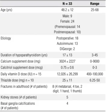
Table 1. Characteristics of patients with hypoparathyroidism
N = 33 Range
Age (yrs) 48.2 ± 12 25-68 Male: 9
Female: 24 (Premenopausal: 14 Postmenopausal: 10) Etiology Postoperative: 18
Autoimmune: 13 DiGeorge: 2
Duration of hypoparathyroidism (yrs) 17 ± 13 3-45 Calcium supplement dose (mg) 3024 ± 2227 0-9000 Calcitriol supplement dose (mcg) 0.75 ± 0.6 0-3 Daily vitamin D dose (IU) n = 15 12,026 ± 26,299 400-100,000 Thiazide dose (mg) n = 10 25 ± 11 6.25-50 Fractures in adulthood (# of patients) 8 (4 metatarsal, 4 toe, 2
digit, 1 hand, 1 thumb) Kidney stones (# of patients) 5 Basal ganglia calcifications
(# of patients)
4
Table 2. Baseline hypoparathyroid bone mineral density (mean ± SD). Each subject measured twice
Cohort range
Lumbar spine BMD T-score
1.21 ± 0.21 1.35 ± 1.88
(0.794, 2.028) (-2.541, 8.912) Femoral neck BMD
T-score
0.93 ± 0.17 0.51 ± 1.44
(0.554, 1.390) (-2.659, 4.370) Total hip BMD
T-score
1.06 ± 0.16 0.72 ± 1.22
(0.654, 1.455) (-2.362, 4.206) Distal 1/3 radius BMD
T-score
0.73 ± 0.06 0.04 ± 1.01
Cop
yright
© ABE&M t
odos os dir
eit
os r
eser
vados
.
3-dimensional analysis applying µCT to the same biop-sies. Appropriate age and sex-matched normal subjects without hypoparathyroidism served as controls. Hypo-parathyroid subjects had signiicantly greater
trabecu-lar bone volume (BV/TV: 26.98 ± 10 vs. 15.39 ± 4%
[mean ± SD]; p < 0.001), trabecular number (Tb.N:
1.831 ± 0.49 vs. 1.355 ± 0.24 mm-1; p < 0.001),
trabec-ular thickness (Tb.Th: 0.193 ± 0.05 vs. 0.148 ± 0.02 mm;
p < 0.001) and connectivity density (Conn.D: 15.899 ±
18.40 vs. 4.958 ± 2.02/mm3; p = 0.001) in comparison
to controls, while trabecular separation (Tb.Sp: 0.642
± 0.10 vs. 0.781 ± 0.13 mm; p < 0.001) and estimation
of the plate-rod characteristic (SMI: -0.457 ± 1.52 vs.
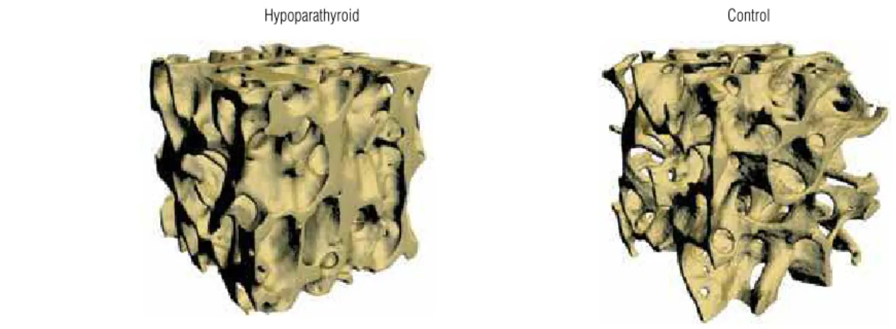
0.742 ± 0.51; p < 0.001) were signiicantly lower, the latter observation implying a more plate-like trabecular structure (Figure 1B). These indings support the data obtained by 2-dimensional histomorphometric analysis indicating that trabecular bone structure in hypopara-thyroidism is abnormal.
Skeletal microstructure in hypoparathyroidism
In order to obtain more information on bone quality in hypoparathyroidism, we have obtained transiliac bone biopsies on most of our patients (14-15). They were matched to age- and sex-matched control subjects who do not have any abnormalities in parathyroid function.
Histomorphometric assessment. Subjects with hypo-parathyroidism had greater trabecular bone volume
(mean ± SD; BV/TV: 23.5 ± 8 vs. 19.7 ± 5%, p = 0.02),
trabecular width (Tb.Wi: 136.1 ± 37 vs. 119.3 ± 21 μm,
p = 0.03), and cortical width (Ct.Wi: 923.4 ± 420 vs.
753.5 ± 246 μm, p = 0.05) than control subjects (Fig-ure 1A). Dynamic skeletal indices, including
mineral-izing surface (MS: 0.85 ± 1.58 vs. 4.27 ± 3.32%, p <
0.0001) and bone formation rate (BFR/BS: 0.006 ±
0.014 vs. 0.032 ± 0.028 μm3/μm2/d, p < 0.0001), were
profoundly suppressed in the hypoparathyroid subjects.
Microcomputed tomography (µCT). We extended these 2-dimensional histomorphometric analyses to a
Figure 1A. A representative photomicrograph of an iliac crest biopsy from a normal control (left) as compared to a hypoparathyroid subject (right).
Figure 1B. 3-D µCT image of iliac crest biopsy in a representative hypoparathyroid subject (left) and a normal control (right).
Normal Hypoparathyroid
Cop
yright
© ABE&M t
odos os dir
eit
os r
eser
vados
.
Material properties of bone in hypoparathyroidism
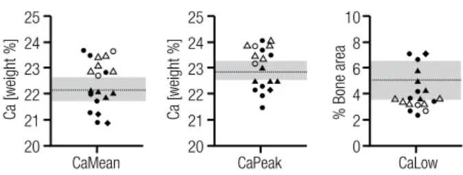
To understand material properties of the hypoparathy-roid skeleton, we subjected the transiliac biopsies to quantitative backscattered electron imaging (qBEI) and Fourier transform infrared imaging (FTIRI − Figures 2A and 2B). Bone mineralization density distribution (BMDD) outcomes as determined by qBEI showed a positive correlation with duration of the disease (Ca-Peak, r = 0.48, p = 0.04; CaHigh, r = 0.55, P = 0.02), as well as increased inter-individual variation (CV +92%) in the hypoparathyroid subjects. On average, the data in the hypoparathyroid individuals did not differ from nor-mal controls. In contrast, by FTIRI, the collagen cross-link ratio was signiicantly higher (p < 0.001) in the hypoparathyroid subjects as compared to normal. Col-lagen cross-link ratio correlated signiicantly with qBEI outcomes that were related to the amount of mineral, as characterized by BMDD parameters (CaMean, r = 0.69, p = 0.001; and CaPeak, r = 0.66, p = 0.002). These data are consistent with the low turnover state of these indi-viduals and suggest a skeleton that is in a low turnover state and has become mature, if not hypermature.
MANAGEMENT OF HYPOPARATHYROIDISM
Conventional management of hypoparathyroidism con-sists of oral calcium and 1,25-dihydroxyvitamin D in relatively large amounts. While this approach can usu-ally maintain a normal serum calcium level in these indi-viduals, it can also be a challenge. Often there are major swings in either direction leading to symptomatic hyper-calcemia or hypohyper-calcemia. Some patients demonstrate heightened sensitivity to small changes in the serum cal-cium even without overtly wide biochemical swings. In addition, the use of relatively high amounts of oral calci-um and vitamin D can lead to worsening hypercalciuria. This, in turn, can lead in some cases to nephrocalcinosis, nephrolithiasis or renal insuficiency (16-18).
A recent alternative therapeutic approach has be-come of interest in view of the availability of PTH for the treatment of osteoporosis. Since it is the parathy-roid hormone that is missing in this disorder, it seems most reasonable to consider this therapeutic approach. In fact, hypoparathyroidism is the sole remaining en-docrine deiciency disease for which the missing hor-mone is not available as a therapy. Moreover, reducing calcium and calcitriol requirements in hypoparathy-roidism with the use of PTH could also potentially lessen the risks of hypercalcemia and hypercalciuria. An additional possible advantage is that PTH use could reduce the risk of soft tissue deposition of cal-cium (nephrocalcinosis, nephrolithiasis and possibly in other soft tissues).
Teriparatide, a PTH molecule foreshortened to an amino terminus region [PTH (1-34)], has been shown to maintain serum calcium in the normal range and to reduce urinary calcium excretion in hypoparathyroid subjects (19-21). Use of the native, full length mol-ecule, PTH(1-84), has recently been investigated by us as another approach to the therapy of hypoparathyroid-ism. A dosing study was irst performed to determine how much and with what frequency PTH(1-84) could be used to raise bone turnover levels in a robust manner but not higher than upper limits of normality. In our preliminary dosing study, we observed that a regimen of 100 µg of PTH(1-84) administered every other day, met this goal better than daily or every third day use of 100 µg of PTH(1-84). The results of a 2-year study are summarized here (22).
Throughout the 2-year clinical trial, requirements for supplemental calcium fell signiicantly from 3.03 ± Figure 2A. Scatter plots of the five BMDD parameters measured in 19
hypoparathyroid patients. The patients are distinguished by categories, gender, and etiology. Grey band and dotted line in the background indicate SD and mean of the reference values.
25
24
23
22
21
20
Ca [weight %]
CaMean CaPeak CaLow
25
24
23
22
21
20
Ca [weight %] % Bone area
10
8
6
4
2
0 Female, postoperative
Female, autoimmune
Female, DiGeorge Male, postoperative
Male, autoimmune
Figure 2B. FTIRI measurements in biopsies from the hypoparathyroid patients (n = 19). Collagen cross link ratio (pyr/deH-DHLNL) were assessed in trabecular bone (HypoPT_collx) and compared with a normal reference population (NL_collx).
4
3
2
1
0
pyr/deH-DHLNL
Cop
yright
© ABE&M t
odos os dir
eit
os r
eser
vados
.
2.3 to 1.66 ± 1.3 g/day (p < 0.05; Figure 3A). The number of subjects on calcium supplementation that was greater than 1.5 g/d decreased from 22 (73%) at study entry to 12 (40%) at study conclusion. 1,25-di-hydroxyvitamin D supplementation declined from the baseline mean of 0.68 ± 0.5 to 0.40 ± 0.5 µg/day (p < 0.05; Figure 3B). The number of subjects on a dose of 1,25-dihydroxyvitamin D that was greater than 0.25 µg/d fell from 25 (83%) at study entry to 15 (50%) at study conclusion. The number of subjects who did not require any 1,25-dihydroxyvitamin D in-creased from 1 to 8. The reductions in the amounts of calcium and vitamin D supplementation were similar regardless of the etiology of the hypoparathyroidism. The number of subjects on hydrochlorothiazide for hy-percalciuria decreased from 10 at baseline to 3 at study conclusion. Those who were on thiazides did not differ from the group in terms of baseline calcium and cal-citriol supplementation.
SERUM AND URINARY CALCIUM LEVELS AND
OTHER INDICES OF MINERAL METABOLISM
Serum calcium concentration was maintained in the lower half of the normal range and during months 9 to 24 was not different from baseline values. Except for a minimal increase in serum calcium during the irst 6 months, serum calcium values did not differ from base-line levels. Of the many determinations of serum cal-cium, only 4% of all values were above the normal range with the highest value recorded in one subject once at 3.2 mmol/L. 24-hour urinary calcium excretion was generally unchanged.
Serum phosphate levels fell from 1.44 ± 0.2 to 1.29 ± 0.2 mmol/L (p < 0.05). 25-hydroxyvitamin D lev-els decreased signiicantly from baseline (150.0 ± 165 to 81.1 ± 45 nmol/L; p < 0.05). 1,25-dihydroxyvita-min D levels did not change (97.2 ± 52 to 111.2 ± 60 pmol/L). Total alkaline phosphatase activity in-creased signiicantly from 66 ± 18 U/L at baseline to 85 ± 22 U/L by month 3 and remained signiicantly above baseline at month 24 (73 ± 21 U/L; p < 0.05). The new steady state level of the alkaline phosphatase activity was well within normal limits. Magnesium fell minimally (0.74 ± 0.1 to 0.72 ± 0.1 mmol/L; p < 0.05). Serum creatinine did not change signiicantly (88.4 ± 796 to 79.6 ± 796 µmol/L; p = NS); nor did GFR as calculated by MDRD (74.9 ± 21 to 76.4 ± 20
mL/min/1.73 m2; p = NS).
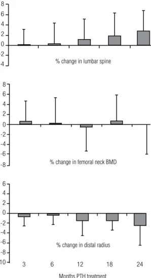
BONE MINERAL DENSITY
Lumbar spine BMD increased from baseline by 2.9 ±
4% (p < 0.05) from 1.24 ± 0.3 to 1.27 ± 0.3 g/cm2 (T
score: +1.7 ± 2 to +1.9 ± 2; Figure 4). Femoral neck BMD did not change signiicantly from baseline (+0.95
± 0.2 g/cm2; T score 0.7 ± 2; Figure 4), while the
dis-tal 1/3 radius BMD decreased by 2.4 ± 4% (p < 0.05)
(+0.72 ± 0.1 to 0.70 ± 0.1 g/cm2; T score -0.03 ± 2 to
-0.26 ± 1; Figure 4).
DISCUSSION
It is clear from our experience that PTH(1-84) repre-sents a promising approach to the management of pa-tients with hypoparathyroidism. Requirements for sup-plemental calcium and 1,25-dihydroxyvitamin D were signiicantly reduced while maintaining normal, stable serum calcium concentrations. In line with the actions Figure 3. Changes in Calcium (3A) and 1,25-dihydroxyvitamin D (3B)
Supplementation.Calcium requirements decreased at 3, 9, 12, 18 and 24 months from baseline, while 1,25-dihydroxyvitamin D requirements decreased by 1 month. Data are mean ± SD; * p < 0.05 as comparison to baseline.
3.5
3
2.5
2
1.5
1
0.5
0
Calcium supplementation (gm/d)
Months PTH treatment
0 1 3 6 9 12 18 24
*
*
* * *
0.8
0.7
0.6
0.5
0.4
0.3
0.2
0.1
0
Months PTH treatment
1,25-dihydroxyvitamin D Supplementation (µg/d)
0 1 3 6 9 12 18 24
* *
*
* *
Cop
yright
© ABE&M t
odos os dir
eit
os r
eser
vados
.
Figure 4. BMD Changes with PTH (1-84) therapy of hypoparathyroidism.
8 6 4 2 0 -2 -4
3 6 12 18 24
8 6 4 2
0 -2 -4 -6
-8
6 4 2 0
-2 -4 -6 -8 -10
% change in lumbar spine
% change in femoral neck BMD
% change in distal radius
Months PTH treatment
of PTH to be anabolic at cancellous bone and catabolic at cortical bone, BMD increased at the lumbar spine and decreased at the distal 1/3 radius. Femoral neck BMD did not change.
Treatment of hypoparathyroidism with PTH is ap-pealing because it provides the hormone that is missing in this disease. Replacement therapy with PTH should lead to a restoration of, or improvement in calcium homeostasis. Illustrating this point, we were able to maintain the serum calcium within the normal range, despite signiicantly reducing the intake of supplemen-tal calcium and vitamin D. The observation that PTH is not associated with hypercalciuria has been seen also when PTH(1-34) was used in hypoparathyroidism (20). With another dosing regimen, perhaps, urinary calcium excretion might decrease, a inding that would take advantage of the physiologic actions of PTH to conserve urinary calcium excretion.
It is dificult to compare our results with the data of Winer and cols. (20) with PTH(1-34), because of the different dosages and frequencies of administration. In the study of Winer and cols. (19), there were no
differ-encesin BMD when the PTH group was compared to
those who were treated with 1,25-dihydroxyvitamin D
alone (19).Our results differ in that the lumbar spine
BMD increased signiicantly. Since PTH is known to be anabolic for cancellous bone, these indings could indicate that new, younger bone is being formed as a result of PTH treatment. More detailed examination of skeletal features, using high resolution imaging or bone biopsy, would be necessary to elucidate which changes in microarchitectural parameters contribute to the increase in trabecular BMD. Such results, which are ongoing, will also be of great interest in terms of a comparison between effects of PTH as a therapy for os-teoporosis or as replacement therapy for hypoparathy-roidism. Along with the increase in lumbar spine BMD, we observed a decrease in the distal 1/3 radius, a site of cortical bone. Again, these results speak to the effects of PTH to cause endosteal resorption, but they do not imply that bone is weakened, because salutary effects on microarchitecture and bone size could well provide bio-mechanical advantages despite the reduction in BMD. Again, more detailed skeletal assessment would be re-quired to answer this question. Overall, these changes in trabecular and cortical skeletal compartments recall the pattern seen with PTH treatment of osteoporosis in individuals who do not have hypoparathyroidism (23).
CONCLUSIONS
Chronic hypoparathyroidism is associated with mark-edly abnormal skeletal microstructure, despite marked increases in bone mineral density. It is also met with a therapeutic challenge to maintain calcium levels with worsening hypercalciuria. Parathyroid hormone prom-ises to improve abnormalities in the skeleton and to provide better biochemical control of this disease.
Acknowledgements: Funded, in part, by NIH grant: NIH
DK069350.
Disclosure: John P. Bilezikian works as a Research Grant and Consultant for NPS Pharmaceuticals.
REFERENCES
Cop
yright
© ABE&M t
odos os dir
eit
os r
eser
vados
.
2. Bilezikian JP. Primary hyperparathyroidism and hypoparathyroi-dism. In: Rakel R, Bope E, editors. Conn’s Current Therapy 2008. 1st ed. Philadelphia: Saunders-Elsevier; 2008. p. 634-7.
3. Rubin MR, Levine M. Hypoparathyroidism and pseudohypopara-thyroidism. In: Rosen C, editors. Primer on the metabolic bone diseases and disorders of mineral metabolism. Washington, DC: ASBMR; 2008. p. 354-61.
4. Shoback D. Clinical practice. Hypoparathyroidism. N Engl J Med. 2008;359:391-403.
5. Sunthornthepvarakul T, Churesigaew S, Ngowngarmratana S. A novel mutation of the signal peptide of the prepropara-thyroid hormone gene associated with autosomal recessive familial isolated hypoparathyroidism. J Clin Endocrinol Metab. 1999;84:3792-6.
6. Sullivan KE. DiGeorge syndrome/velocardiofacial syndrome: the chromosome 22q11.2 deletion syndrome. Adv Exp Med Biol. 2007;601:37-49.
7. Brown EM. Anti-parathyroid and anti-calcium sensing receptor antibodies in autoimmune hypoparathyroidism. Endocrinol Me-tab Clin North Am. 2009;38:437-45.
8. Abugassa S, Nordenstrom J, Eriksson S, Sjoden G. Bone mineral density in patients with chronic hypoparathyroidism. J Clin Endo-crinol Metab. 1993;76:1617-21.
9. Fujiyama K, Kiriyama T, Ito M, et al. Attenuation of postmenopau-sal high turnover bone loss in patients with hypoparathyroidism. J Clin Endocrinol Metab. 1995;80:2135-8.
10. Seeman E, Wahner HW, Offord KP, Kumar R, Johnson WJ, Riggs BL. Differential effects of endocrine dysfunction on the axial and the appendicular skeleton. J Clin Invest. 1982;69:1302-9.
11. Touliatos JS, Sebes JI, Hinton A, McCommon D, Karas JG, Pal-mieri GM. Hypoparathyroidism counteracts risk factors for osteo-porosis. Am J Med Sci. 1995;310:56-60.
12. Rubin MR, Dempster DW, Zhou H, Shane E, Nickolas T, Sliney J Jr, et al. Dynamic and structural properties of the skeleton in hypo-parathyroidism. J Bone Miner Res. 2008;23:2018-24.
13. Silverberg SJ, Shane E, de la Cruz L, et al. Skeletal disease in primary hyperparathyroidism. J Bone Miner Res. 1989;4:283-91. 14. Rubin MR, Dempster DW, Zhou H, Shane E, Nickolas T, Sliney J Jr,
et al. Dynamic and structural properties of the skeleton in hypo-parathyroidism. J Bone Miner Res. 2008;23:2018-24.
15. Rubin MR, Dempster DW, Kohler T, Stauber M, Zhou H, Shane E, et al. Three-dimensional cancellous bone structure in hypopara-thyroidism. Bone 2010;46(1):190-5. [Epub 2009 Sep 25]
16. Christiansen C, Rodbro P, Christensen MS, Hartnack B, Transbol I. Deterioration of renal function during treatment of chronic renal failure with 1,25-dihydroxycholecalciferol. Lancet. 1978;2:700-3. 17. Kurokawa K. Calcium-regulating hormones and the kidney.
Kid-ney Int. 1987;32:760-71.
18. Litvak J, Moldawer MP, Forbes AP, Henneman PH. Hypocalcemic hypercalciuria during vitamin D and dihydrotachysterol therapy of hypoparathyroidism. J Clin Endocrinol Metab. 1958;18:246-52. 19. Winer KK, Ko CW, Reynolds JC, Dowdy K, Keil M, Peterson D,
et al. Long-term treatment of hypoparathyroidism: a randomized controlled study comparing parathyroid hormone-(1-34) versus calcitriol and calcium. J Clin Endocrinol Metab. 2003;88:4214-20. 20. Winer KK, Yanovski JA, Cutler GB Jr. Synthetic human para-thyroid hormone 1-34 vs calcitriol and calcium in the treatment of hypoparathyroidism. JAMA. 1996;276:631-6.
21. Winer KK, Yanovski JA, Sarani B, Cutler GB Jr. A randomized, cross-over trial of once-daily versus twice-daily parathyroid hor-mone 1-34 in treatment of hypoparathyroidism. J Clin Endocrinol Metab. 1998;83:3480-6.
22. Rubin MR, Sliney Jr J, McMahon DJ, Silverberg SJ, Bilezikian JP. Therapy of hypoparathyroidism with intact parathyroid hormone. Osteoporosis Int. 2009. [In press]