Lack of association between
iron status at birth and growth
of preterm infants
Ausência de associação entre
indicadores de anemia ao
nascimento e crescimento de
prematuros
ABSTRACT
OBJECTIVE: To assess the association between iron status at birth and growth of
preterm infants.
METHODS: Ninety-five premature babies (26 to 36 weeks of gestational age) born
from July 2000 to May 2001 in a public hospital in Rio de Janeiro, Southeastern Brazil, were followed up for six months, corrected by gestational age. Iron measurements at birth were available for 82 mothers and 78 children: hemoglobin, hematocrit, mean corpuscular volume and plasma iron. All children received free doses of iron supplement (2 mg/kg/day) during the follow-up period and up to two years of age. Multivariate linear regression analyses with repeated measurements were performed to assess factors associated to linear growth.
RESULTS: Growth was more pronounced up to 40 weeks of gestational age,
increasing about 1.0 cm/week and then slowing down to 0.75 cm/week. The multivariate analysis showed growth was positively associated with birth weight (0.4 cm/100 g; p≤0.001) and negatively associated with gestational age at birth (-0.5 cm/week; p≤0.001). There was no association between cord iron and mother iron measurements and growth (p>0.60 for all measures). Only two children had anemia at birth, whereas 43.9% of mothers were anemic (hemoglobin <11 g/dl). Also, there was no correlation between anemia indicators of mothers and children at birth (r<0.15; p>0.20).
CONCLUSIONS: Maternal anemia was not associated with anemia in preterm
infants and iron status of mothers and children at birth was not associated with short-term growth of preshort-term infants.
KEYW O RD S: Anemia. Infant, premature, growth & development. Iron, blood.
RESU M O
OBJETIVO: Avaliar a associação entre indicadores de anemia no nascimento e o
crescimento de prematuros.
MÉTODOS: Crianças prematuras (26-36 semanas de idade gestacional) (n=95),
nascidas de julho de 2000 a maio de 2001, em hospital público do Rio de Janeiro, foram seguidas por seis meses, corrigidos pela idade gestacional. Foram obtidos em 82 mães e 78 crianças os indicadores de anemia: hemoglobina, hematócrito, volume
Rosely SichieriI
Vania M atos FonsecaI
D aniel H offmanII
N adia M aria F TrugoIII
Aníbal Sanchez M ouraIV
I Instituto de Medicina Social. Universidade
Estadual do Rio de Janeiro. Rio de Janeiro, RJ, Brasil
I I Department of Nutritional Sciences.
Rutgers, the State University of New Jersey. New Brunswick, NJ, EUA
I I IDepartamento de Bioquímica. Instituto de
Química. Universidade Federal do Rio de Janeiro. Rio de Janeiro, RJ, Brasil
I V Departamento de Fisiologia. Instituto de
Biologia. Universidade Estadual do Rio de Janeiro. Rio de Janeiro, RJ, Brasil
Correspondence:
Rosely Sichieri
Instituto de Medicina Social - UERJ Rua São Francisco Xavier, 524 7º andar Bloco D
20550-900 Rio de Janeiro, RJ, Brasil E-mail: [email protected]
corpuscular médio e ferro plasmático. Os prematuros receberam suplemento de ferro (2 mg/kg/dia) durante o seguimento. Análises de regressão linear multivariadas, com medidas repetidas, avaliaram os fatores associados ao crescimento linear.
RESULTADOS: O crescimento dos prematuros foi mais acentuado até as 40 semanas
de idade gestacional, com aumento de aproximadamente 1,0cm/semana. Após essa fase, o crescimento foi de 0,75 cm/semana. Na análise multivariada o crescimento associou-se positivamente com o peso ao nascer (0,4 cm/100 g de peso ao nascer; p≤0,001) e negativamente com a idade gestacional (-0,5 cm/semana; p≤0,001). Não se observou associação entre os indicadores de anemia, tanto da mãe quanto das crianças (p>0,60 para todos indicadores) e o crescimento. Somente duas crianças apresentavam anemia no nascimento, enquanto 43,9% das mães apresentavam anemia (hemoglobina<11 g/dl). Os indicadores de anemia da mãe e da criança no nascimento também não apresentaram correlação importante (r<0,15; p>0,20).
CONCLUSÕES: A anemia materna não se associou com a anemia dos prematuros
no nascimento e os indicadores de anemia das mães e das crianças não influenciaram o crescimento das crianças nascidas prematuras.
D ESCRITO RES: Anemia. Prematuro, crescimento e desenvolvimento. Ferro, sangue.
INTRODUCTION
Anemia is a highly prevalent disease in Brazil.15
Al-though enriching cereal products with iron became mandatory in 2002, pregnant women from low socio-economic groups still lack iron supplements on a regular basis. A reduced dietary iron intake is the most likely explanation for the high prevalence of iron deficiency in both pregnant women and infants and may contribute to slow catching up of children born
with low birth weight (LBW).19 Despite the abundant
literature on maternal anemia and birth weight, show-ing that iron and folate deficiencies durshow-ing pregnancy contribute to increased incidence of preterm births
and fetal growth retardation,13 there are limited data
on the consequences of iron status on infant growth. During early postnatal period a positive association between infant anemia or iron deficiency with lack of appetite and higher risk of infection has been
shown,9 therefore iron status at birth may explain the
catching up of preterm infants. Also, disease burden among those LBW infants depends on their growth
pattern.12 In a population-based cohort study
con-ducted in Southern Brazil comprising 3,582 children examined at birth, 20 and 42 months of age, catch-up growth from zero to 20 months was related to subse-quent risks of hospital admissions and mortality, and those children who were small-for-gestational-age but
presented substantial weight gain (≥0.66 z-score) up
to the age of 20 months had 65% fewer subsequent hospital admissions than the other
small-for-gesta-tional-age babies.23 These rapid-growing children had
admission and mortality rates similar to those ob-served for children born with adequate birth weight
for their gestational age. These findings suggest that growth promotion efforts for preterm babies may have at least short-term benefits.
Despite the fact that anemia is the most worldwide prevalent nutritional disease, the majority of studies investigating the relationship between maternal and child iron deficiency have focused on birth weight as the endpoint and not growth of preterm children, a high risk group for anemia. Thus, it was tested the hy-pothesis that mother’s and children’s iron status at birth, measured by hemoglobin, hematocrit and plasma iron, would be associated with these children’s growth.
M ETH O D S
for hemoglobin, 34 for plasma iron, and 65 for both hematocrit and hemoglobin. For maternal iron status measurements there were 82 samples and plasma iron was measured in 34 of them.
A sample size of 50 infants with a type I error of 0.05 and a type II error of 0.20 allows to measure changes greater than 4 cm in length.
Maternal venous blood samples were collected in heparin tubes. Umbilical cords were clamped at birth and blood samples were collected into heparinized tubes by nurses previously trained. Whole blood sam-ples were used for the testing of hemoglobin, hematocrit and mean corpuscular volume in an auto-mated system. All blood samples were centrifuged within two hours of sampling and plasma samples were stored at -70°C before being tested. Plasma
sam-ples (300 ml) were mixed with 1,050 µl of 0.5% nitric
acid and 150 µl internal standard (10 ppm scandium in
100 ppm Triton) for iron measurement by inductively coupled plasma-atomic emission spectrometry using Plasma 1000 Emission Spectrometer (Perkin-Elmer).
Gestational age was assessed by mothers’ report of their last menstrual period (87 newborns), early ultra-sound dating (two newborns) and New Ballard Score
examination (six newborns).2 Anthropometric
meas-urements of the infants were taken two hours after delivery by nurses previously trained. Measures dur-ing follow-up were taken by two research assistants previously trained. Weight was measured using a 5 g precision electronic scale. Length was obtained us-ing a portable stadiometer, 1 mm precision. Demo-graphic and socioeconomic data were collected be-fore discharge.
Mothers who volunteered to participate in the study were invited to attend visits on a monthly basis or at any time their children were sick. At the scheduled appointments anthropometric measures were taken and mothers were counseled on feeding practices. The
study maternity hospital promotes breastfeeding and nurses and medical staff are trained to support moth-er’s decision to breastfeed. In the analysis, breastfeed-ing duration was categorized into months regardless of their frequency.
All children received free doses of iron supplement (2 mg/kg/day) during the follow-up period and up to two years of age.
Sex differences in the baseline anthropometric data and hemoglobin were tested by Student’s t-test. Pos-sible confounding variables in the association be-tween hemoglobin at baseline and anthropometric changes, such as mothers’ height, socioeconomic fac-tors, newborn morbidity, and breastfeeding practice
were analyzed using ANOVA, Student’s t-test, or χ2.
Factors associated with growth were modeled using a hierarchical framework. Criterion for inclusion in the model was p<0.20 in the univariate analysis. Family income and mother schooling were entered first. The second level variables included mothers’ height, new-borns’ gestational age, sex, birth weight, morbidity (need for assisted ventilation and sepsis). The third level included days of hospitalization during the first six months and breastfeeding.
The effect of iron measurements at birth on growth was analyzed based on temporal changes of infant’s length examined through repeated-measures random regres-sion analysis using SAS software program, verregres-sion 8.0, and PROC MIXED. The term of interest was the asso-ciation between iron measures and the rate of change in length, evaluated by iron-time interaction. Since children attended visits in the outpatient clinic on an irregular basis, with different numbers of follow-up waves for each child, analysis was carried out by means of linear random regression models with random inter-cept and slope. For inclusion of the covariates models were compared by Akaike’s information criterion. Af-ter inclusion of confounding variables in the model its
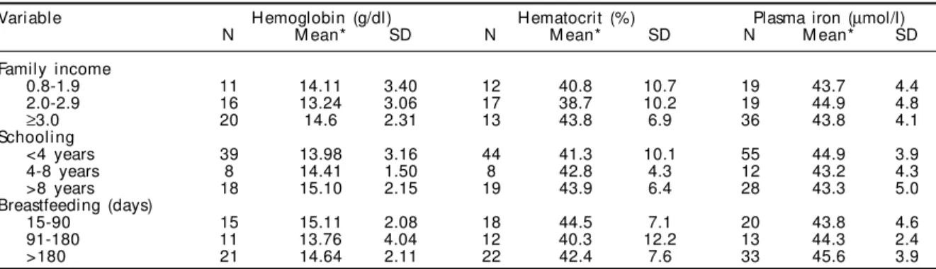
Table 1 - Mean and standard deviation (SD) of cord hemoglobin, hematocrit and plasma iron in preterm infants according to socioeconomic baseline factors and breastfeeding duration evaluated during follow-up. Rio de Janeiro, Brazil, 2000-2001.
Variable Hemoglobin (g/dl) Hematocrit (%) Plasma iron (µmol/l)
N M ean* SD N M ean* SD N M ean* SD
Family income
0.8-1.9 11 14.11 3.40 12 40.8 10.7 19 43.7 4.4
2.0-2.9 16 13.24 3.06 17 38.7 10.2 19 44.9 4.8
≥3.0 20 14.6 2.31 13 43.8 6.9 36 43.8 4.1
Schooling
<4 years 39 13.98 3.16 44 41.3 10.1 55 44.9 3.9
4-8 years 8 14.41 1.50 8 42.8 4.3 12 43.2 4.3
>8 years 18 15.10 2.15 19 43.9 6.4 28 43.3 5.0
Breastfeeding (days)
15-90 15 15.11 2.08 18 44.5 7.1 20 43.8 4.6
91-180 11 13.76 4.04 12 40.3 12.2 13 44.3 2.4
>180 21 14.64 2.11 22 42.4 7.6 33 45.6 3.9
covariance structure was tested by the likelihood ratio test. The final model included only random intercept, which is similar to an exchangeable covariance struc-ture (compound symmetry).
The variable age was centered at 40 weeks (expected duration of gestation) and was treated as a continuous variable. Models accounted for a discontinuity in time by the inclusion of two variables: time 1=minimum (ges-tational age, 40 weeks) and time 2=maximum (0, age in
weeks +40) as suggested by Bryson5 (2003). These two
variables estimates change from birth up to 40 weeks of gestational age and after 40 weeks. Upon examination of residual plots of the models, length was square root transformed, which was effective in linearizing the
rela-tionship and stabilizing the variance.16
Family income and mother’s schooling were catego-rized as in Table 1. Only eight children developed late-onset sepsis. Models were remained unchanged with the inclusion of the variable morbidity.
The study was approved by the Research Ethics Com-mittee of Instituto Fernandes Figueira (Fiocruz) and mothers signed an informed consent.
RESU LTS
Male and female babies showed no differences in anthropometric measures or cord iron markers. How-ever, females had a statistically significant lower ges-tational age (Table 2). Mothers’ hematocrit and hemoglobin measures were much lower than those observed among infants (Table 2), but there was no correlation between hemoglobin (r=0.04; p=0.74), hematocrit (r=0.15; p=0.24) or plasma iron (r=0.21; p=0.27) measures of mothers and infants. Only one female baby and one male baby had anemia at birth (cord blood hemoglobin level <10.5 g/dl), whereas among mothers anemia was highly prevalent, as well
as inadequate plasma iron (<10.7 µmol/l), which was
seen in 32% of them (Table 2).
Factors that could affect child growth were explored for their association with iron status. Cord hemoglobin, hematocrit, and plasma iron were not associated with family income, mother’s level of edu-cation and breastfeeding duration throughout the follow-up (Table 1). Mother’s height was also assessed but only four mothers were below 1.50 m.
At birth it was found no association between mark-ers of iron status and gestational age, weight, and length. The effect of mother’s and infant’s iron status at birth on infant’s growth was tested for hemoglobin, hematocrit and plasma iron as continuous variables and also by classifying mothers as anemic and non-anemic according to the classification shown in Ta-ble 2. Both crude and adjusted analyses showed no effect of iron status on children growth. Data for the multivariate analysis with cord hemoglobin is shown in Table 3.
Results based on hematocrit were quite similar to those for hemoglobin. For that reason data is shown only for hemoglobin and plasma iron.
An overall comparison of iron effect on length mean, adjusted for birth weight, gestational age and sex, are shown for the models: 1) with the overall cohort (N=95); 2) adjusted for mother hemoglobin (N=82); and 3) adjusted for cord iron (N=43) (Figure). Only estimated measures for anemic mothers and iron cord below median are shown because lines cannot be distinguished.
D ISCU SSIO N
There is strong evidence that severe iron deficiency
causes growth impairment,18 but it remains unclear to
what extent iron status is related to growth. Prema-ture infants are at risk for early postnatal iron defi-ciency because they accrue less iron during gesta-tion, grow more rapidly after birth, and are typically
undertreated with enteral iron.17 Regardless of that,
Table 2 - Anthropometric and iron status measurements of preterm infants and their mothers at birth. Rio de Janeiro, Brazil, 2000-2001.
Variable M ale babies Female babies M other
N M ean 95% CI N M ean 95% CI N M ean 95% CI
Gestational age (w eeks)* 39 33.7 31.5-35.8 56 32.6 29.8-35.4 Birth weight (g) 39 2,087 1,457-2,717 56 1,924 1,223-2,625
Length (cm) 39 44.7 40.5-48.8 56 43.5 38.9-48.0
H ematocrit (%) 31 42.3 33.7-50.9 40 41.9 33.0-50.8 82 34.2 33.1-35.2
Hemoglobin (g/dl) 27 14.3 11.5-17.0 38 14.3 11.6-17.1 82 11.2 10.8-11.5 Plasma iron (µmol/l) 16 33.0 24.9-41.0 18 33.0 26.3-39.7 34 15.7 12.9-18.5
MCV (µm3) 80 87.6 82.2-92.8
Prevalence of anemia (%) %
Hemoglobin <11 g/dl 82 43.9 34.6-53.1
Hemoglobin <9.5 g/dl 82 9.8 4.5-15.1
MCV <84 µm3 80 22.0 13.9-30.0
in the population of premature infants, some catch-up to normal growth patterns is expected, making them an interesting group to explore the association be-tween iron status and growth. On the other hand, at least in developing countries, preterm infants may suffer from infectious diseases that could impair their development and they may not receive adequate nu-tritional care to ensure healthy growth. Therefore, it was chosen a neonatal unit with strong support for breastfeeding, caring mainly for low-income mothers and with an approach for anemia prevention on in-fancy that includes iron supplementation for all preterm infants. Thus, the effect of both mother’s and their premature infant’s iron status on health and growth patterns, in relation to other factors that affect growth, such as socioeconomic factors, was studied. A limitation of the study was not to include a meas-ure of compliance to iron supplementation.
The study sample was homogeneous with respect to feeding practices and socioeco-nomic parameters, as shown in Table 1. Also, an effort was made to follow up the entire cohort up to their sixth month of life, but only 75% were followed up to the fifth month and only 50% up to the end of the study period. In addition, the repeated measurement analysis used proved to be an effective way of dealing with missing observations. Also, after comparing baseline values of the losses to follow-up with those followed up for six months of corrected age there was no differ-ence on gestational age, birth weight, hemoglobin, family income, and mother schooling. Any residual difference due to incomplete follow-up would be towards the association between iron status and growth, because anemic children would come more frequently to the care service. It was also found no association at baseline between birth weight, length and iron status. In a
sys-tematic review, Allen1 (2001) also found no
evidence to support a relationship between
iron deficiency as cause of premature birth and low birth weight. Recent studies on iron supplementa-tion showed that the effect is affected by the initial maternal iron status, which allows placenta and fetus
to compete for iron.6
The study data also showed a non-significant tendency to a negative correlation of blood cord hematocrit, hemoglobin, and iron with birth weight. A possible explanation for these negative associations at base-line can be that fetal oxygen saturation is only 45% and it is associated with high erythropoietin levels
and rapid red blood production.21 It is only during the
last two months of pregnancy that hemoglobin F, more
saturated with O2, is replaced with the adult
hemoglobin A. The most important factors regulating hemoglobin-oxygen affinity are changes related to the concentrations of red blood cell
2,3-diphosphogly-Table 3 - Longitudinal linear regression analysis of cord blood hemoglobin, and covariates on length of preterm infants during six-month follow-up for corrected age. Rio de Janeiro, Brazil, 2000-2001.
Regression coefficient Standard error p-val ue
Sex (Female/male) -0.34 0.61 0.57
Gestational age (w eeks) -0.56 0.18 0.002
Birth weight (100 g) 0.34 0.062 <0.0001
Age weeks till 40 weeks - AGE1* 1.08 0.31 0.0008
Age after 40 weeks - AGE2* 0.63 0.06 <0.0001
Hemoglobin (HB) (g/dl) -0.128 0.825 0.74
HB - AGE1* 0.0016 0.021 0.93
HB - AGE2* -0.0027 0.0042 0.52
*Interaction between age in weeks until 40 weeks AGE1 (expected term date) and after 40 weeks -AGE2. If hemoglobin had an effect on growth a statistically significant association would be expected. AGE1 and AGE2 allowed a discontinuity in time. AGE1 = minimum (gestational age, 40 weeks) and AGE2 = maximum (0, age in weeks +40) as suggested by Singer & Willett20 (2003)
Figure - Estimated means of length (cm) during six-month follow-up of the overall cohort of preterm children (N=95), those preterm children from anemic mothers (hemoglobin <11 mg/dl) (N=82) and those with cord hemoglobin below the median (N=65; median=14.7 mg/dl). Rio de Janeiro, Brazil, 2000-2001.
All cohort (Ntotal =95) 9 51 92 75 71 64 54 47
Mother Hb (Ntotal=82) 7 43 75 64 60 49 46 34
Cord Hb (Ntotal=65) 5 34 62 51 49 43 33 30
Gestational age Postnatal age cm
30 35 40 45 50 55 60 65 70
anemic all cohort iron<median
cerate (2,3-DPG).21 The decline in fetal hemoglobin in
the newborn period appears to be strictly regulated. The switchover from fetal hemoglobin to adult hemoglobin synthesis follows a sigmoid curve with a crossover point about 30 to 32 weeks post-concep-tion. Consistent with these findings, the present study found higher levels of hemoglobin and hematocrit in children compared to mothers.
Several studies,10,11,14 but not all,3,4,22 have shown a
positive association between cord and maternal iron status. In general, these associations are present when mothers with severe anemia are included in the analy-ses or when newborns from mothers with severe anemia are compared with those of non-anemic moth-ers. Otherwise when maternal moderate anemia is
present no associations are found.4,24 The data from
the present study showed no association of hematocrit, hemoglobin and plasma iron measures between moth-ers and infants. These results are consistent with the absence of severe anemia in the mothers studied and with a previous study of Brazilian mothers and in-fants in early postpartum (1-5 days), which showed no association between mothers and infants
regard-ing these variables, and also in ferritin values.22
Ma-ternal iron status at the end of pregnancy or in early
postpartum may also affect infant status. De Benaze8
et al (1989) also found a positive association between ferritin levels of mothers and those of two-month-old children. Among children born to anemic mothers, the odds ratio for anemia at two months age was 5.7 compared to children born from non-anemic moth-ers; even after controlling for food intake, morbidity,
and socioeconomic level.7
Lack of power due to a small sample size is not a probable explanation for the lack of association be-tween iron status and growth, since a difference in length of 4 cm, taken for sample size calculation,
have been reported in other studies.18
To the authors’ knowledge few studies have explored the association between iron status at the time of birth for preterm infants and their subsequent growth to the gestational age of 40 weeks. This aspect of fetal/infant growth appears to be an important step in the analysis in order to adjust for the rapid growth of more prema-ture children. Based on the results of this study, it is concluded that maternal anemia is not associated with anemia in preterm infants and that iron status of preterm infants and their mothers, at the time of birth, is not associated with short-term growth of preterm infants.
REFEREN CES
1. Allen LH. Biological mechanisms that might underlie iron’s effects on fetal growth and pre-term birth. J Nutr. 2001;131(2 Suppl 2):581-9.
2. Ballard JL, Khoury JC, Wedig K, Wang L, Eilers-Walsman BL, Lipp R. New Ballard score, expanded to include extremely premature infants. J Pediatr. 1991;119:417-23.
3. Barton DP, Joy MT, Lappin TR, Afrasiabi M, Morel JG, O’Riordan J, et al. Maternal erythropoietin in singleton pregnancies: a randomized trial on the effect of oral hematinic supplementation. Am J Obstet Gynecol. 1994;170:896-901.
4. Bhargava M, Kumar R, Iyer PU, Ramji S, Kapani V, Bhargava SK. Effect of maternal anemia and iron depletion on foetal iron stores, bithweight and gestation. Acta Paediatr Scand. 1989;78:321-2. 5. Bryson L. Doing data analysis with the multilevel
model for change. In: Singer JD, Willett JB. Applied longitudinal data analysis: modeling chance and event occurrence. Oxford: University Press; 2003. p. 75-137. 6. Cogswell ME, Parvanta I, Ickes L, Yip R, Brittenham
GM. Iron supplementation during pregnancy, anemia, and birth weight: a randomized controlled trial. Am J Clin Nutr. 2003;78:773-81.
7. Colomer J, Colomer C, Gutierrez D, Jubert A, Nolasco A, Donat J, et al. Anaemia during pregnancy as a risk factor for infant iron deficiency: report from the Valencia Infant Anaemia Cohort (VIAC) study. Paediatr Perinat Epidemiol. 1990;4:196-204.
8. De Benaze C, Galan P, Wainer R, Hercberg S. Prevention de l’anemie ferriprive au cours de la grossesse par une supplementation martiale précoce: un essai controlé. Rev Epidemiol Sante Publique. 1989;37:109-18.
9. Dewey KG. The challenges of promoting optimal infant growth. J Nutr. 2001;131:1879-80. 10. Gaspar MJ, Ortega RM, Moreiras O. Relationship
between iron status in pregnant women and their babies: investigation in a Spanish population. Acta Obstet Gynecol Scand. 1993;72:534-7.
11. Hokama T, Takenaka S, Hirayama K, Yara A, Yoshida K, Itokazu K, et al. Iron status of newborns born to iron deficient mothers. J Trop Pediatr. 1996;42:75-7. 12. Kilsztajn S, Rossbach A, Carmo MS, Sugahara GT.
13. King JC. The risk of maternal nutritional depletion and poor outcomes increases in early or closely spaced pregnancies. J Nutr. 2003;133(5 Suppl 2):1732-6. 14. Lao TT, Loong EP, Chin RK, Lam CW, Lam YM.
Relationship between newborn and maternal iron status and haematological indices. Biol Neonate. 1991;60:303-7.
15. Monteiro CA, Szarfarc SC, Mondini L. Tendência secular da anemia na infância na cidade de São Paulo (1984-1996). Rev Saúde Pública. 2000;34(6 Supl):26-40.
16. Neter J, Wasserman W, Kutner M. Applied linear statistical models: regression, analysis of variance, and experimental designs. 2nd ed. Homewood (IL): R. D. Irwin; 1985.
17. Rao R, Georgieff MK. Perinatal aspects of iron metabolism. Acta Paediatr Suppl. 2002;91:124-9. 18. Rivera JA, Hotz C, Gonzalez-Cossio T, Neufeld L,
Garcia-Guerra A. The effect of micronutrient deficiencies on child growth: a review of results from community-based supplementation trials. J Nutr. 2003;133(11 Suppl 2):4010-20.
19. Salsbury DC. Anemia of prematurity. Neonatal Netw. 2001;20:13-20.
20. Singer JD, Willett JB. Applied Longitudinal data analysis: modeling change and event occurrence. New York: Oxford University press; 2003. 21. Stockman JA. Anemia of prematurity: current
concepts in the issue of when to transfuse. Pediatr Clin North Am. 1986;33(1):111-28.
22. Trugo NMF, Donangelo CM, Koury JC, Freitas LA, Feldheim W. Evaluation of iron and folate status in urban Brazilian mothers of low socioeconomic status and their infants. Colloq INSERM. 1990;197:95-8. 23. Victora CG, Barros FC, Horta BL, Martorell R.
Short-term benefits of catch-up growth for small-for-gestational-age infants. Int J Epidemiol. 2001;30(6):1325-30.
24. Viteri FE. The consequences of iron deficiency and anemia in pregnancy. Adv Exp Med Biol.
1994;352:127-40.