CASE REPORT
DOI: 10.1590/1516-3180.2013.1315441Tuberous sclerosis complex diagnosed from oral lesions
Complexo esclerose tuberosa diagnosticado a partir de lesões orais
Leonardo de Jesus Araújo
I, Guilherme Braga Muniz
II, Edmilson Santos
III, João Paulo Versiani Ladeia
IV, Hercílio Martelli Júnior
V,
Paulo Rogério Ferreti Bonan
VUniversidade Estadual de Montes Claros (Unimontes), Montes Claros, Minas Gerais, Brazil
ABSTRACT
CONTEXT: Tuberous sclerosis complex (TSC) is a genetic disease in the group known as neurocutane-ous syndromes, with dominant autosomal inheritance. It is characterized by skin and adnexal lesions and central and peripheral nervous system tumors, with neurological and psychiatric indings. It may afect the heart, kidneys, eyes, face, bones, lungs, stomach and dentition.
CASE REPORT: We present the case of a 66-year-old man with dermatological signs that included hy-popigmented maculae, confetti-like lesions, shagreen plaque, angioibromas on nasolabial folds, neck and back, nail dystrophy and periungual ibromas on ingers and toes. An electroencephalogram produced normal results, but magnetic resonance imaging showed a nodular image measuring 1.2 x 1.0 cm close to the Monro foramen, which was similar to cerebral parenchyma and compatible with a subependymal giant-cell astrocytoma. A conservative approach was taken, through control imaging examinations on the lesion for seven years, with absence of any expansive process or neurological symptoms. Abdomi-nal ultrasonography revealed a solid, heterogenic and echogenic mass with a calciied focus, measuring 4.6 x 3.4 cm, in the right kidney, compatible with angiomyolipoma. The patient was treated by means of complete nephrectomy because of malignant areas seen on histopathological examination and died one month after the procedure. This case report illustrates the importance of oral clinical indings such as dental enamel pits and angioibromas in making an early diagnosis of TSC, with subsequent screening examinations, treatment and genetic counseling.
RESUMO
CONTEXTO: O complexo esclerose tuberosa é uma doença genética pertencente ao grupo das facoma-toses, de herança autossômica dominante, caracterizada por lesões acometendo pele e anexos, além de tumores do sistema nervoso central e periférico, com presença de achados neurólogicos e psiquiátricos, podendo acometer coração, rins, olhos, dentes, mucosa oral e outros órgãos.
RELATO DE CASO: Apresentamos o caso de um paciente do sexo masculino, 66 anos de idade, que apre-sentava diversos sinais dermatológicos, como máculas hipopigmentadas, lesões em confete, placas tipo
shagreen, angioibromas nas regiões cervical, nasolabial e dorsal, distroias ungueais e ibromas nos dedos das mãos e pés. Embora o encefalograma não tenha mostrado alterações, a ressonância nuclear magnéti-ca apresentou imagem nodular com aspecto semelhante ao parênquima cerebral, medindo 1.2 x 1.0 cm e próxima ao forame de Monro, compatível com astrocitoma subependimário de células gigantes. Aborda-gem conservadora foi escolhida por meio de controle imaginológico da lesão por sete anos, com ausência de sintomas neurológicos ou processos expansivos. A ultrassonograia de abdômen total revelou massa medindo 4.6 x 3.4 cm, com áreas ecogênicas e heterogêneas, apresentando focos de calciicação no rim direito, com padrão semelhante ao de um angiomiolipoma. O paciente foi submetido a nefrectomia total, devido à presença de áreas de malignidade ao exame histopatológico, e evoluiu para óbito um mês após o procedimento. Este relato de caso ilustra a importância de achados clínicos orais, tais como lesões no esmalte dentário e angioibromas, para o diagnóstico precoce dessa doença e posterior rastreamento, tratamento e aconselhamento genético.
IMSc. Researcher, Postgraduate Program on
Health Sciences, Universidade Estadual de Montes Claros (Unimontes), Montes Claros, Minas Gerais, Brazil.
IIMedical Student. Scientiic Initiation Program,
Institute of Health Sciences, Faculdades Unidas do Norte de Minas (Funorte), Montes Claros, Minas Gerais, Brazil.
IIIMD. Full Professor, Scientiic Initiation Program,
School of Medicine, Institute of Health Sciences, Faculdades Unidas do Norte de Minas (Funorte), Montes Claros, Minas Gerais, Brazil.
IVMBA. Researcher, Institute of Exact and Biological
Sciences, Universidade Federal de Ouro Preto (UFOP), Ouro Preto, Minas Gerais, Brazil.
VPhD. Full Professor, Postgraduate Program
on Health Sciences, Universidade Estadual de Montes Claros (Unimontes), Montes Claros, Minas Gerais, Brazil.
KEY WORDS: Diagnosis. Tuberous sclerosis. Neurocutaneous syndromes. Pathology.
Phenotype.
PALAVRAS-CHAVE: Diagnóstico. Esclerose tuberosa. Síndromes neurocutâneas. Patologia.
C A S E R E P OR T | Araújo LJ, Muniz GB, Santos E, Ladeia JPV, Martelli Júnior H , Bonan PRF
352 Sao Paulo Med J. 2013; 131(5):351-5 INTRODUCTION
Tuberous sclerosis complex (TSC) or Bourneville’s disease, is a neurocutaneous syndrome (registered in the Online Mendelian Inheritance in Man as MIM 191100) characterized by autosomal dominant inheritance, with high prevalence of de novo muta-tions and variable expression.1-3 Among the reported indings, lesions of the skin, adnexa, central nervous system, heart, kid-neys and other organs stand out.1-4 he prevalence is estimated to be 1:6000 live births,2,5 and two thirds of all cases do not have any familial history.2,4 he phenotypic expression of TSC is highly variable and, in some cases, it may be diicult to establish a dein-itive clinical diagnosis.3 Generally, the diagnosis is made based on multiple clinical criteria that are categorized into major and minor features. At the Tuberous Sclerosis Consensus Conference of 1998, the clinical diagnostic criteria of TSC were revised and a new classiication system based on major and minor indings was established.6 he presence of two major features, or one major and two minor features, is suicient for a deinitive diagnosis.1-4 Over recent years, mutation analysis has become an additional diagnostic tool in cases of familial as well as sporadic TSC.7
Two disease-determining genes have been deined, named TSC1 and TSC2, on chromosomes 9q34 and 16p13, respectively. he TSC1 gene has 23 exons and its protein product is hamartin. he TSC2 gene has 41 exons in the coding region and a leader exon with a transcript that exists as multiple isoforms; its protein prod-uct is tuberin. Both hamartin and tuberin are tumor-suppressor proteins that are involved in cell growth and diferentiation.7
Considering the rarity of diagnosing TSC based on oral lesions, the aim of this paper was to present a case report with emphasis on the importance of identifying of dental enamel pits and oral angioibromas for diagnosing this disease.
CASE REPORT
A 66-year-old non-Caucasian man was referred to the Stomatology Clinic of the State University of Montes Claros (Universidade Estadual de Montes Claros, Unimontes), state of Minas Gerais, Brazil, due to the presence of ibrous prolifera-tive lesions in the oral mucosa. Intraoral examination revealed hypoplastic defects on the vestibular face of the upper central incisor and canines and ibrous proliferations in the lower-lip mucosa, without apparent traumatic factors. he patient reported that he had been a tobacco user since his teenage years (Figure 1). he labial lesion was excised, and microscopic analy-sis using hematoxylin and eosin staining revealed a lesion com-patible with oral angioibroma (Figure 2). he association of dental enamel pits and gingival angioibromas led to the clinical hypothesis of TSC and a search for skin lesions was performed. he patient’s medical history included intestinal ulcers and hematemesis, which had been treated using omeprazole. here were positive reports of convulsive crises, with the last episode in
1999, which had been controlled using phenobarbital. he fam-ily’s history included occurrences of hypertension, diabetes mel-litus, cancer and TSC.
General clinical examination showed hypopigmented mac-ulae, confetti-like lesions, shagreen plaque, angioibromas on nasolabial folds, neck and back, nail dystrophy and periun-gual ibromas on hands and feet (Figure 1), seen under Wood’s light. On physical examination, the patient did not show any signs or symptoms of cardiovascular, endocrine, respiratory, immune or musculoskeletal disorders.
Brain magnetic resonance imaging was performed in a 1.5-T scanner including T1, T2, and FLAIR (luid acquisition inversion recovery), and showed a nodular image similar to cerebral paren-chyma, measuring 1.2 x 1.0 cm, with projection of the anterior cornu of the let lateral ventriculum, close to the Monro foramen (Figure 2). A conservative approach was chosen for this case, with control imaging examinations over a seven-year period, without any expansive process. hese indings were compatible with a subependymal giant-cell astrocytoma. An electroenceph-alogram produced normal results and no evidence of signs or symptoms of psychopathological processes.
Abdominal ultrasonography was also requested. Although this showed that the viscera were normal, there was a solid, het-erogenic and echogenic mass with a calciied focus inside, mea-suring 4.6 x 3.4 cm, in the right kidney, which was compat-ible with angiomyolipoma. Kidney function was normal and asymptomatic, and urine analyses did not show proteinuria. Echocardiography, additional routine laboratory tests and fun-doscopy examinations were normal.
he patient was admitted to the Santa Casa Hospital of Montes Claros, Minas Gerais, Brazil, for laparoscopic examina-tion and biopsies on his right kidney. Histopathological exam-ination on the material obtained was compatible with angio-myolipoma, but also revealed important areas of malignancy consistent with renal cell carcinoma. he patient then underwent medial longitudinal laparotomy and complete nephrectomy of right kidney. He died one month ater the procedure. Permission to perform an autopsy was not granted.
Prior approval to report on this case was obtained from the Institutional Ethics Committee (protocol no. 1047/08) and the patient signed an informed consent statement. We followed the eval-uation and management recommendations put forward by Roach et al.6 his case was compatible with the diagnostic criteria estab-lished for TSC, which were conirmed by the clinical, imaging and histopathological indings and by the presence of a family history.
DISCUSSION
Tuberous sclerosis complex diagnosed from oral lesions | CASE REPORT
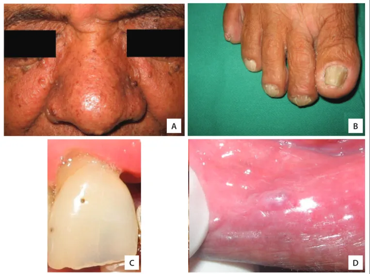
Figure 1. Clinical features identii ed in the patient aff ected by tuberous sclerosis complex. (A) Multiple facial papules aff ecting the
nasolabial folds and the nose, compatible with angioi bromas; (B) non-traumatic ungual and periungual i bromas on the right foot; (C) enamel pitting in the permanent upper central incisor tooth; and (D) i brous proliferation in the lower-lip mucosa, without apparent traumatic factors (angioi broma).
A
B
C
D
Figure 2. Neurological tuberous sclerosis complex features detected by means of magnetic resonance imaging (MRI) in a 1.5-T scanner.
(A) Coronal and (B) axial FLAIR (l uid acquisition inversion recovery) magnetic resonance imaging (MRI), demonstrating the Monro foramen nodule, compatible with subependymal giant-cell astrocytoma, with and without administration of gadolinium contrast (GdDTPA), respectively; (C) sagittal T1 MRI after GdDTPA, showing a hyperintense signal from the tumor.
CASE REPORT | Araújo LJ, Muniz GB, Santos E, Ladeia JPV, Martelli Júnior H , Bonan PRF
354 Sao Paulo Med J. 2013; 131(5):351-5
Sturge-Weber syndrome, ataxia-telangiectasia and von Hippel-Lindau disease.2,3,8 he term TSC refers to multiple sclerotic masses scattered throughout the cerebrum. TSC was irst described by von Recklinghausen in 1862 and in more recent times was reported by Bourneville, Pringle and Vogt.9-11 he most common features of TSC include facial angioibromas, hypopigmented cutaneous mac-ulae, shagreen patches in the lumbar area, cerebral cortical tubers, subependymal nodules, subependymal giant-cell astrocytomas, cardiac rhabdomyomas and renal angiomyolipomas. Its minor fea-tures may include hamartomatous rectal polyps, non-renal hamar-tomas and multiple renal and bone cysts.2-4,6
Our patient presented classical cutaneous indings, including multiple angioibromas on his face. hese hamartomatous lesions are preferentially located on the nasolabial folds, malar regions (where they usually present symmetrically, in the form of “butter-ly wings”) and the nose and chin.2-4,6,12 he diferential diagnosis includes acne vulgaris and dermatosis papulosa nigra3,13,14 (Figures
1 and 2). Several cosmetic treatments have been described includ-ing curettage, surgical excision, cryosurgery, electrosurgery, derm-abrasion, pulsed dye laser and CO2 laser. However, the recurrence rates are considered to be high.2,13,15
Central nervous system involvement in TSC invariably presents with cortical tubers, subependymal glial nodules, white matter ham-artomas and subependymal giant-cell astrocytoma. Neurological manifestations range from slight or even nonexistent to extremely severe symptoms. he most common neurological inding is sei-zures. However, other manifestations such as mental retardation of difering degrees and ob structive hydrocephaly secondary to tumor growth are frequently seen. One of the most important complica-tions in patients with TSC is the development of subependymal giant-cell astrocytomas close to the Monro foramen. Imaging tech-niques, especially brain magnetic resonance, are now well estab-lished for diagnosing and following up TSC patients.3,16
Kidney involvement frequently occurs in TSC cases. he main manifestations are angiomyolipomas, cysts and renal malignan-cies.2-4,6 Renal angiomyolipoma is a benign tumor composed of three diferent types of tissue: fatty, smooth muscle and vascular.17 Angiomyolipoma oten occurs in association with tuberous sclero-sis between the second and third decades of life in isolated form.17,18 It primarily afects women between the fourth and seventh decade of life. he most common signs and symptoms are abdominal pain, palpable abdominal mass, hematuria and other consequences of intra-tumoral hemorrhage.17,19 he latter occurs in approximate 25% of the patients, and 10% of these patients may present hypovolemic shock in the acute phase. he symptoms and complications of angi-omyolipoma are related to its size and rapidity of growth. Lesions greater than 4 cm indicate a greater risk of complications, such as hemorrhage. According to the literature, the treatment depends on the lesion size.17 Between 40% and 80% of individuals with TSC present renal angiomyolipoma with multiple bilateral asymptomatic
tumors, which may be associated with cysts and, less frequently, with renal carcinoma.17,18,20 Total nephrectomy should be used very rarely; it is only justiied in cases of uncontrollable bleeding that is a risk to the patient’s life, or in cases of central tumors, extensive necrosis, inlammation of the renal tissue or presence of renal carcinoma in the same kidney, as in the present case.17
Oral manifestations of TSC are quite frequent and are charac-terized mainly by ibrous hyperplasia (angioibromas) and dental enamel pitting.2,15 Angioibromas are frequently located in the ante-rior portions of the gingiva, but they are not rare on the lips, tongue, and palate. In addition to TSC, other syndromes may include facial and oral papules/nodules in their clinical spectrum, including Cowden syndrome, Birt-Hogg-Dubé syndrome and multiple endo-crine neoplasia type 1.2,3,21 Dental enamel pitting is observed in up to 100% of the patients with TSC. Dental pits can also be observed in the general population, but at lower frequency and with fewer lesions than in TSC cases. Enamel pits are also observed in cases of pitted hypoplastic amelogenesis imperfecta, vitamin D-dependent rickets, pseudohypoparathyroidism and junctional epidermolysis bullosa.2,22 A systematic survey of indexed articles in the Medline/PubMed, Embase, Lilacs, Scirus, SciELO and Cochrane Library databases revealed that, to date, few articles have emphasized the importance of oral lesions such as concomitant dental enamel pits and oral angioibromas in diagnosing this disease. he papers surveyed are described in Table 1.
Table 1. Results from systematic search of indexed articles in
medical databases performed on April 9, 2012, using descriptors
for the main clinical indings observed in this case report*
Database* Search strategy Results
PubMed
((“Tuberous Sclerosis”[MeSH]) OR (Tuberous Sclerosis))
AND
((“Dental Enamel” [MeSH]) OR (Dental Enamel) OR (Dental Enamel Pits) OR (“Angioibroma” [MeSH]) OR Angioibroma)
179
Lilacs or SciELO
((Tuberous Sclerosis) OR (Esclerose Tuberosa) OR (Esclerose Tuberosa) OR (Esclerosis Tuberosa) OR (Doença de Bourneville Epiloia)
OR (Facomatose de Bourneville) AND
((Dental Enamel) OR (Dental Enamel Pits) OR (Esmalte Dentário) OR (Esmalte Dental) OR (Cutícula Dentária Esmalte) OR (Cutícula de
Esmalte) OR Angioibroma)
3
Embase
((Tuberous Sclerosis )) AND
((Dental Enamel) OR (Dental Enamel Pits) OR Angioibroma)
62
Scirus
((Tuberous Sclerosis)) AND
((Dental Enamel) OR (Dental Enamel Pits) OR Angioibroma)
610
Tuberous sclerosis complex diagnosed from oral lesions | CASE REPORT
CONCLUSION
his report on a case of TSC illustrates that proper identiication of clinical oral features such as dental enamel pits and angioibro-mas may help towards achieving early diagnosis of this disease and, ultimately, towards starting appropriate screening examina-tions, treatment and genetic counseling.
REFERENCES
1. Roach ES, Miller VS. Neurocutaneous disorders. Cambridge: University Press; 2004.
2. De Jesus Araújo L, Yamamoto De Almeida L, Santos Lima J, Martelli-Júnior H, Ferreti Bonan PR. Evaluation of MMP-1, MMP-10, TIMP-1, a-SMA and TGF-b1 in angioibromas of tuberous sclerosis. Minerva Stomatol. 2011;60(1-2):25-33.
3. Araujo L de J, Lima LS, Alvarenga TM, et al. Oral and neurocutaneous phenotypes of familial tuberous sclerosis. Oral Surg Oral Med Oral Pathol Oral Radiol Endod. 2011;111:87-94.
4. Narayanan V. Tuberous sclerosis complex: genetics to pathogenesis. Pediatr Neurol. 2003;29(5):404-9.
5. Osborne JP, Fryer A, Webb D. Epidemiology of tuberous sclerosis. Ann N Y Acad Sci. 1991;615:125-7.
6. Roach ES, Gomez MR, Northrup H. Tuberous sclerosis complex consensus conference: revised clinical diagnostic criteria. J Child Neurol. 1998;13(12):624-8.
7. Apak A, Haliloğlu G, Köse G, et al. Mutation analysis of TSC2 gene in 33 Turkish familial cases with tuberous sclerosis. Turk J Pediatr. 2003;45(1):1-5.
8. Lin DD, Barker PB. Neuroimaging of phakomatoses. Semin Pediatr Neurol. 2006;13(1):48-62.
9. Bourneville DM. Sclérose tubéreuse des circonvolutions cérébrales: idiotie et épilepsie hémiplégique. Arch Neurol (Paris).1881;1:81-91. 10. Pringle JJ. A case of congenital adenoma sebaceum. British Journal
of Dermatology. 1890;2:1-14. Available from: http://www2.biusante. parisdescartes.fr/livanc/?cote=epo0074&p=1&do=page. Accessed in 2012 (Jan 7).
22. Vogt H. Zur Pathologie und pathologischen Anatomie der versclriedenen Idiotieform. Monatsschr Psychiatr Neurol. 1908; 24:106-50.
12. López-López J, Rodríguez de Rivera Campillo E, Marques-Soares MS, et al. Esclerosis tuberosa y manifestaciones orales. Caso clínico [Tuberous sclerosis and its oral manifestations. A clinical case]. Med Oral Patol Oral Cir Bucal. 2004;9(3):216-23.
13. Stables G, Sheehan-Dare R. The dermatological features of tuberous sclerosis and their treatment. Scan Facts. Available from: http:// www.tuberous-sclerosis.org/publications/fs25derm.pdf. Accessed in 2012 (Aug 23).
14. Wee SA, Fangman B. Tuberous sclerosis. Dermatol Online J. 2007;13(1):22.
15. Scully C. Orofacial manifestations in tuberous sclerosis. Oral Surg Oral Med Oral Pathol. 1977;44(5):706-16.
16. Carvalho-Neto A, Bruck I, Antoniuk SA, Marchiori E, Gasparetto EL. Espectroscopia de prótons por RM da região do forame de Monro em pacientes com complexo esclerose tuberosa [Proton MR spectroscopy of the foramen of Monro region in patients with tuberous sclerosis complex]. Arq Neuro-psiquiatr. 2008;66(2B):303-7. 17. Schneider-Monteiro ED, Lucon AM, Figueiredo AA, Rodrigues Junior
AJ, Arap S. Bilateral giant renal angiomyolipoma associated with hepatic lipoma in a patient with tuberous sclerosis. Rev Hosp Clin Fac Med Univ São Paulo. 2003;58(2):103-8.
18. van Baal JG, Smits NJ, Keeman JN, Lindhout D, Verhoef S. The evolution of renal angiomyolipomas in patients with tuberous sclerosis. J Urol. 1994;152(1):35-8.
19. O’Donnell M, Fleming S. Angiomyolipoma of kidney--a cause of recurrent retroperitoneal haemorrhage. Br J Urol. 1995;76(4):521-2. 20. Ewalt DH, Sheield E, Sparagana SP, Delgado MR, Roach ES. Renal
lesion growth in children with tuberous sclerosis complex. J Urol. 1998;160(1):141-5.
21. Sparling JD, Hong CH, Brahim JS, Moss J, Darling TN. Oral indings in 58 adults with tuberous sclerosis complex. J Am Acad Dermatol. 2007;56(5):786-90.
22. Flanagan N, O’Connor WJ, McCartan B, et al. Developmental enamel defects in tuberous sclerosis: a clinical genetic marker? J Med Genet. 1997;34(8):637-9.
Sources of funding: This study was supported by grants from the Minas Gerais State Research Foundation (FAPEMIG), Brazil (APQ-00898-08). H Martelli-Júnior was supported by the National Council for Scientiic and Technological Development (CNPq), Brazil, and PRF Bonan was supported by Fapemig
Conlict of interest: None
Date of irst submission: December 29, 2011
Last received: December 14, 2012
Accepted: January 7, 2012
Address for correspondence:
Leonardo de Jesus Araújo Rua Calcita, 162
Monte Carmelo — Montes Claros (MG) — Brasil CEP 39402-025
Tel. (+55 38) 3223-6167