J Bras Pneumol. 2007;33(3):343-346
343
Case Report
Pulmonary amyloidosis: radiographic finding
of nodular opacities in a heavy smoker*
Jorge Montessi1, Edmilton Pereira de Almeida2, João Paulo Vieira3, Cândida Maria Horta4,
Marcus da Matta Abreu5, Carlos Eduardo Dainezzi Bolognani6,Sandra Márcia Carvalho Ribeiro Costa7
Abstract
Pulmonary amyloidosis is a rare disease, characterized by extracellular deposition of fibrillary protein in the lungs. Amyloidosis is a generic term for a heterogeneous group of diseases, including Alzheimer’s disease and type 2 diabetes mellitus. In the respiratory system, it appears in various forms: tracheobronchial; nodular pulmonary; and alveolar septal (diffuse parenchymal). We present the case of a woman who was a 20 pack-year smoker and had nodular pulmonary amyloidosis, as diagnosed through tests performed prior to laparoscopic cholecystectomy.
Keywords: Amyloidosis/diagnosis; Lymphoproliferative disorders; Neoplasms; Lung.
* Study carried out at the Monte Sinai Hospital, Juiz de Fora (MG) Brazil.
1. PhD in Thoracic Surgery. Universidade Federal de Juiz de Fora – UFJF, Federal University of Juiz de Fora – Juiz de Fora (MG) Brazil. 2. Specialist in Thoracic Surgery. Universidade Federal de Juiz de Fora – UFJF, Federal University of Juiz de Fora – Juiz de Fora (MG) Brazil. 3. Masters in Thoracic Surgery. Juiz de Fora School of Medical and Health Sciences – SUPREMA – Juiz de Fora (MG) Brazil.
4. Masters in Pulmonology. Juiz de Fora School of Medical and Health Sciences – SUPREMA – Juiz de Fora (MG) Brazil.
5. Resident in the Department of Thoracic Surgery. University Hospital of the Federal University of Juiz de Fora, Juiz de Fora (MG) Brazil. 6. Fifth-year Medical Student. Universidade Federal de Juiz de Fora – UFJF, Federal University of Juiz de Fora – Juiz de Fora (MG) Brazil.
7. Specialist in Pathological Anatomy. Chief of the Department of Pathological Anatomy of the Hospital Monte Sinai – HMS, Monte Sinai Hospital – Juiz de Fora (MG) Brazil.
Correspondence to: Jorge Montessi. Centro de Estudos Hospital Monte Sinai, Rua Vicente Beguelli, 315, Dom Bosco, CEP 36035-550, Juiz de Fora, MG, Brasil. Phone 55 32 3239-4194. E-mail: [email protected]
Submitted: 11 October 2005. Accepted, after review: 4 July 2006.
Introduction
Amyloidosis is a disease characterized by extracellular deposition of fibrillary proteins in a variety of organs.(1-3)
It is a generic term for a heterogeneous group of diseases, including Alzheimer’s disease and type 2 diabetes mellitus. It can be hereditary or acquired, potentially fatal or a merely accidental finding.(3) In a simplified way, the disease can
be subdivided into localized or systemic form. It can also be classified as primary or secondary.(4) Amyloidosis of the
respiratory tract was first described in 1877 by Lesser.(1) Since
then, a number of classifications, based on radiological, bronchoscopic, and clinical findings, have been proposed.(1,3)
The objective of the present study was to report the clin-ical case of a patient with pulmonary amyloidosis (nodular type) diagnosed at the Serviço de Pneumologia e Cirurgia Torácica do Hospital Monte Sinai de Juiz de Fora
(SPCT-HMS-JF, Pulmonology and Thoracic Surgery Clinic of the Juiz de Fora Monte Sinai Hospital).
Case report
A 57-year-old Caucasian female (born in the city of Juiz de Fora, in the state of Minas Gerais, married, and a retired teacher) was referred to the SPCT-HMS-JF because the X-ray (Figure 1) prior to cholecystectomy revealed a nodule (of unknown stability and with irregular borders) in the right pleural cavity. The patient, who was totally asymptomatic, was a smoker (20 pack-years) and had a family history of cutaneous amyloidosis.
344 Montessi J, Almeida EP, Vieira JP, Horta CM, Abreu MM, Bolognani CED, Costa SMCR
J Bras Pneumol. 2007;33(3):343-346
segment of the middle lobe. There were no enlarged mediastinal lymph nodes (Figure 2).
Due to the high likelihood of malignancy, the patient was submitted to right anterolateral thora-cotomy with wedge resection of the lesion, and the surgical sample was sent directly (not frozen) to the pathological anatomy laboratory.
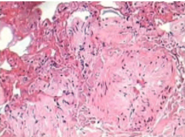
Macroscopic examination revealed brownish-yellowish, elastic, nodular fragments measuring 2.0 × 1.3 cm and 1.0 × 0.7 cm, and cut surfaces presenting a thick mucoid aspect. Analysis of the frozen section identified no malignancy. Microscopic evaluation of the histopathological paraffin sections showed amorphous eosinophilic material surrounded by vacuolated macrophages and foci of negative images of cholesterol crystals, together with thick septa and blood vessels (Figure 3). Congo red staining revealed deposition of amyloid material in loose patches, as well as on the walls of the blood vessels and septa, consistent with a histopatholog-ical diagnosis of amyloidosis (Figure 4).
The postoperative evolution was favorable and uneventful, and the water-sealed chest tube was removed on postoperative day 2. The patient was discharged on postoperative day 3 and entered a phase of clinical and radiological outpatient evalua-tion. In the most recent evaluation, the patient was totally asymptomatic.
Discussion
Amyloidosis is a generic term for a hetero-geneous group of diseases associated with the
Figure 1 - Anteroposterior chest X-ray. Figure 2 - Computed tomography scan.
Figure 3 - Hematoxylin and eosin staining showing the presence of amyloid material.
Pulmonary amyloidosis: radiographic finding of nodular opacities in a heavy smoker
J Bras Pneumol. 2007;33(3):343-346
345
deposition of abnormal fibrillary proteins on tissues and organs.(2,5,6) There are various different forms of
amyloidosis.(1)
Amyloidosis can be classified, through immuno-histochemistry, into three types: reactive (AA type); primary idiopathic (AL type); and transthyretin-related amyloidosis (ATTR type). The AA type of amyloidosis, which is derived from the serum amyloid A protein, can be hereditary or can result from chronic infectious processes, having been associated with familial Mediterranean fever and tuberculosis. This form of amyloidosis rarely manifests as a respira-tory disease. The AL type of amyloidosis (light-chain amyloidosis), which is the most common type,(1) is
considered a systemic process and, due to the large
deposition of cleaved products of immunoglobulin, manifests as heart failure, renal insufficiency, or nephropathy.(1,7) The deposition-related impairment
caused by idiopathic amyloidosis is also found in the non-systemic form, with the isolated involvement of organs, for example, in the nervous system or in the respiratory tract.(1) The involvement of thelung
parenchyma caused by the AL type occurs in 28% of the patients and does not affect survival.(5) The
ATTR type of amyloidosis is associated withsenile systemic amyloidosis and most frequently manifests as cardiovascular dysfunction, polyneuropathy or renal impairment.(1)
There have been few reported cases of systemic AA amyloidosis affecting the lungs. However, the AL type has been a unanimous finding in studies of fibrillary protein sequencing. Autopsy series have confirmed that diffuse amyloidosis of the lung parenchyma is the most common histological type of systemic AL amyloidosis. The clinical characteris-tics rarely lead to confusion with pulmonary edema or fibrosis. Advanced lung disease is not related to hereditary amyloidosis.(3) In the primary systemic
form, amyloid fibrils can occur at other sites, such as in the heart, kidneys, tongue, gastrointestinal tract, blood vessel walls, nerves, skin, muscles, and peri-articular structures. Of the patients with this form of the disease, 10-15% present concomitant multiple myeloma.(4) The great majority of patients
with pulmonary impairment present the primary form of the disease.(1,4)
The many classification systems proposed, all of which are based on the location, clinical aspects, and chemical characteristics of the amyloid, show a limited understanding of the disease. This is true of
even the most recent proposals. In a simplified way, the disease can be subdivided into localized and systemic forms. It can also be classified as primary or secondary. The secondary systemic form is generally related to neoplastic, infectious, or chronic inflam-matory processes, including tuberculosis, chronic kidney disease (especially pyelonephritis), syphilis, leprosy, inflammatory bowel disease, osteomyelitis, parasitic infections, rheumatoid arthritis, and bron-chiectasis.(4,8) In the secondary form, the incidence
of clinically or radiologically significant pulmo-nary impairment is very low. The great majority of patients with pulmonary impairment have the primary form of the disease. Primary pulmonary amyloidosis is a rare disease that presents focal or systemic characteristics.(4)
In the case described here, immunohistochem-istry was not performed to define the type of amyloidosis, since it had already been clinically defined as primary.
Regarding the distribution of the lesions in the respiratory system, amyloidosis can be divided, didactically, into the following forms: tracheobron-chial; nodular pulmonary; and alveolar septal (also known as diffuse parenchymal).(4,7) The
tracheo-bronchial and nodular pulmonary forms present focal characteristics, whereas the alveolar septal form presents systemic characteristics.(4)
A diagnosis of tracheobronchial amyloidosis is rare.(1,3) Tracheobronchial amyloidosis is associated
with tracheobronchopathia osteoplastica, which is characterized by the presence of calcification and cartilage nodules within the airways.(3)
The presence of amyloid nodules located in the lung parenchyma is a finding that needs to be distinguished from neoplasia. These nodules are typically peripheral and subpleural; occur more frequently in the lower lobe, can be bilateral, and range from 0.4 to 15 cm in diameter. They grow slowly and frequently calcify and form a cavity.(3)
346 Montessi J, Almeida EP, Vieira JP, Horta CM, Abreu MM, Bolognani CED, Costa SMCR
J Bras Pneumol. 2007;33(3):343-346
primary or metastatic neoplasia and granulomatous diseases, especially the hyalinizing granulomas. In the nodular form, masses of amyloid, surrounded by plasma cells, lymphocytes, and giant cells, are the most common findings.(4)
The histopathological diagnosis is made by the finding of amyloid, which is an inert, proteinaceous, homogeneous, acellular, and eosinophilic material that, when subjected to histochemical staining with Congo red, shows green birefringence under polar-ized light.(1,2,4,9)
The present case was one of nodular pulmo-nary amyloidosis, which happened to be diagnosed through tests performed prior to cholecystectomy. The clinical and radiological evidence indicated a nodule of neoplastic origin since the radiological finding revealed a nodule with irregular borders, and the patient had a history of smoking. Consequently, we chose to perform an exploratory thoracotomy that allowed the correct diagnosis, and that, in case of malignancy, would make it possible to perform a potentially curative procedure. Therefore, the X-ray finding of a nodule with irregular borders in the right pleural cavity can lead to the inclusion of
pulmonary amyloidosis in the radiological differen-tial diagnosis.
References
1. Silva LMC, Bellicanta J, Marques RD, Silva LCC. Amiloidose traqueobrônquica. J Pneumol. 2004;30(6):581-4.
2. Falk RH, Comenzo RL, Skinner M. The systemic amyloidoses. N Engl J Med. 1997; 337 (13):898-909.
3. Gillmore JD, Hawkins PN. Amyloidosis and the respiratory tract. Thorax 1999;54(5):444-51.
4. Marchiori E, Souza Jr AS, Ferreira A, Azevedo KC, Fialho SM, Crespo SJV. Amiloidose pulmonar: aspectos na tomografia computadorizada. Radiol Bras. 2003;36(2):89-94.
5. Berk JL, Keane J, Seldin DC, Koyama J, Sanchorawala V, Dember LM, et al. Persistent pleural effusions in primary systemic amyloidosis: etiology and prognosis. Chest. 2003;124(3):969-77.
6. Monroe AT, Walia R, Zlotecki RA, Jantz MA. Tracheobronchial amyloidosis: a case report of successful treatment with external beam radiation therapy. Chest. 2004;125(2):784-9. 7. Lim JK, Lacy MQ, Kurtin PJ, Kyle RA, Gertz MA. Pulmonary
marginal zone lymphoma of MALT type as a cause of localised pulmonary amyloidosis. J Clin Pathol. 2001;54(8):642-6. 8. Utz JP, Swensen SJ, Gertz MA. Pulmonary amyloidosis. The
Mayo Clinic experience from 1980 to 1993. Ann Intern Med. 1996;124(4):407-13.