Rev Odontol UNESP. 2015 Sept-Oct; 44(5): 251-256 © 2015 - ISSN 1807-2577
ORIGINAL ARTICLE
Doi: http://dx.doi.org/10.1590/1807-2577.1079
Periodontal health in patients under conventional
and lingual orthodontic therapies
Saúde periodontal em pacientes sob tratamentos ortodônticos convencional e lingual
José Gonzalo TAPIA-RIVERA
a, Andréia COTRIM-FERREIRA
b, Laurindo BORELLI-NETO
c,
Marcos Gabriel PRIETO
d, Rívea Inês FERREIRA-SANTOS
e*
aUNICID – Universidade da Cidade de São Paulo, São Paulo, SP, Brazil bInstitutoVellini, São Paulo, SP, Brazil
cFACIG – Faculdade de Ciências de Guarulhos, Guarulhos, SP, Brazil dInstituto Prieto & Prieto, Campo Grande, MS, Brazil
eUNIP – Universidade Paulista, Campinas, SP, Brazil
Resumo
Objetivo: Alguns parâmetros clínicos de saúde periodontal foram avaliados comparativamente em pacientes que utilizavam braquetes convencionais e linguais. Material e método: Um examinador treinado registrou as frequências de placa bacteriana visível (PB) e sangramento à sondagem (SS), bem como dos índices higiene oral simplificado
(IHO-S) e gengival modificado (IGM), em 83 pacientes de duas clínicas. Os efeitos dos tratamentos ortodônticos na saúde periodontal foram analisados por regressão logística (α=0,05). Resultado: No grupo convencional, a frequência de placa bacteriana foi significativamente mais elevada nas superfícies vestibulares dos dentes anteriores (OR = 12,5) e posteriores superiores (OR = 3,6), p < 0,01. O SS nos dentes posteriores também foi mais frequente neste grupo, p < 0,05. O grupo lingual apresentou frequência mais alta de placa bacteriana nas superfícies linguais dos dentes anteriores (OR = 4,3; p = 0,0034). O grupo convencional apresentou frequências significativamente elevadas de gengivite leve nas regiões vestibulares dos dentes anteriores (OR = 9,0) e posteriores superiores (OR = 16,7), p < 0,05, e de papilas anteriores (OR = 9,0 p = 0,0003). Por outro lado, o grupo lingual evidenciou gengivite leve mais frequentemente nas superfícies linguais dos dentes anteriores (OR = 54,5), p < 0,01. Conclusão: Com base nos resultados deste estudo, as condições clínicas de saúde periodontal podem ser consideradas razoáveis em pacientes que utilizavam braquetes convencionais e linguais.
Descritores: Braquetes ortodônticos; placa dentária; índice periodontal.
Abstract
Objective: Some clinical periodontal health parameters were assessed comparatively in patients using conventional and lingual brackets. Material and method: A trained examiner registered the frequencies of visible plaque (VP), bleeding on probing (BOP), as well as the simplified oral hygiene (OHI-S) and modified gingival (MGI) indices in 83 subjects from two clinics. The effects of orthodontic treatments on periodontal health were analyzed using logistic regression (α = 0.05). Result: In the conventional group, the frequency of visible plaque was significantly higher on the buccal surfaces of anterior (OR = 12.5) and maxillary posterior (OR = 3.6) teeth, p < 0.01. BOP in posterior teeth was also more frequent in this group, p < 0.05. The lingual group presented higher frequency of visible plaque on the lingual surfaces of anterior teeth (OR = 4.3; p = 0.0034). The conventional group had significantly higher frequencies of mild gingivitis in the buccal regions of anterior (OR = 9.0) and maxillary posterior (OR = 16.7) teeth, p < 0.05, and anterior papillae (OR = 9.0; p = 0.0003). On the other hand, the lingual group evidenced mild gingivitis more often in the lingual regions of anterior teeth (OR = 54.5), p < 0.01. Conclusion: Based on the results of this study, the clinical periodontal health conditions may be considered acceptable for patients using both conventional and lingual brackets.
INTRODUCTION
Orthodontic treatment, when not well-monitored, may induce adverse periodontal efects1,2. here may be associated increases
in the quantity, composition, metabolic activity and pathogenicity of the oral microbiota3. Following tooth-banding, an increase in
pocket probing depth may be observed. A statistically signiicant increase of black-pigmented bacteroides has been found3.
Longitudinal assessment of patients demonstrated that placement of ixed orthodontic appliances inluenced clinical periodontal and microbial parameters, which were only partially normalized three months ater bracket removal2.
Although lingual therapy represents the most esthetic orthodontic treatment option4,5 because brackets are not visible and the lips are
not protruded5, some discomfort6, speech alteration7 and diiculty
in oral hygiene8-10 have been reported. Evidence comparing clinical
periodontal parameters associated with dental plaque accumulation in patients using lingual and conventional brackets is scarce. Moreover, few studies registered data on the periodontal status of patients using lingual brackets11,12.
his comparative study analyzed some parameters that indicate changes in the periodontal health (visible plaque, simpliied oral hygiene index, modiied gingival index and bleeding on probing) in patients under conventional and lingual orthodontic treatments.
MATERIAL AND METHOD
Subjects
Ater approval by an Institutional Review Board (protocol n. 13580292), 83 patients of both genders were selected from two private clinics, one in São Paulo city, state of São Paulo (SP clinic); the other in Campo Grande city, state of Mato Grosso do Sul (MS clinic). Patients were included in the sample according to the following criteria: 1. good general health and no systemic diseases, investigated by a structured questionnaire; 2. no extensive caries lesions or severe alveolar bone loss; and, 3. no use of medicine that may alter oral physiology up to 4 months before the study, e.g. antibiotics and anti-epileptic drugs.
Patients undergoing lingual orthodontic treatment in both clinics were predominantly female (71.4% in SP and 77.3% in MS). he mean ages varied in SP (conventional: 19.6 years ± 8.9; lingual: 34 years ± 12.1) and MS (conventional: 28.8 years ± 14.2; lingual: 29.2 years ± 10.5).
he mean time (months) since beginning lingual orthodontic treatment was shorter (SP: 20.7 months ± 16.6; MS: 16.8 months ± 9.3) compared to conventional treatment (SP: 28.9 months ± 20.0; MS: 18.4 months ± 11.1), although this diference was not statistically signiicant1 (SP: p = 0.1055; MS: p = 0.7814). It should be highlighted
that the minimum time of two months was observed for a single patient under conventional treatment. he remaining individuals had been using ixed appliances for periods longer than 3 months.
he stainless steel brackets used in conventional therapy were designed for the MBT® (MBT system, 0.022” Slot, 3M Unitek,
1 Mann-Whitney test, α = 0.05
Monrovia, CA, USA) and Roth® (Roth prescription, 3M Unitek , Monrovia, CA, USA) techniques; the stainless steel lingual brackets included the STB® (STB Lingual System, Ormco Corp., Glendora, CA, USA), Kurz® (Kurz appliance 7th generation, Ormco Corp.,
Glendora, CA, USA), ORJ® (ORJ Lingual bracket 0.018”, Hangzhou, Zhejiang, China) and PSWb® (PSWb lingual bracket 0.018”,
Monoblock, Tecnident, São Carlos, SP, Brazil) systems. Orthodontic bands were cemented onto the maxillary and mandibular irst molars in 100% of patients under conventional treatment in the SP clinic and in 90% of patients in the MS clinic. he percentages of patients undergoing lingual treatment who had cemented bands on the molars were 0% (SP clinic) and 20% (MS clinic).
he following exclusion criteria were deined: pregnancy, chronic smoking (only three individuals were smokers and used to smoke 2-4 cigarettes/day), use of esthetic (non-metallic) brackets and history of systemic diseases (e.g., Diabetes Mellitus). Considering that patients who reported the daily use of dental loss, regardless of the frequency, would be at lower risk for gingivitis13, the structured
questionnaire included questions related to the use of dental loss and its daily frequency. In the total sample, 25 patients did not use dental loss (30.1%), 7 used an interproximal toothbrush (8.5%) and 51 used dental loss 1-4 times/day (61.4%).
Clinical Evaluation
For training, after receiving practical instructions from an experienced periodontist, the examiner conducted clinical assessments twice in 15 orthodontic patients not included in the sample. he examiner registered the presence of visible plaque (VP), the simpliied oral hygiene index (OHI-S), bleeding on probing (BOP) and the modiied gingival index (MGI) using sterilized dental mirrors, dental probes and World Health Organization/WHO periodontal probes (Hu-Friedy®, Chicago, Il, USA).
he oral hygiene was clinically assessed using the OHI-S14, which
records the visible plaque and dental calculus on the buccal surfaces of the maxillary right irst molar and central incisor, maxillary let irst molar and mandibular let central incisor, and on the lingual surfaces of the mandibular right and let irst molars. In this study, the examiner also analyzed the opposite free surfaces of the same teeth. he scores deining oral hygiene are: 0.0-1.2 (adequate), 1.3-3.0 (acceptable) and 3.1-6.0 (poor).
Evaluation of periodontal health conditions included recording the MGI15 based on examination of the maxillary right irst molar
and central incisor, maxillary let irst molar, mandibular right irst molar, and the let central incisor and irst molar. Six sites were assessed on the gingiva: distal buccal papilla, buccal margin, mesial buccal papilla, distal lingual papilla, lingual margin and mesial lingual papilla. Each site was scored according to the following criteria: 0 (no inlammation), 0.1-1.0 (mild gingivitis), 1.1-2.0 (moderate gingivitis) and 2.1-3.0 (severe gingivitis).
To evaluate BOP16, the WHO probe was gently inserted in the
Statistical Analyses
To analyze the efects of treatment type on the frequencies of VP, BOP, OHI-S and MGI, logistic regression models were adjusted (α = 0.05 and the standard power of 80%)17,18. he reference
subgroup was conventional treatment. Patients were dichotomized as having adequate OHI-S/ acceptable OHI-S. Based on MGI data, this variable was also dichotomized as adequate/ mild gingivitis. he analyses were performed using the R sotware version 2.15.1 (he R Foundation for Statistical Computing, Wien, Austria).
RESULT
Patients under conventional treatment would have more chances of presenting dental plaque (in variable amounts, from the cervical to the incisal/ occlusal aspect) on the buccal surfaces of anterior teeth (OR = 1/0.08 = 12.5) and maxillary posterior teeth (OR = 1/0.28 = 3.6) than patients under lingual therapy (Table 1). he opposite was observed for the lingual surfaces of anterior teeth (OR = 4.32; p < 0.01). No efect of any covariable on the OHI-S was found.
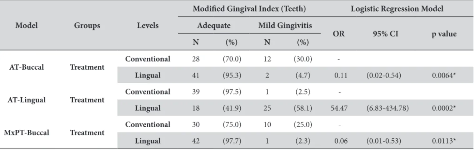
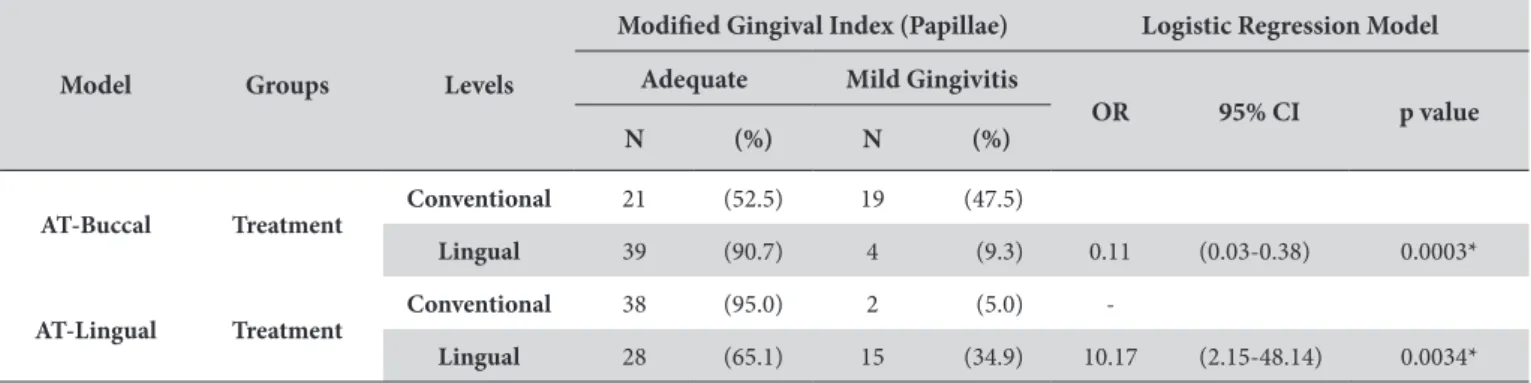
Based on registered MGI data, all patients had scores varying from 0 (adequate) to 0.7 (mild gingivitis). In patients having some kind of alteration, the scores varied from 0.1-0.7 (this relatively high score was registered for only one patient). herefore, the MGI had to be dichotomized as adequate/ mild gingivitis. As shown in Table 2, patients under conventional treatment would also have higher chances of presenting mild gingivitis on the buccal surfaces of anterior (OR = 1/0.11 = 9.0; p = 0.0064) and maxillary posterior (OR = 1/0.06 = 16.7; p = 0.0113) teeth. Furthermore, this group evidenced signiicantly higher frequency of mild gingivitis on the buccal surfaces of anterior interproximal papillae (Table 3). Conversely, patients using lingual brackets would have signiicantly higher chances of developing mild gingivitis on the lingual surfaces (OR = 54.47, p = 0.0002) and interproximal papillae (OR = 10.17; p = 0.0034) of the anterior teeth (Tables 2 and 3).
Patients under conventional treatment had signiicantly higher frequencies of BOP in the free surfaces of maxillary and mandibular posterior teeth (Table 4), resulting in corresponding greater chances of demonstrating BOP (OR = 1/0.29 = 3.4 and OR = 1/0.26 = 3.8, respectively). No statistically signiicant diference was found in the interproximal papillae.
Table 1. Multiple logistic regression for analyzing the efect of treatment type on the presence of dental plaque, registered on the buccal and lingual surfaces of anterior and posterior teeth
Model Groups Levels
Presence of Plaque Logistic Regression Model no yes
OR 95% CI p value N (%) N (%)
AT-Buccal Treatment
Conventional 14 (35.0) 26 (65.0)
-Lingual 37 (86.0) 6 (14.0) 0.08 (0.03-0.25) <0.0001*
AT-Lingual Treatment
Conventional 21 (52.5) 19 (47.5)
-Lingual 9 (20.9) 34 (79.1) 4.32 (1.62-11.51) 0.0034*
MxPT-Buccal Treatment
Conventional 17 (42.5) 23 (57.5)
-Lingual 31 (72.1) 12 (27.9) 0.28 (0.11-0.71) 0.0072*
AT: Anterior Teeth (11 and 31); MxPT: Maxillary Posterior Teeth (16 and 26); OR: odds ratio; 95% CI: 95 per cent conidence interval. *Signiicant at level of 1% (p value < 0.01).
Table 2. Multiple logistic regression for analyzing the efect of treatment type on the Modiied Gingival Index (dichotomized as adequate and mild gingivitis), registered on the buccal and lingual surfaces of anterior and posterior teeth
Model Groups Levels
Modiied Gingival Index (Teeth) Logistic Regression Model Adequate Mild Gingivitis
OR 95% CI p value N (%) N (%)
AT-Buccal Treatment
Conventional 28 (70.0) 12 (30.0)
-Lingual 41 (95.3) 2 (4.7) 0.11 (0.02-0.54) 0.0064*
AT-Lingual Treatment
Conventional 39 (97.5) 1 (2.5)
-Lingual 18 (41.9) 25 (58.1) 54.47 (6.83-434.78) 0.0002*
MxPT-Buccal Treatment
Conventional 30 (75.0) 10 (25.0)
-Lingual 42 (97.7) 1 (2.3) 0.06 (0.01-0.53) 0.0113*
DISCUSSION
he chances of VP (Table 1) and mild gingivitis (Tables 2 and 3) occurring on the buccal surfaces of anterior and maxillary posterior teeth, as well as BOP occurring on maxillary and mandibular posterior teeth (Table 4) would be signiicantly greater for patients under conventional treatment. Nevertheless, the lingual etching of brackets does not prevent the adverse efect of dental plaque formation during orthodontic treatment1,11. he higher frequencies of dental
plaque and mild gingivitis on the lingual surfaces of anterior teeth (Tables 1, 2 and 3) may be related to the limitation of mechanical self-cleaning imposed by the lingual brackets. Moreover, the design of some lingual brackets, with hooks on the cervical region, may favor plaque accumulation1. Oral hygiene was signiicantly impaired
in patients using pre-adjusted lingual brackets in comparison to those using customized lingual brackets19. In this study, all patients
used pre-adjusted brackets.
Placement of biomaterials and orthodontic appliances inluences
in situ dental plaque formation1,20. However, the initial thickness of
bioilm is smaller on the lingual surfaces20, presumably due to the
action of the tongue and salivary low promoting a self-cleaning mechanism. Although oral hygiene is even more important for lingual therapy, since plaque accumulation and gingivitis are not detected by the patient4, the lingual brackets would supposedly
not necessarily induce microbial and periodontal alterations at the same level as that of conventional treatment21.
he present study was conducted in two clinics located in diferent Brazilian states and with patients who used distinct types of brackets. No clinically signiicant efect was observed for the origin. Based on anamnestic data, 70% of the total sample reported daily hygiene of the proximal surfaces, which is important to prevent not only periodontal disease but also caries lesions. Despite the possible inluence of diferent types of lingual brackets, adequate oral hygiene instructions and monitoring seem to be the clinical key measures for preventing gingival inlammation in orthodontic patients.
Clinical periodontal parameters were evaluated in 83 orthodontic patients, most of whom had been using orthodontic appliances for more than 3 months. he maximum increase in periodontal and microbial parameters was registered at 3 months of orthodontic treatment22,23. Nevertheless, bacteria forming dental plaque,
associated with orthodontic treatment, may be quantiied at one and ive weeks ater bonding24. Concerning lingual therapy, a
signiicant increase in the dental plaque index was observed ater one month of treatment9 and gingivitis was diagnosed in 7 out of
every 10 patients ater 3 months of treatment10. Greater dental plaque
formation is expected ater placement of orthodontic appliances, in both conventional23,24 and lingual treatments11,12, which in turn
Table 3. Multiple logistic regression for analyzing the efect of treatment type on the Modiied Gingival Index (dichotomized as adequate and mild gingivitis), registered on the buccal and lingual interproximal papillae of anterior and posterior teeth
Model Groups Levels
Modiied Gingival Index (Papillae) Logistic Regression Model Adequate Mild Gingivitis
OR 95% CI p value N (%) N (%)
AT-Buccal Treatment
Conventional 21 (52.5) 19 (47.5)
Lingual 39 (90.7) 4 (9.3) 0.11 (0.03-0.38) 0.0003*
AT-Lingual Treatment
Conventional 38 (95.0) 2 (5.0)
-Lingual 28 (65.1) 15 (34.9) 10.17 (2.15-48.14) 0.0034*
AT: Anterior Teeth (11 and 31); OR: odds ratio; 95% CI: 95 per cent conidence interval. *Signiicant at level of 1% (p value < 0.01).
Table 4. Multiple logistic regression for analyzing the efect of treatment type on the presence of bleeding on probing, registered on the buccal or lingual surfaces of anterior and posterior teeth
Model Groups Levels
Bleeding on Probing (Teeth) Logistic Regression Model no yes
OR 95% CI p value N (%) N (%)
MxPosterior Treatment
Conventional 24 (60.0) 16 (40.0)
-Lingual 36 (83.7) 7 (16.3) 0.29 (0.10-0.81) 0.0184*
MdPosterior Treatment
Conventional 23 (57.5) 17 (42.5)
-Lingual 36 (83.7) 7 (16.3) 0.26 (0.09-0.73) 0.0106*
is associated with an increase in probing depth and BOP11,12,25.
Increasing dental plaque formation and BOP in patients under lingual therapy were registered between 4 weeks11 and 3 months12
ater beginning orthodontic treatment.
Patients under conventional therapy exhibited higher frequencies of dental plaque and mild gingivitis on the buccal surfaces of maxillary posterior teeth (Tables 1 and 2). hese indings may be related to the presence of orthodontic bands inserted into the irst molars3,20,26. Seating these accessories may compromise the health of
the surrounding periodontal tissues and may be associated with the occurrence of periodontopathogenic bacteria3. Orthodontic bands
may also be associated with the signiicantly higher frequency of BOP in maxillary and mandibular posterior teeth (Table 4).
Dental plaque levels in orthodontic patients would be 2 to 3 times higher than are observed in adults without ixed appliances27.
he indings of this study are in agreement with those of previous investigations11,12,22-24, since it clearly demonstrates that patients
under both conventional and lingual treatment are prone to VP accumulation and the development of mild gingivitis. As a result of microbiological changes ater bracket etching, increased metabolic activity and pathogenicity of the oral microlora may be reported11.
his can be conirmed by speciic microbial analyses. Nevertheless,
the unique laboratory microbial parameter is not a direct measure of gingival disease. Host salivary features and immunological defenses should be taken into account. hat is why clinical evaluation is mandatory in every study of periodontal health. Gingivitis may be completely treated in one week by adequate tooth brushing and use of dental loss. Even BOP may be solved without intervention, if there is no bone loss. Hence, if monitoring and motivation for maintaining proper oral hygiene are frequently carried out, controlled levels of VP and mild gingivitis will not have important clinical repercussions on periodontal health.
CONCLUSION
Orthodontic brackets ixed on any tooth surface contribute to dental plaque retention. Signiicantly higher frequencies of dental plaque and mild gingivitis were recorded on the buccal surfaces of patients under conventional therapy, as well as on the anterior lingual surfaces of patients under lingual therapy. Patients under conventional therapy also presented signiicantly higher frequency of bleeding on probing in posterior teeth. Despite the indings of this study, it may be concluded that clinical periodontal health conditions were acceptable for both types of treatment.
REFERENCES
1. Sfondrini MF, Debiaggi M, Zara F, Brerra R, Comelli M, Bianchi M, et al. Influence of lingual bracket position on microbial and periodontal parameters in vivo. J Appl Oral Sci. 2012 May-June;20(3):357-61. http://dx.doi.org/10.1590/S1678-77572012000300011. PMid:22858704. 2. van Gastel J, Quirynen M, Teughels W, Coucke W, Carels C. Longitudinal changes in microbiology and clinical periodontal variables after
placement of fixed orthodontic appliances. J Periodontol. 2008 Nov;79(11):2078-86. http://dx.doi.org/10.1902/jop.2008.080153. PMid:18980516. 3. Diamanti-Kipioti A, Gusberti FA, Lang NP. Clinical and microbiological effects of fixed orthodontic appliances. J Clin Periodontol. 1987
July;14(6):326-33. http://dx.doi.org/10.1111/j.1600-051X.1987.tb00979.x. PMid:3509967.
4. Hohoff A, Stamm T, Kühne N, Wiechmann D, Haufe S, Lippold C, et al. Effects of a mechanical interdental cleaning device on oral hygiene in patients with lingual brackets. Angle Orthod. 2003 Oct;73(5):579-87. PMid:14580027.
5. Caniklioglu C, Oztürk Y. Patient discomfort: a comparison between lingual and labial fixed appliances. Angle Orthod. 2005 Jan;75(1):86-91. PMid:15747820.
6. Hohoff A, Stamm T, Goder G, Sauerland C, Ehmer U, Seifert E. Comparison of 3 bonded lingual appliances by auditive analysis and subjective assessment. Am J Orthod Dentofacial Orthop. 2003 Dec;124(6):737-45. http://dx.doi.org/10.1016/j.ajodo.2003.08.022. PMid:14666090. 7. Hohoff A, Seifert E, Fillion D, Stamm T, Heinecke A, Ehmer U. Speech performance in lingual orthodontic patients measured by sonagraphy
and auditive analysis. Am J Orthod Dentofacial Orthop. 2003 Feb;123(2):146-52. http://dx.doi.org/10.1067/mod.2003.12. PMid:12594420. 8. Hohoff A, Fillion D, Stamm T, Goder G, Sauerland C, Ehmer U. Oral comfort, function and hygiene in patients with lingual brackets. A
prospective longitudinal study. J Orofac Orthop. 2003 Sept;64(5):359-71. http://dx.doi.org/10.1007/s00056-003-0307-6. PMid:14692050. 9. Sinclair PM, Cannito MF, Goates LJ, Solomos LF, Alexander CM. Patient responses to lingual appliances. J Clin Orthod. 1986
June;20(6):396-404. PMid:3461002.
10. Årtun J. A post treatment evaluation of multibonded lingual appliances in orthodontics. Eur J Orthod. 1987 Aug;9(3):204-10. http://dx.doi. org/10.1093/ejo/9.3.204. PMid:3311772.
11. Demling A, Demling C, Schwestka-Polly R, Stiesch M, Heuer W. Short-term influence of lingual orthodontic therapy on microbial parameters and periodontal status: a preliminary study. Angle Orthod. 2010 May;80(3):480-4. http://dx.doi.org/10.2319/061109-330.1. PMid:20050740. 12. Demling A, Demling C, Schwestka-Polly R, Stiesch M, Heuer W. Influence of lingual orthodontic therapy on microbial parameters and
periodontal status in adults. Eur J Orthod. 2009 Dec;31(6):638-42. http://dx.doi.org/10.1093/ejo/cjp064. PMid:19687149.
13. Zanatta FB, Moreira CH, Rösing CK. Association between dental floss use and gingival conditions in orthodontic patients. Am J Orthod Dentofacial Orthop. 2011 Dec;140(6):812-21. http://dx.doi.org/10.1016/j.ajodo.2011.06.028. PMid:22133946.
14. Greene JC, Vermillion JR. The simplified oral hygiene index. J Am Dent Assoc. 1964 Jan;68(1):7-13. http://dx.doi.org/10.14219/jada. archive.1964.0034. PMid:14076341.
16. Ainamo J, Bay I. Problems and proposals for recording gingivitis and plaque. Int Dent J. 1975 Dec;25(4):229-35. PMid:1058834. 17. Hosmer DW, Lemeshow S. Applied logistic regression. 2nd ed. New York: Wiley; 2000.
18. Demidenko E. Sample size determination for logistic regression revisited. Stat Med. 2007 Aug;26(18):3385-97. http://dx.doi.org/10.1002/ sim.2771. PMid:17149799.
19. Stamm T, Hohoff A, Ehmer U. A subjective comparison of two lingual bracket systems. Eur J Orthod. 2005 Aug;27(4):420-6. http://dx.doi. org/10.1093/ejo/cji034. PMid:16043479.
20. Auschill TM, Hellwig E, Sculean A, Hein N, Arweiler NB. Impact of the intraoral location on the rate of biofilm growth. Clin Oral Investig. 2004 June;8(2):97-101. http://dx.doi.org/10.1007/s00784-004-0255-6. PMid:14986070.
21. Demling A, Heuer W, Elter C, Heidenblut T, Bach FW, Schwestka-Polly R, et al. Analysis of supra- and subgingival long-term biofilm formation on orthodontic bands. Eur J Orthod. 2009 Apr;31(2):202-6. http://dx.doi.org/10.1093/ejo/cjn090. PMid:19304761.
22. Ristic M, Svabic MV, Sasic M, Zelic O. Clinical and microbiological effects of fixed orthodontic appliances on periodontal tissues in adolescents. Orthod Craniofac Res. 2007 Nov;10(4):187-95. http://dx.doi.org/10.1111/j.1601-6343.2007.00396.x. PMid:17973685.
23. Naranjo AA, Triviño ML, Jaramillo A, Betancourth M, Botero JE. Changes in the subgingival microbiota and periodontal parameters before and 3 months after bracket placement. Am J Orthod Dentofacial Orthop. 2006 Sept;130(3):275.e17-22. http://dx.doi.org/10.1016/j. ajodo.2005.10.022. PMid:16979483.
24. Pellegrini P, Sauerwein R, Finlayson T, McLeod J, Covell DA Jr, Maier T, et al. Plaque retention by self-ligating vs elastomeric orthodontic brackets: quantitative comparison of oral bacteria and detection with adenosine triphosphate-driven bioluminescence. Am J Orthod Dentofacial Orthop. 2009 Apr;135(4):426.e1-9, discussion 426-7. http://dx.doi.org/10.1016/j.ajodo.2008.08.018. PMid:19361723.
25. Alexander SA. Effects of orthodontic attachments on the gingival health of permanent second molars. Am J Orthod Dentofacial Orthop. 1991 Oct;100(4):337-40. http://dx.doi.org/10.1016/0889-5406(91)70071-4. PMid:1927984.
26. Erbe C, Hornikel S, Schmidtmann I, Wehrbein H. Quantity and distribution of plaque in orthodontic patients treated with molar bands. J Orofac Orthop. 2011 Mar;72(1):13-20. http://dx.doi.org/10.1007/s00056-010-0001-4. PMid:21484542.
27. Klukowska M, Bader A, Erbe C, Bellamy P, White DJ, Anastasia MK, et al. Plaque levels of patients with fixed orthodontic appliances measured by digital plaque image analysis. Am J Orthod Dentofacial Orthop. 2011 May;139(5):e463-70. http://dx.doi.org/10.1016/j.ajodo.2010.05.019. PMid:21536188.
CONFLICTS OF INTERESTS
he authors declare no conlicts of interest.
*CORRESPONDING AUTHOR
Rívea Inês Ferreira-Santos, Departamento de Radiologia, UNIP – Universidade Paulista, Campus Swit, Av. Comendador Enzo Ferrari, 280, Swit, 13045-770 Campinas – SP, Brazil, e-mail: [email protected]