Fibers Obtained from Alginate, Chitosan and Hybrid Used in the Development of Scafolds
Daniela Camargo Furuyaa, Silgia Aparecida da Costaa, Rodrigo Cardoso de Oliveirab, Humberto
Gomes Ferrazc, Adalberto Pessoa Juniord, Sirlene Maria da Costaa*
Received: July 07, 2016; Revised: October 21, 2016; Accepted: December 27, 2016
The main aim of this study was to develop scafolds based on alginate, chitosan and hybrid ibers with and without glycerol. The scafolds developed underwent assessments for tensile property, swelling ratio and weight loss, cellular viability, degradation and biomineralization, as well as DSC/TGA thermal analysis. Tenacity values showed that use of glycerol and the interaction between alginate and chitosan as a hybrid iber were associated with increasing tenacity values. In the swelling and weight loss study, the scafolds containing glycerol presented lower weight loss and higher water absorption values in all scafolds, compared to scafolds without glycerol, indicating that glycerol acted as a stabilizer. None of the alginate, chitosan and hybrid scafolds, with or without glycerol, decreased cell viability. On the third day of the biomineralization assay, chitosan without glycerol indicated the presence of apatite crystals. The degradation study showed that glycerol may have acted as a stabilizer.
Keywords: Alginate, chitosan, iber, scafolds, tissue engineering
* e-mail: [email protected]
1. Introduction
Bone tissue engineering is a multidisciplinary ield, focused on designing a scafold to mimic the environment of the extracellular matrix and the study of more eicient signaling molecules and growth factors that stimulate regeneration. Thus, the scafold has to behave as a temporary structure supporting bone tissue regeneration while it degrades and is replaced with the new bone1,2.
Textiles have been used in a wide range of medical devices, i.e., in contact with biological environments, such as vascular grafts, repair meshes, etc.3 Fibers are particularly useful for
producing scafolds due to their inherent properties, such as satisfactory porosity, adjustable elastic modulus and light weight, which promote tissue regeneration4. There are several
diferent methods of producing iber-based scafolds, such as electrospinning, meltspinning, wetspinning, biospinning and microluidic spinning5,6. The wetspinning method was
chosen for this study.
The ideal scafold will have a number of properties, such as biocompatibility, biodegradability, desirable pore size and satisfactory porosity, non-toxic responses, easy handling and an abundant source of raw material7,8. Scafold
porosity does not need to be uniform, because natural bone
is not uniform8, which is another reason for using ibers to
develop these scafolds.
The biopolymers selected for this study were alginate and chitosan. Alginate is a polysaccharide derived from brown algae and it comprises 1/4 linked β-D-mannuronic acid (M) and its α-L-guluronic acid (G). The properties of alginate are related to the proportion and dimension of the guluronic blocks (G) in the chain9. It is biocompatible,
biodegradable, and has the ability to absorb 200-300 times its own weight, thus showing potential for use as a Drug Delivery System10. Alginate scafolds have the ability to
support tissue regeneration, including bone, skin, liver, etc7. Chitosan is a natural polymer derived from the partial
deacetylation of chitin, obtained from the exoskeleton of crustaceans. It is the most abundant cationic polysaccharide and the second most abundant in nature, behind only cellulose. It comprises (1-4)-2-acetamido-2-deoxy-ß-D-glucan (N-acetyl D-glucosamine) and (1-4)-2-amino-2-deoxy-ß-D-glucan (D-glucosamine) units. It is also a biocompatible and biodegradable polymer and it is osteoconductive, as well as hemostatic and antimicrobial11. Chitosan scafolds
are biodegradable and tissue biocompatible. However, this polymer is mechanically weak, which is why it needs to be used in conjunction with another polymer12.
a School of Arts, Sciences and Humanities, Textile and Fashion Course, University of São Paulo, Arlindo Béttio, 1000, 03828-000, Jardim Keralux, SP, Brazil
b Department of Biological Sciences, Bauru School of Dentistry, University of São Paulo, Alameda Dr.
Octávio Pinheiro Brisolla, 9-75, Vila Universitária, 1701290, Bauru, SP, Brazil
c Department of Pharmacy, School of Pharmaceutical Sciences, University of São Paulo, Av. Prof. Lineu Prestes, 580, Cidade Universitária, 05508-000, Butantã, SP, Brazil
d Department of Biochemical and Pharmaceutical Technology, School of Pharmaceutical Sciences,
The interaction between alginate and chitosan generates a polyelectrolyte complex that can be obtained by precipitating cationic polymers when mixed with anionic polymers in aqueous solutions, leading to mutual precipitation, which occurs in this case, because alginate and chitosan are anionic and cationic polysaccharides, respectively13. Another advantage
of the interaction between its opposite charges is an upgrade in the mechanical strength of the developed structure formed by these two polymers14,15. This combination is signiicant
for scafold development, because while alginate ofers functional groups for cellular regeneration, chitosan supports the scafold structure16. Finally, the interaction of these two
polymers creates a strong connection between them, because alginate is a hydrophilic polysaccharide and promotes low protein adsorption, diferent from chitosan, a less hydrophilic polysaccharide that promotes high protein adsorption17,18.
The aim of this study is to develop and characterize scafolds based on alginate, chitosan and hybrid alginate/ chitosan ibers. All scafolds were produced with and without glycerol in order to identify any positive properties as potential plasticizers.
2. Materials and Methods
2.1. Materials
Sodium alginate (20,000 – 40,000 cps viscosity), crab-extracted chitosan (85 % deacetylation minimum) and all reagents used were acquired from Sigma Co. (St. Louis, ME, USA).
2.2 Methods
2.2.1 Preparation of alginate, chitosan and hybrid ibers with and without glycerol
Alginate, chitosan and hybrid ibers were produced using the wet spinning technique with a syringe (Hypodermic syringes, polypropylene Luer-lock tip, capacity 10 mL, graduated, 25 x 7 mm and needle Ø 0.6 x L 2.5 mm).
Alginate 5 % (m/v) was dissolved in 100 mL of 0.1 M sodium hydroxide by stirring at room temperature overnight. The sodium alginate solution with and without glycerol was injected into a coagulation bath at 30 ºC containing 2 % calcium chloride (m/v). Chitosan 2.2 % (m/v) was dissolved in 200 mL of 2 % acetic acid (v/v) by stirring at room temperature overnight and subsequently adding 250 mL of methanol (v/v). The chitosan gel was iltered using a vacuum pump. The chitosan gel with and without glycerol was injected into a coagulation bath at 30 ºC containing 300 mL of 0.5 M sodium sulfate, 100 mL of 1 M sodium hydroxide and 600 mL of distilled water. The resultant ibers were extracted from this coagulation medium after 24 hours and placed in 50 % methanol for 2 h. Hybrid ibers were produced by
extruding the alginate gel, with and without glycerol, in a solution containing 50 % calcium chloride solution (v/v) and 50 % of 0.02 % chitosan gel (m/v). The ibers remained in this solution for 24 h. Subsequently, the ibers were stored in 50 % (v/v) methanol for 2 h. 2.5 % glycerol (m/m) was added to all gel solutions in order to ascertain its efect as a plasticizer. The resultant alginate, chitosan and hybrid ibers were washed three times in distilled water and part of the ibers were rolled manually on a polypropylene cylindrical support, dried at room temperature conditions for 24 h and characterized according to item 2.2.3. The other part of the ibers were used to produce the scafolds (item 2.2.2).
2.2.2. Preparation of alginate, chitosan and hybrid scafolds with and without glycerol
The alginate, chitosan and hybrid scafolds were developed using the freeze-drying method. After production (item 2.2.1), the ibers previously weighed in a mass of approximately 0.255 g, were placed in a polystyrene mold and stored in a ultrafreezer at -67 °C for 2 h to freeze the structures. Then these structures were lyophilized in a L101 Liotop lyophilizer until dried, a process lasting approximately 60 h at -30 ºC. The scaffolds were produced using two different polystyrene molds, both with external dimensions of 8.54 x 12.76 cm. The mold for the production of large structures corresponded to the 12-well cell culture plate, with a diameter and depth of approximately 15 mm per well. The mold for the production of small structures corresponded to the 96-well plate for cell cultures, with 6.4 mm deep 96-wells. The production process for the scafolds was the same for both molds. The smaller structures were prepared to facilitate the coverage of the samples in microscopy assays. The scafolds produced in the large mold were characterized for cell viability (item 2.2.4.3), DSC / TGA (item 2.2.4.1) and absorption and weight loss (item 2.2.4.2). While the scafolds produced in the small mold were characterized for biomineralization (item 2.2.4.5), degradation with lysozyme (item 2.2.4.4) and SEM/EDS.
2.2.3. Mechanical testing of the ibers
For diameter and linear density determinations in accordance with ISO 5084 (1996)19, dried samples ibers
were stored at 20 ± 2 ºC and relative humidity of 65 ± 4 % for 24 h. After acclimatization, each sample was weighed using an analytical balance (AUW220D, Shimadzu, São Paulo, Brazil). The results obtained represent the ratio between the weight and the iber length20. The assay was performed
based on ISO 2060 (1994)21, ISO 1139 (1973)22 and ISO 139
(2005)23. Tensile properties of the acclimatized ibers (rupture
strength, tenacity, elongation and Young’s modulus) were determined according to ASTM D 3822 (2007)24 using an
Accordingly, in order to determine tenacity (strength value shared by linear density), iber ineness (linear density or count number) was calculated in terms of TEX, deined as the weight in grams per 1,000 m of iber, by weighing a known length of the iber. A load cell of 10 N, gauge length of 200 mm, automatic pre-tension and crosshead speed of 100 mm/ min were used. The tensile parameters were determined when the iber broke immediately after maximum elongation.
2.2.4. Characterization of the scafolds
2.2.4.1 Diferential Scanning Calorimetry – DSC/ Thermogravimetry – TGA
DSC tests were performed in the DSC 7020 (Exstar, SII Nano Technology In., Japan) under an inert nitrogen atmosphere at a low rate of 50 mL/min. Temperatures ranged from 25 – 350 °C with a heating rate of 10 °C/min. A closed aluminum capsule containing 2 mg of sample mass was used. The temperature and fusion heat were calibrated with Indium.
TG analysis was performed in a TG/DTA 7200 (Exstar, SII Nano Technology In., Japan) under a 100 mL/min nitrogen atmosphere. Temperatures ranged from 25 – 600 ºC with heating rate of 10 ºC/min. Closed platinum capsules containing 3.5 mg of sample mass were used. The temperature and heat fusion were calibrated with Calcium oxalate before the assay. Both DSC and TG analyses were performed with alginate and chitosan polymers and of alginate, chitosan and hybrid iber scafolds, with and without glycerol.
The assay was performed at the Department of Pharmacy, School of Pharmaceutical Sciences.
2.2.4.2. Studies on water uptake or swelling and weight loss
Swelling behavior and scafold weight loss were performed in triplicate. The scafolds were weighed and immersed in 10 mL of distilled water. The lask samples (15 mL) were closed with Parailm (Pechiney Plastic Packaging Co., Chicago, IL, USA) and placed in a thermo regulated bath (TEC-420, Tecnal, Piracicaba, Brazil) under agitation (60 rpm), at 37 ºC for 1, 3, 7, 15, 21 and 30 days. For water uptake calculation, an analytical balance (AUW220D, Shimadzu, São Paulo, Brazil)was used to determine the initial scafold mass (mi) and the inal mass (mw), after incubation and removal of the excess solution with ilter paper. Subsequently, the scafolds were dried at 40 ºC until constant weight was attained in order to determine (mf). Water uptake and weight loss are calculated by equations 1 and 2.
2.2.4.3. Cell viability assay
2.2.4.3.1. Cell culture
For this study, NIH3T3 P12 ibroblasts (ATCC – American Type Culture Collection) were used. Cells were cultivated in Dulbecco’s modiied Eagle medium (DMEM) supplemented with 10 % bovine fetal serum (FBS – NUTRICELL) and incubated in an incubator at 37 ºC containing 5 % CO2.
After subconluence, cells were subcultivated with trypsin (0.25 % trypsin, 1mM EDTA – Sigma Aldrich®), in order to
dissociate cells from the culture lasks, and maintained for 5 min in the incubator at 37 ºC with 5 % CO2. Then the trypsin
was inactivated from DMEM using 10 % FBS. Cells were transferred to 15 mL Falcon tubes (TPP® - Techno Plastic
Products) and centrifuged for 5 min at 4 ºC and 1200 rpm. Subsequently, the supernatant was discarded and cells were resuspended in new DMEM with 10 % FBS. Cellular account was made with trypan blue protocol and cells were used for experimental assays of MTT and crystal violet.
According to ISO No. 10.993-5, the concentrations used in this study were 10 mg extract (samples of alginate, chitosan and hybrid scafolds, with and without glycerol) for 1 mL DMEM with 10 % FBS, i.e., proportion of 10 mg/1 mL. For viability assays, 2x10³ cells/well were plated
in 96 wells plates. After the 24-hour incubation period, the medium was replaced with DMEM with 10 % FBS and conditioned with the scafolds. Control groups received only the DMEM medium: with 10 % FBS (positive control) and 1 % FBS (negative control). Each plate was analyzed in an experimental period of 24 and 48 h after the addition of the conditioned medium25.
2.2.4.3.1.1. MTT
After each experimental period (24 and 48 h), cells were washed with PBS and incubated in a solution containing 0.5 mg MTT for 1 mL DMEM without FBS. After this procedure, the plates were maintained at 37 ºC for 4 h, when the solution was removed and the insoluble pigment was extracted in dimethyl sulfoxide (DMSO). Absorbance was measured at 562 nm by Synergy H1 Monochromator based Biotek.
2.2.4.3.1.2. Crystal Violet
After each experimental period (24 and 48 h), cells were washed with PBS and 100% methanol was added for 10 min. After the methanol was removed, 0.2 % crystal violet solution was added to 20 % ethanol for 3 min. Then the solution was removed and the wells were washed twice with PBS to remove the excess dye. Finally, 0.05 mol.L-1
of sodium citrate solution was added with 50 % ethanol for 10 min. Absorbance was determined at 540 nm by Synergy H1 Monochromator based Biotek.
/
( )
Water uptake
=
"
Q
m
W-
m
iV
m
i%
#
100 1
/
( )
2.2.4.3.2. Statistical Analysis
MTT and Crystal violet statistical analyses were performed using GraphPad Prism 4 software selecting statistical test one way ANOVA using Tukey’s test with p < 0.05 indicating statistical signiicance. The cellular viability assay was performed in the Biochemistry Laboratory at the Biological Sciences Department of the Bauru School of Dentistry, University of São Paulo.
2.2.4.4. In vitro degradation study
This study was performed in triplicate in order to determine the inluence of the lysozyme on the scafold properties. The chitosan and hybrid scafolds, with and without glycerol, weighing approximately 0.100 g were immersed in 10 mL of PBS pH 7.4 ± 0.02 containing lysozyme (EC 3.1.2.17, chicken egg white, 40000 U/mg, Sigma), similar to that found in human serum (13 mg/L), and incubated at 37 ºC at diferent time intervals (1, 3, 5, 7 and 10 days) under static conditions. After each incubation time, the scafolds were dried at 40 ºC and subjected to SEM analysis. After these incubation periods, the concentration of reducing sugars were determined by DNS method26. Absorption and weight loss
were determined as outlined above (item 2.2.4.2).
2.2.4.5. In vitro biomineralization study
The biomineralization assay was performed in triplicate. The alginate, chitosan and hybrid scafolds, with and without, glycerol weighing approximately 0.100 g were immersed in 10 mL of 1x simulated body luid (SBF) and then incubated at 37 ºC in closed Falcon tubes for preset time periods of 1, 3, 5, 7, 15, 21 and 30 days. SBF is a solution that contains the necessary minerals to mimic the body luid, prepared according to the method27. After the preset time interval,
the scafolds were removed and dried in 40 ºC. The dried scafolds were subjected to SEM analysis for examination of mineralization.
3. Results and Discussion
3.1 Mechanical testing
Tension testing or material behavior under strength and deformations applied along the axis is one of the most important characteristics related to mechanical properties28.
Fiber tenacity is the quotient of breaking load by the count number. This calculation is necessary in order to normalize the efect of diferent thickness values found in some samples containing several ibers20. Table 1 shows the tenacity values
of all ibers. Tenacity values for alginate, chitosan and hybrid ibers were between 7.25-7.77, 4.47-8.64 and 9.97-10.0 cN/ tex, and the use of glycerol is associated with a trend of
increasing tenacity values. The tenacity values obtained in this study are slightly inferior to those found in literature (14.0-18 cN/Tex)29 and (11.0-18 cN/Tex)30,31. However, it is
worth mentioning that the cited references do not mention the alginate concentrations used. Hybrid ibers presented an increase of 27.3 % and 55.2 % in tenacity values compared to alginate and chitosan, respectively.
3.2 Scafold preparation
The results of scafold production can be observed in Figures 1 and 2. The scafolds presented an interconnected porous structure due to iber structure.
3.3. DSC/TGA
Diferential Scanning Calorimetry is an important study of a material thermal behavior that identiies mass loss, reduction reactions and desorption by the endothermic peaks and crystallization, polymerization reactions and oxidation by the exothermic peaks32. The results of the DSC of the
scafolds with and without glycerol are analyzed in Figure 3. According to the thermogram of the scafolds without glycerol (Figure 3a), endothermic peaks of alginate occurred at 122.4 ºC and 195.6 ºC, of chitosan at 181 ºC and the hybrid at 174.2 ºC. Comparing these peaks, the hybrid endothermic peak is located in a intermediate temperature between alginate and chitosan. The exothermic peaks of alginate occurred at 271.7 ºC, of chitosan at 304.1 ºC and the hybrid at 277.7 ºC. A displacement of the hybrid peaks (both, endothermic and exothermic) was noted, with regard to the polymers themselves, indicating the formation of new chemical bonds and the interaction of alginate and chitosan. In the thermogram of scafolds with glycerol (Figure 3b), a displacement of the hybrid endothermic and exothermic peaks occurred, with regard to the alginate and chitosan peaks, which indicates the same behavior noted in the thermogram of the scafolds without glycerol (Figure 3a). The hybrid endothermic peak occurred at 173.9 ºC and the exothermic peak at 277.5 ºC. In all scafolds with glycerol, a displacement of the endothermic and exothermic peaks was noted, compared to the results of scafolds without glycerol, indicating the interaction between the polymers and the glycerol.
Through thermogravimetry, the relationship between material mass and its temperature could be detected. The decomposition of the polymers can be analyzed, indicating weight loss of the sample or water evaporation33. The TGA
results are analyzed in Figure 4.
Table 1. Tenacity values of alginate, chitosan and hybrid ibers without and with glycerol
Fibers Linear density (tex) Breaking Load (N) Tenacity (cN/tex) Elongation (%) Young’s Modulus (N/tex)
Alginate 22.3 ± 0.98 1.63 ± 0.11 7.25 ± 0.51 4.89 ± 2.43 4.77 ± 0.49 Alginate with glycerol 17.1 ± 0.63 1.35 ±0.25 7.77 ± 1.39 5.41 ± 0.45 5.02 ± 0.45 Chitosan 17.2 ± 0.7 0.77 ± 0.05 4.47 ± 0.28 1.45 ± 0.97 4.16 ± 1.01 Chitosan with glycerol 17.2 ± 0.65 1.49 ± 0.08 8.64 ± 0.51 4.25 ± 2.83 5.55 ± 0.41 Hybrid 23.4 ± 1.8 2.23 ± 0.24 9.97 ± 0.99 6.86 ± 1.32 5.64 ± 0.94 Hybrid with glycerol 20.9 ± 2.01 2.09 ± 0.12 10 ± 0.80 6.58 ± 1.91 5.73 ± 0.64
Figure 1: Scafolds of (a) alginate without glycerol, (b) alginate with glycerol, (c) chitosan without glycerol, (d) chitosan with glycerol,
(e) hybrid without glycerol and (f) hybrid with glycerol produced in mold of 15 mm per well in digital image and optical microscopy with 20x magniication.
and deacetylated units of chitosan and depolymerization34.
The scafolds with glycerol behaved in a similar fashion to the scafolds without glycerol.
3.4. Swelling studies and weight loss
In tissue engineering, swelling ability is responsible for the polymeric matrix expansion related to scafold morphology
and cell-nutrient transmission15,35. The swelling ratio and the
Figure 2. Optical microscopy of (a) alginate without glycerol, (b) chitosan withoutglycerol, (c) hybrid without glycerol, (d), alginate with
glycerol, (e) chitosan with glycerol and (f) hybrid with glycerol scafolds produced in mold of 6.4 mm per well with 20x magniication.
Figure 3: DSC peaks of scafolds based on alginate, chitosan and hybrid (a) without and (b) with glycerol.
Figure 5: Results of (a) swelling ratio and (b) weight loss of alginate, chitosan and hybrid scafolds with glycerol.
with glycerol values were higher than the scafold without glycerol, indicating that the presence of glycerol had increased the swelling behavior of the scafolds.
The highest weight loss corresponds to the alginate scafold with glycerol, accounting for 16 % weight loss compared with 15 % from the hybrid scafold with glycerol and 11.1 % from the chitosan scafold with glycerol on the thirtieth day. Compared to the scafolds without glycerol, the scafolds containing glycerol presented less weight loss in all scafolds, suggesting that the presence of glycerol worked as a stabilizer of the scafold structures36,37.
3.5. Cell viability assay
Cell viability and proliferation on a material are indications of material compatibility, suggesting potential tissue engineering applications. None of the samples decreased cell viability.
After the 24 and 48 h, all scafolds with and without glycerol presented absorbance values above the negative control (Figure 6). After 48 h, chitosan scafolds with and without glycerol presented 0.46 % and 3.11 % higher absorbance compared to the positive control, respectively, indicating the possibility of cell proliferation in these scafolds. The hybrid scafold without glycerol presented absorbance values close to the positive control (3.67 % above) after 48 h, also indicating possible cell proliferation.
3.5.1. Crystal violet
After the 24-hour period, all scaffolds presented results above the positive control values, and the
chitosan scaffold with glycerol presented 45.2 % greater absorbance than the negative control (Figure 7); after the 48-hour period, the greater absorbance rate was the hybrid scaffold without glycerol, presenting a rate 37.6 % higher than the positive control, indicating possible cell proliferation. In this assay, it could be concluded that the blend of alginate and chitosan improved the action of the polymers, compared to the results for the pure substances, because the hybrid scaffold presented a relevant result after 48 h.
Analyzing the two assays (MTT and violet crystal), the results of the hybrid scafold showed satisfactory biological results in both experiments, with absorbance values near and above the positive control, respectively. This indicates good compatibility and potential as a biomaterial.
3.6. In vitro degradation study
Degradation of chitosan and hybrid scafolds with and without glycerol was studied by immersing the scafolds in PBS solution containing lysozyme. The degradation rate was analyzed by SEM (Figure 8).
Figure 6: Cellular viability assay using MTT after 24h and 48 h
of the alginate, chitosan and hybrid scafolds without and with glycerol. *Signiicant statistic diference (p<0.05) in related to the positive control.
Figure 7: Cellular viability assay using crystal violet after 24h
and 48 h of the alginate, chitosan and hybrid scafolds without and with glycerol. *Signiicant statistic diference (p<0.05) in related to the positive control.
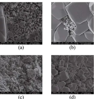
Figure 8: SEM of (a) chitosan without glycerol, (b) chitosan with
glycerol, (c) hybrid without glycerol and (d) hybrid with glycerol scafolds in the seventh day of assay.
Figure 9: Concentration of reduced sugars of the hybrid and chitosan
scafolds without and with glycerol.
3.7. In vitro biomineralization study
Biomineralization of alginate, chitosan and hybrid scafolds with and without glycerol was studied by immersing the scafolds in the SBF solution. The mineral deposition in the scafolds was analyzed by SEM (Figure 10).
Chitosan with and without glycerol presented an incidence of mineral deposition compared to the other
scafolds after three days of assay (Figure 10c; 10d). The presence of apatite crystals could be conirmed by the EDS (Figure 10cl; 10dl), indicated by the presence of Ca, P, Mg,
K. In the hybrid scafold without glycerol, apatite crystals also appeared on the third day (Figure 10a; 10b). On the seventh day, the incidence of hydroxyapatite increased in the hybrid scafold with and without glycerol (Figure 11a; 11b), and on the ifteenth day the chitosan scafold with and without glycerol presented evidence of mineralization with the apatite crystals (Figure 11c; 11d).
4. Conclusions
Alginate, chitosan and hybrid scafolds with and without glycerol were developed and characterized. Tensile tests presented results indicating that the use of glycerol and the interaction between alginate and chitosan as a hybrid iber were associated with a trend of increasing tenacity values. Hybrid ibers presented an increase of 27.3 % and 55.2 % in tenacity values, compared to chitosan and alginate ibers, respectively. DSC/TG indicated an interaction between the polymers in the thermal analysis. The swelling study and weight loss demonstrated that the scafolds containing glycerol presented less weight loss and even higher water absorption values in all scafolds, compared to the scafolds without glycerol, indicating that glycerol is a stabilizer. None of the alginate, chitosan and hybrid scafolds with and without glycerol decreased cell viability. In the MTT assay, chitosan with and without glycerol presented higher absorbance than the positive control, indicating cell proliferation. On the third day of the biomineralization assay, chitosan with and without glycerol and the hybrid scafold without glycerol presented apatite crystals. The degradation study showed that glycerol may have worked as a stabilizer.
Figure 10: SEM and EDS of (a;al) alginate without glycerol, (b;bl) alginate with glycerol, (c;cl) chitosan without glycerol, (d;dl) chitosan with glycerol, (e;el) hybrid without glycerol and (f;fl) hybrid with glycerol scafolds in third day of assay.
Figure 11. SEM of (a) chitosan without glycerol and (b) chitosan
with glycerol scafolds in the seventh day of assay; (c) hybrid without glycerol and (d) hybrid with glycerol scafolds in the tenth day of assay.
This attests to the fact that the combination of the two polymers can be considered an advance in the development of biomaterial. All tests showed promising results, but other
essays are needed for a better evaluation for its application in bone tissue engineering.
5. Acknowledgements
The authors gratefully acknowledge University of São Paulo, CAPES, FAPESP (2013/08617-7), CNPq Brazil for the inancial support. The authors are grateful to Centro Integrado de Pesquisa (CIP) – Bauru School of Dentistry – USP for providing some work facilities.
6. References
1. Chen Q, Liang S, Thouas GA. Elastomeric biomaterials for tissue engineering. Progress in Polymer Science. 2013;38(3-4):584-671. 2. Vacanti JP, Vacanti CA. The History and Scope of Tissue
Engineering. In: Lanza R, Langer R, Vacanti J, eds. Principles of Tissue Engineering. London: Elsevier; 2013. p. 3-8. 3. Ayaz HGS, Perets A, Ayaz H, Gilroy KD, Govindaraj M, Brookstein
D, et al. Textile-templated electrospun anisotropic scafolds for regenerative cardiac tissue engineering. Biomaterials. 2014;35(30):8540-8552.
4. Doser M, Planck H. Textiles for implants and regenerative medicine. In: Bartels VT, ed. Handbook of Medical Textiles. Cambridge: Woodhead Publishing; 2011. p. 132-152. 5. Tamayol A, Akbari M, Annabi N, Paul A, Kadhemhosseini A,
Juncker D. Fiber-based tissue engineering: Progress, challenges and opportunities. Biotechnology Advances. 2013;31(5):669-687. 6. Braghirolli DI, Stefens D, Pranke P. Electrospinning for regenerative
7. Venkatesan J, Bhatnagar I, Manivasagan P, Kang KH, Kim SK. Alginate composites for bone tissue engineering: A review. International Journal of Biological Macromolecules. 2015;72:269-281.
8. Bose S, Roy M, Bandyopadhyay A. Recent advances in bone tissue engineering scaffolds. Trends in Biotechnology. 2012;30(10):546-554.
9. Wang P, Tawiah B, Tian A, Wang C, Zhang L, Fu S. Properties of alginate iber spun-dyed with luorescent pigment dispersion.
Carbohydrate Polymers. 2015;118:143-149.
10. Venkatesan J, Bhatnagar I, Kim SK. Chitosan-alginate biocomposite containing fucoidan for bone tissue engineering. Marine Drugs. 2014;12(1):300-316.
11. Croisier F, Jérôme C. Chitosan-based biomaterials for tissue engineering. European Polymer Journal. 2013;49(4):780-792. 12. Li Z, Ramay HR, Hauch KD, Xiao D, Zhang M. Chitosan-alginate hybrid scafolds for bone tissue engineering. Biomaterials. 2005;26(18):3919-3928.
13. Simsek-Ege FA, Bond GM, Stringer J. Polyelectrolyte complex formation between alginate and chitosan as a function of pH.
Journal of Applied Polymer Science. 2003;88(2):346-351. 14. Baysal K, Aroguz AZ, Adiguzel Z, Baysal BM. Chitosan/
alginate crosslinked hydrogel: Preparation, characterization and application for cell growth purposes. International Journal of Biological Macromolecules. 2013;59:342-348.
15. Lee K, Ahn S, Choi CH, Lee D, Jung WK, Kim G. Functionallized alginate/chitosan biocomposites consisted of cylindrical struts and biologically designed for chitosan release. Current Applied Physics. 2014;14(8):1105-1106.
16. Iwasaki N, Yamane ST, Majima T, Kasahara Y, Minami A, Harada K, et al. Feasibility of polysaccharide hybrid materials for Scafolds in cartilage tissue engineering: evaluation of chondrocyte adhesion to polyion complex ibers prepared from alginate and chitosan. Biomacromolecules. 2004;5(3):828-833.
17. Hu WW, Yu HN. Coelectrospinning of chitosan/alginate ibers by dual-jet system for modulating material surfaces. Carbohydrate Polymers. 2013;95(2):716-727.
18. Hyland LL, Taraban MB, Hammouda B, Yu YB. Mutually reinforced multicomponent polysaccharide networks. Biopolymers. 2011;95(12):840-851.
19. Maluf E, Kolbe W. Textile ibers. In: Technical Textile Industry
Data. 2nd ed. São Paulo: Institute for Technological Research and Brazilian Textile and Confection Industry Association (ABIT); 2003. p. 1-64.
20. International Organization for Standardization (ISO). ISO 5084:1996. Determination of thickness of textiles and textile products. Geneva: International Organization for Standardization; 1996. 21. International Organization for Standardization (ISO). ISO
2060:1994. Textiles -- Yarn from packages -- Determination of linear density (mass per unit length) by the skein method.
Geneva: International Organization for Standardization; 1994.
22. International Organization for Standardization (ISO). ISO 1139:1973. Textiles - Designation of yarns. Geneva: International Organization for Standardization; 1973.
23. International Organization for Standardization (ISO). ISO 139:2005. Textiles - Standard atmospheres for conditioning and testing.
Geneva: International Organization for Standardization; 2005. 24. ASTM International. ASTM D3822. Standard Test Method for
Tensile Properties of Singles Textile Fibers. West Conshohocken: ASTM International; 2007.
25. Volpato LE, Oliveira RC, Epinosa MM, Bagnato VS, Machado MAAM. Viability of ibroblasts cultured under nutritional stress irradiated with red laser, infrared laser, and red light-emitting diode. Journal of Biomedical Optics. 2011;16(7):075004.
26. Ghose TK. Measurement of cellulase activities. Pure and Applied Chemistry. 1987;59(2):257-268.
27. Kokubo T, Takadama H. How useful is SBF predicting in vivo
bone bioactivity? Biomaterials. 2006;27(15):2907-2915.
28. Hearle JWS, Morton WE. Physical Properties of Textile Fibres.
4th ed. Cambridge: Woodhead Publishing; 2008.
29. Niekraszewicz B, Niekraszewicz A. The structure of alginate, chitin and chitosan ibres. In: Eichhorn SJ, Hearle JWS, Jafe M, Kikutani T, eds. Handbook of Textile Fibre Structure Volume 2: Natural, Regenerated, Inorganic and Specialist Fibres. Cambridge: Woodhead Publishing; 2009. p. 266-306.
30. Anandjwala RD. Role of advanced textile materials in healthcare. In: Anand S, Kennedy JF, Miraftab M, Rajendran S, eds.
Medical Textiles and Biomaterials for Healthcare. Cambridge: Woodhead Publishing; 2006. p. 90-98.
31. Kaswell ER. Wellington Sears Handbook of Industrial Textiles.
New York: Wellington Sears Company; 1963.
32. Wendlandt WW. Thermal Analysis. New York: John Wiley & Sons; 1986.
33. Price DM, Hourston DJ, Dumont F. Thermogravimetry of Polymers. In: Meyers RA, ed. Encyclopedia of Analytical Chemistry. Chichester: John Wiley & Sons; 2000. p. 8094-8105. 34. Lima PA, Resende CX, Soares GD, Anselme K, Almeida LE.
Preparation, characterization and biological test of 3D-scafolds
based on chitosan, fibroin and hydroxyapatite for bone
tissue engineering. Materials Science and Engineering: C. 2013;33(6):3389-3395.
35. Valente JFA, Valente TAM, Alves P, Ferreira P, Silva A, Correia, IJ. Alginate based scafolds for bone tissue engineering. Materials Science and Engineering: C. 2012;32(8):2596-2603. 36. Bradbury SL, Jakoby WB. Glycerol as an enzyme-stabilizing
agent: efects on aldehyde dehydrogenase. Proceedings of the National Academy of Sciences of the United States of America. 1972;69(9):2373-2376.
37. Vagenende V, Yap MGS, Trout BL. Mechanisms of protein stabilization and prevention of protein aggregation by glycerol.