Role of endoglin and VEGF family expression in colorectal
cancer prognosis and anti-angiogenic therapies
Sandra F Martins, Rui M Reis, Antonio Mesquita Rodrigues, Fátima Baltazar, Adhemar Longatto Filho
Sandra F Martins, Rui M Reis, Fátima Baltazar, Adhemar, Rui M Reis, Fátima Baltazar, AdhemarRui M Reis, Fátima Baltazar, Adhemar, Fátima Baltazar, AdhemarFátima Baltazar, Adhemar, AdhemarAdhemar Longatto Filho,, Life and Health Sciences Research Institute, School of Health Sciences,University of Minho, Portugal, Cam-pos of Gualtar, 4710-057 Braga, Portugal
Sandra F Martins, Antonio Mesquita Rodrigues,, Antonio Mesquita Rodrigues,Antonio Mesquita Rodrigues, Coloproc-tology Unit, Hospital Braga, Portugal
Rui M Reis,, Molecular Oncology Research Center, Barretos Cancer Hospital, CEP 14784-400, Barretos, S. Paulo, Brazil Adhemar Longatto Filho,Laboratory of Medical Investigation (LIM) 14, Faculty of Medicine, University of São Paulo, Brazil Author contributions: Martins SF, Reis RM, Baltazar F, Mesquita Rodrigues A, Longatto-Filho A designed the structure of the review. Martins FS and Longatto-Filho A wrote the ini-tial draft of the manuscript. Martins SF, Reis RM, Baltazar F, Mesquita Rodrigues A, Longatto-Filho A wrote the inal version of the manuscript.
Correspondence to:Adhemar Longatto Filho,Professor, Laboratory of Medical Investigation (LIM) 14, Faculty of Med-icine, University of São Paulo,
Brazil. longatto@ecsaude.uminho.pt
Telephone: + 351-969690729 Fax: + 351-253-604847 Received: February 9, 2011
Revised: March 2, 2011 Accepted: April 5, 2011
Published online: June 10, 2011
Abstract
Colorectal cancer (CRC) is one of the cancer models and most of the carcinogenic steps are presently well understood. Therefore, successful preventive measures are currently used in medical practice. However, CRC is still an important public health problem as it is the third most common cancer and the fourth most frequent cause of cancer death worldwide. Nowadays, pathologic stage is a unique and well-recognized prognostic indica-tor, however, more accurate indicators of the biologic behavior of CRC are expected to improve the speciicity of medical treatment. Angiogenesis plays an important role in the growth and progression of cancer but its role as a prognostic factor is still controversial. Probably the most important clinical implication of tumor
angiogen-REVIEW
World J Clin Oncol 2011 Jun� 10� 2���� 2�2�2�0 10� 2���� 2�2�2�00� 2���� 2�2�2�0 ISSN 221��4333 �onlin�� © 2011 Baishid�ng. All rights r�s�rv�d.
Online Submissions: http://www.wjgnet.com/2218-4333ofice
wjco@wjgn�t.com doi�10.530�/wjco.v2.i�.2�2
World Journal of
Clinical Oncology
W J C O
esis is the development of anti-angiogenic therapy. The goal of this review is to critically evaluate the role of angiogenic markers, assessed by either endoglin-related microvessel density or expression of vascular endothelial growth factor family members in the CRC setting and discuss the role of these angiogenic markers in anti-angiogenic therapies.
© 2011 Baishideng. All rights reserved.
Key words: Angiogenesis; Colorectal cancer; Colorectal cancer treatment; Endoglin; Prognosis; Vascular endo-thelial growth factor
Peer reviewer: Murielle Mimeault, PhD, Department of Biochemistry and Molecular Biology, College of Medicine, Eppley Cancer Institute, 7052 DRC, University of Nebraska Medical Center, 985870 Nebraska Medical Center, Omaha, NE 68198-5870, United States; Paolo Chiefi, MD, PhD, Associate Professor, Department of Experimental Medicine, Second Uni-versity of Naples, Via Costantinopoli, 16, 80138 Naples, Italy
Martins SF, Reis RM, Mesquita Rodrigues A, Baltazar F, Lon-gatto Filho A. Role of endoglin and VEGF family expression in colorectal cancer prognosis and anti-angiogenic therapies.
World J Clin Oncol 2011; 2(6): 272-280 Available from: URL:
http://www.wjgnet.com/2218-4333/full/v2/i6/272.htm DOI: http://dx.doi.org/10.5306/wjco.v2.i6.272
COLORECTAL CANCER EPIDEMIOLOGY
sults were reported by Abdalla et al and Choti et al, with
a 5-year overall survival rate of 58% following resec-tion[21] and a rate of 67% described by de Haas et al[22].. These higher survival rates likely relect improvements in patient selection, perioperative and postoperative care, multidisciplinary treatment, and an appropriately ag-gressive approach to safe hepatic resection[21].Therefore, early diagnosis is critical to improve survival rates in CRC[23] andowing to its typically slow growth, there is a large potential for reducing the burden of the disease by early detection and removal of precancerous lesions or early cancer stages[24].
On the other hand, the pathologic clinical stage is currently the single most well-established prognostic indicator, but it does not fully predict individual clinical outcome[7, 25, 26]; also, the response of clinically-identical tu-mors to the same treatment may be vastly different[1]. This is particularly contentious for those tumors with interme-diate stage disease (Stage Ⅱ, T3-T4N0M0)[7], where one third of patients with tumor-free lymph nodes have recur-rences, and therefore, adjuvant chemotherapy may be ben-eicial[27]
. In this group, carcinoma cells are not detected in lymph nodes by conventional staging methods in 24% of patients. Surgical technique and specific pathological staining may improve staging accuracy and the appropriate selection of patients for chemotherapy[27].Furthermore, the identiication of cancer penetration or perforation is particularly important in deining CRC aggressiveness[14]
. Accordingly, identiication of prognostic molecular mark -ers capable of categorizing those patients at high-risk, would be very helpful for improving treatment strategies mainly in lymph node negative patients, determining the characteristics of patients’ outcome, predicting cancer dis-semination and recognizing which patients might beneit most from adjuvant chemotherapy and those unlikely to benefit thus sparing them the toxicities of treatment[14, 27-29]
.
Molecular markers may improve clinicopathologic staging and provide a basis to guide novel therapeutic strategies which target speciic tumor-associated molecules according to individual tumor biology[1, 2, 7, 14], however, so far, no ideal molecular marker has been found to predict disease progression[29].
HIGHLIGHTS OF THE ANGIOGENESIS
PHENOMENON
Angiogenesis plays a key role in tumorigenesis and meta-static processes[1, 28, 30]. It consists of the formation of new blood vessels from the endothelium of pre-existing vas-culature[2, 30]. Sprouting from existing blood vessels is the principal process of angiogenesis and involves prolifera-tion of activated endothelial cells, migraprolifera-tion of endothelial cells to reach remote targets, assembly of endothelial cells into new capillary tubes, followed by synthesis of a new basement membrane and maturation of vessels with for-mation of a vascular lumen[30]. However, recruitment and
in situ differentiation of bone marrow-derived endothelial Martins SF et al. Endoglin and VEGF colorectal cancer
2�3 Jun� 10, 2011|Volum� 2|Issu� �|
WJCO|www.wjgn�t.com
the second most common cause of death from malignant disease, and despite improvements in treatment mortality remains high with metastatic spread to the liver occurring in about 50% of patients[7].
European countries rank highest in the global statis-tics, both in terms of CRC incidence and mortality. From 1998 to 2002, the incidence of CRC in Europe for men and women was 38.5 and 24.6 (world age standardization (ASR-W)) per 100 000 inhabitants and mortality over the same period was 18.5 and 10.7 (ASR-W) per 100 000 in-habitants, respectively[8]. However, over the past twenty-ive years, mortality rates among Caucasians have steadily declined[9]. Data from the World Health Organization (WHO), between 1997 and 2007 have revealed that mor-tality from CRC declined by around 2% per year from 19.7 to 17.4/100 000 for men (world standardized rates), and from 12.5 to 10.5/100 000 for women, and these re-cent decreases in CRC mortality rates in several European countries are likely due to improvement in earlier diagno-sis and treatment, with a consequent higher survival[10].
CRC incidence is generally higher in men, and the risk increases with age, as the majority of cases are diag-nosed in patients older than 50 years[1, 3, 8], with only 5% of cases recorded in patients younger than 40 years[1]. A large nationwide study identiied CRC as one of the 10 most commonly diagnosed cancers among men and women aged 20-49 years[11]. The prevalence of advanced CRC also increases with age and is higher among men than women[12].
COLORECTAL CANCER PROGNOSIS AND
DISEASE PROGRESSION
The main prognostic factors in CRC are tumor size (T), lymph node involvement (N), grade of differentiation (G) and distant disease spread (M)[1-3,9,13,14]. Other important factors include invasion of blood and/or lymphatic ves-sels and penetration or perforation of the bowel wall[14].
Long-term survival correlates with stage of the dis-ease[9, 15-17],and this is the most important predictor of mortality. The ive-year survival rate for localized disease is 90.4%, but only 39% of CRC is diagnosed at this early stage[9, 16].Approximately 15-20% of patients die as a consequence of CRC in early stages compared with 40-80% in advanced stages[15]. The overall 5-year survival rate varies among studies but is approximately 60%[9, 15, 16]. Stage-speciic survival rates are 96%, 87%, 55%, and
5% for TNM stage Ⅰ, Ⅱ, Ⅲ, and Ⅳ, respectively[9,17,18]. One third of the patients submitted to curative intent surgery die of local and/or distant tumoral recurrence [15]
. Among the sites of metastasis, liver is the organ most frequently involved (38%-60% of cases), followed by abdominal lymph nodes (38%), lung (38%) and peri-toneum (28%)[14].Of those diagnosed with metastatic disease, less than 10% are still alive after 5 years[16]. The 5-year overall survival rates for patients in whom hepatic resection was technically feasible and who had metastasis conined to the liver was only 25%-40%[7,19,20]
re-progenitor cells are also involved[30].
Tumor angiogenesis is essential to allow neoplastic mass development favoring access to the blood com-ponents, and also strengthening the vascular routes in the metastatic process[25, 31-33]. Neovascularization as a whole promotes tumor growth by supplying nutrients, oxygen and releasing growth factors that promote tumor cell proliferation[25, 30, 34-36]. Hypoxia in solid tumors occurs at a distance of ≥ 70 μm from functional blood vessels and it is generally accepted that tumors do not exceed a volume of 1-2 mm3 without induction of angiogenesis[36]. Intra-tumoral vasculature density is believed to be associated directly with cancer cell entrance into the systemic blood circulation, with the ability of cancer cells to invade locally normal anatomic structures, and the establishment of blood-borne metastases in distant organs[32, 37]. Regulation of tumor angiogenesis is the result of a complex balance between many stimulatory and inhibitory factors, which are secreted by both tumor cells and host-iniltrating cells as well as by tumoral stroma-cells activity[2, 30, 34]. Malignant neoplastic cells promote angiogenesis by secreting growth factors such as vascular endothelial growth factor (VEGF), hepatocyte growth factor (HGF) and platelet-derived growth factor (PDGF), among others that stimulate endo-thelial migration and proliferation[2,25,31,33,37,38].
The role of angiogenesis as a prognostic factor, how-ever, is still controversial[13, 39]. Weidneretal irst reported a
direct correlation between the incidence of metastasis and the number and density of blood vessels in invasive breast cancers. Similar studies have endorsed this correlation in gastrointestinal cancers[33] and in a variety of malignancies[2, 7, 13, 25, 35, 37]
. An association between increased angiogenesis and an increased incidence of metastases and a subsequent decrease in survival curve rates was observed for the vast majority of solid tumors[2, 7, 3, 25, 35, 37].
Several studies revealed high angiogenic activity in CRC, which was more likely correlated with aggressive histopathological features that included parietal invasion, tumor stage, grade of tumor differentiation, metastatic potential and poor patient survival[1, 13, 32]. Tanigawa[35]etal conirmed this premise, although a signiicant variation in patient populations and techniques was used, which can explain, in part, the inverse relationship between tumor vascularity and patient survival observed by these authors. Gurzu[13]et al added that augmented angiogenesis in CRC
was higher in early-stages of tumoral proliferation but was not a progressively increasing process, having rather an oscillating character.
However, other studies revealed that angiogenesis does not provide any significant information[13, 28, 30]. These controversial statements may be credited to the lack of standardization of the different methods of counting tumoral blood vessels and to the different cut-offs used to define relevant parameters to consolidate the results and, lastly, to the different antibodies used to highlight the blood vasculature[13, 28, 30].
Despite the debates, assessment of tumor angiogen-esis may be particularly useful in prognostic classiication
of patients with apparent early cancer by conventional tu-mor staging, some of which may still develop early recur-rence or metastasis (despite being staged as having early cancers by conventional parameters such as tumor size)[30]. De Vita[37]etal observed that highly angiogenic tumors
were associated with the presence of lymph node invasion . Nevertheless, a higher percentage of patients with node-positive colon cancer than those without will experience recurrence and might beneit from anti-angiogenic adju -vant therapy. Thus, angiogenesis can be used to identify a subset of patients at high risk for recurrence regardless of their lymph node involvement[35].
There is evidence that blood vessel density is also im-portant in predicting cancer response to chemotherapy or radiotherapy[20]. Angiogenic tumors have a more aggressive phenotype and the degree of intra-tumoral microvessels is signiicantly predictive of poor response to platinum-based chemotherapy in terms of complete response, as seen in two studies, one in squamous cell carcinoma patients[40] and the other in patients with epithelial ovarian cancers[41]. In addition, Takagi[42]et al observed that blood vessel density
was a valid predictor of the effects of intra-arterial targeted carboplatin chemotherapy and concurrent radiotherapy for treating human oral and oropharyngeal squamous cell carcinomas. Zhang[43]etal, trying to identify reliable
predic-tive factors for local control of hypopharyngeal cancer (HPC) treated by radiotherapy, observed that microvessel density (MVD) in biopsy specimens was closely correlated with local control of HPC treated by radiotherapy.In one study of 28 patients with advanced gastric cancer treated by paclitaxel and carboplatin, tumors with medium MVD showed a signiicantly higher response rate compared with those with either a high or low MVD[44].Long course of radiotherapy signiicantly decreased angiogenesis in rectal cancer tissue. MVD have been found to be a favorable marker for tumor behavior during radiotherapy and a pre-dictor of overall survival after a long course of radiothera-py. Further investigations are now needed to determine the changes in angiogenesis during a shorter course of radio-therapy[1].However, the most important clinical implication of tumor angiogenesis is probably the development of anti-angiogenic therapy, targeting tumor vessels instead of cancer cells[30].
ENDOGLIN AND ASSESSMENT OF
MI-CROVESSEL DENSITY AS ANGIOGENIC
MARKERS
Microvessel density (MVD) assessment is the most com-mon technique used to quantify intratumoral and peritu-moral angiogenesis in cancer[2, 7, 28, 30, 39]. It was irst devel -oped by Weidner et alin 1991 who used pan-endothelial
immunohistochemical staining of blood microvessels, mainly with Factor VIII related antigen (F. VIII Ag or von Willebrand’s factor), CD31 or CD34, and rarely CD105[2].
fact that it is a dynamic process. Intra-tumoral micro-vessels can be identiied by immunostaining of endothelial cells by two categories of human endothelial cell-speciic antibodies: the pan-endothelial cell markers and speciic antibodies that bind selectively to proliferating endotheli-um[44, 45].CD31 is utilized as the pan-endothelial marker of choice; it is characterized by equal intensity of staining for small and large vessels. The disadvantages associated with staining for CD31 antigen include co-staining of inlam -matory cells. The selective antibodies, such as endoglin, distinguish quantitatively between tumor neovasculariza-tion and pre-existing vessels with no or poor staining of lymphatics and normal quiescent blood vessels[46]. Most studies revealed that high MVD predicts occurrence of metastatic disease[2, 7, 13, 25, 32, 35, 37], and although tumor an-giogenesis is unlikely to be the only factor responsible, it provides large numbers of leaking blood vessels for vas-cular invasion[25].
Endoglin (CD105) is a receptor for the TGF-β1 mol-ecule that is up-regulated in tumor angiogenesis [13, 25, 29]. Its secretion is induced by hypoxia[29] and, as it is present mainly in new vessels, it is very useful in the assessment of newly formed vessels in malignant neoplasms[13, 25, 29]. It is also currently accepted as a potential target for anti-angiogenic therapy, especially in cancer patients at risk of developing metastases[29]. The endoglin antibody binds preferentially to the activated endothelial cells that partici-pate in tumor angiogenesis, however, endoglin expression is weak/or negative in vascular endothelium of normal tissues; accordingly, it is a more specific and sensitive marker of tumor angiogenesis than the others commonly used such as pan-endothelial markers[25, 29]. Intra-tumoral MVD determined by immunohistochemical staining for endoglin has been reported to be an indicator of poor prognosis in many types of solid neoplasia such as breast carcinoma, cervical cancer, endometrial carcinoma, gastric carcinoma, melanoma, some testicular tumors, non-small cell lung cancer, prostate cancer, renal cell carcinoma and squamous cell carcinoma[29].
In CRC, many reports indicate that endoglin assessed immunohistochemically correlates not only with MVD, but also with survival curves, and it has also been identi-ied as a valuable parameter for predicting increased risk of developing metastatic disease[25, 29,42]. Yan[47]etal
report-ed that MVD was higher in CRC patients with metastases than in those without and observed that the specificity and sensitivity of MVD in predicting metastatization in CRC was 66.22% and 51.72%, respectively. In other stud-ies, the presence of endoglin also had a prognostic mean-ing, showing a positive correlation with the presence of angio-lymphatic invasion, lymph node metastases, tumor stage and hepatic metastases, reinforcing the premise that endoglin might be considered for further therapeutic trials as anti-angiogenic therapy[25, 29].
Endoglin is not only expressed on the cell surface but its soluble form can also be detected in the blood[29, 48]
.Myśliwiec[29]et al demonstrated an apparent
continu-ous endoglin rise in plasma from patients with metastatic
colorectal cancer, and Li[48] et al reported that circulating
endoglin levels positively correlated with CRC Dukes’ stage and survival; patients with a high MVD, above the median 3.10 × 250, showed the worst prognosis. Taka-hashi[49] et al observed that increased serum endoglin was
associated with metastasis in patients with solid tumors including colorectal and breast carcinomas; and, in CRC patients, the difference in endoglin levels between the metastasis-negative patients and the metastasis-positive patients was statistically significant. Conversely, it was recently demonstrated that assessment of endoglin in plasma is not a useful maker of CRC, but might be help-ful in selecting patients with metastatic diseas[29].
V A S C U L A R E N D O T H E L I A L G R O W T H
FACTOR FAMILY AND CRC
Quantiication of angiogenic factors in solid malignant tumors provides an alternative to MVD evaluation in as-sessing tumor angiogenic activity[28, 30]. Numerous studies have demonstrated that tumor overexpression of vas-cular endothelial growth factor (VEGF) correlates with high tumor MVD and is associated with advanced tumor stage or tumor invasiveness in various common human cancers[30, 37, 50, 51] and, its overexpression in colon cancer tissue indicates poor prognosis[51]; although paradoxically, some data showed that MVD might have a signiicant prognostic value in colon cancer tissue, whilst VEGF has not[52].
VEGF is the most widely studied angiogenic factor; it increases vascular permeability and is the most potent, direct acting, angiogenic protein known[28, 29, 36, 37, 52]. Nor-mally, VEGF is weakly expressed in a wide variety of hu-man and animal tissues; however, high levels of VEGF expression can be detected at sites where physiologic angiogenesis is required, such as fetal tissue or placenta, or in the vast majority of human tumors and other diseases i.e., chronic inlammatory disorders, diabetes mellitus, and ischemic heart disease[37]. Furthermore, both VEGF and its receptors are expressed at high levels in metastatic hu-man colon carcinomas and in tumor-associated endotheli-al cells, respectively[37].Consequently, VEGF is recognized as a prominent angiogenic factor in colon carcinoma and the assessment of VEGF expression may be useful for predicting metastasis from CRC[37].In fact, VEGF expres-sion was found to be higher in patients with metastatic tumors than in those with non-metastatic tumors[37,38], and high levels of VEGF expression were associated with advanced cancer stage and related with unfavorable prog-nosis[51-53].
De Vita etal [37] reported that preoperative serum
VEGF levels might be useful for predicting the outcome of colon cancer patients following surgery. After surgery, VEGF levels tend to decrease compared with preopera-tive concentrations[30, 37]. Conversely, elevated VEGF levels after surgery may indicate signiicant residual disease, even
2�5 Jun� 10, 2011|Volum� 2|Issu� �|
if it is not evident macroscopically[37].
Other studies have shown that VEGF is also a use-ful marker for prognosis by signiicantly correlating with angio-lymphatic invasion, lymph node status and depth of invasion, notwithstanding it was not an independent prognostic factor[25, 29].
Although numerous publications dealing with the measurement of circulating VEGF for diagnostic and therapeutic monitoring have been published, the relation-ship between the production of tissue VEGF and its con-centration in blood is still unclear[31]. Some of the contro-versies regarding the clinical value of VEGF serum level measurement are related to the well-known fact that cir-culating VEGF is largely found in platelets, and as a con-sequence an open debate is ongoing to clarify if VEGF serum levels truly reflect tumor expression of VEGF or whether there are other potential sources of circulat-ing VEGF, such as blood cells[30]. Cressey[31]et al noted
that the cell-associated isoform (VEGF189), but not the soluble isoforms (VEGF121 and VEGF165) appear to play an important role in tumor progression. In addition, Serum VEGF protein levels are a prognostic parameter for progression-free and overall survival in CRC. Patients with high soluble VEGF levels might have a more aggres-sive disease, and the improved outcome observed in their series might be a relection of the disease biology[54,55]
. The effect of VEGF depends not only on tumor cell expression of VEGF, but also on the VEGF recep-tors in the endothelial cells[30]. The ligands of the VEGF family include VEGF-A, VEGF-B, VEGF-C, VEGF-D and VEGF-E; and the receptors are VEGFR-1, R-2 and R-3[56].
VEGF-Ais commonly overexpressed by a wide variety of humantumors, and this overexpression has been corre-lated with progression,invasion and metastasis, MVD, and poorer survivaland prognosis[56].In CRC, VEGF-A is the ligand of the VEGF family most abundantly expressed[29]. VEGF-A promotes angiogenesis through enhancement of permeability, activation, survival, migration, invasion, and proliferation of endothelial cells[57].VEGF-A and VEGF-B play a role in early tumor development at the stage of ad-enoma formation[7, 58].
Myśliwiec[29]et al
found a strong positive association with VEGF-A plasma concentrations assessed post-operatively and the presence of distant metastases. Zlo-bec[59]et al also correlated high VEGF expression with
re-sponse to preoperative radiotherapy in patients with rectal tumors.
VEGF-C and -D are glycoproteins structurally similar and sharing areas of sequence homology with VEGF-A. In CRC, augmented VEGF-C expression has been found to correlate with lymphatic invasion and lymph node metastasis[60]. Elevated levels of serum VEGF-C have been found in patients with breast cancer, lung cancer and cervical cancer and it appears to be an independent marker for early diagnosis of cancer metastasis. Moreover, increased VEGF-C mRNA expression in tumor tissues
correlates positively with lymphatic metastasis and poor prognosis[61]. A correlation between VEGF-D expression levels in the primary tumor and lymph node metastasis is still disputable, with controversial data reported[62].
Another important fact is that through the deve-lopment of anti-angiogenic therapy, CRC prognosis is improving[30, 63-65]. Median survival of patients with meta-static CRC (mCRC) treated with best supportive care is approximately 6 mo. Palliative chemotherapy considerably improves treatment outcome, with luorouracil (FU) plus irinotecan and/or oxaliplatin extending median overall survival to approximately 20 mo[66].Thus, in the past de-cade, the median overall survival of patients with mCRC has increased from 12 mo to approximately 20 mo, mainly due to the development of new combinations with stan-dard chemotherapy[67]. Currently, anti-angiogenic treat-ment can prolong the survival time by some months, how-ever, the results are not reproducible for all cases[13].There have been clinical trials which show as many as 94% of invasive carcinomas and 88% of in situ carcinomas having
a complete response[68].Unfortunately, there are no tumor characteristics or molecular markers at present that help to identify patients who are likely to benefit from anti-angiogenic treatment[69].
Bevacizumab (BV) is a monoclonal antibody against VEGF with anti-angiogenic properties, and several clini-cal trials supported the use of BV in the irst-line treat -ment of mCRC[70].BV is typically used in combination with other chemotherapeutic agents such as oxaliplatin, irinotecan, leucovorin, and 5-luorouracil (5-FU) for treat -ment of patients with mCRC[70, 71].In addition to its direct anti-angiogenic effects, BV may also improve the delivery of chemotherapy by changing tumor vasculature and decreasing the elevated interstitial pressure in tumors[69]. When combined with standard chemotherapy regimens, it has been associated with significant improvements, compared with chemotherapy alone, in the eficacy end points of overall survival, progression-free survival, and response rates in patients with mCRC, and for some fa-cilitates secondary resections[72].Jubb[73]etal demonstrated
that in patients with mCRC, the addition of BV to irinote-can, 5-FU/leucovorin (IFL) improves survival regardless of the level of VEGF expression, or MVD.In a review by Tappenden[74]etal, the addition of BV to IFL resulted in a statistically signiicant increase in median overall sur -vival (OS) of 4.7 mo, and in a median progression-free survival (PFS) of 4.4 mo. An overall tumor response rate of 44.8% was reported for BV plus IFL compared with 34.8% for IFL plus placebo within one study.In a pivotal, placebo-controlled, phase III trial in patients with mCRC (Genentech Study 2107), the addition of BV to IFL re-sulted in a signiicantly longer survival time (20.3 vs 15.6
mo) and progression-free survival time (10.6 vs 6.2 mo)
patients with mCRC who were unsuitable candidates for first-line therapy with irinotecan (9.2 vs 5.5 mo). There
was also a trend towards a longer survival time in patients receiving 5-FU, LV, and BV (16.6 vs 12.9 mo)[77].BV was
also tested in mCRC combined with an oxaliplatin-based regimen in the second-line setting. In this randomized phase Ⅲ trial (E3200), patients with previously treated CRC were randomized into 3 arms: FOLFOX4 plus BV, FOLFOX4 and BV only. Results showed superior sur-vival and progression-free sursur-vival in the FOLFOX4 plus BV arm. In this study, BV was equally effective with the oxaliplatin-based regimen[78].
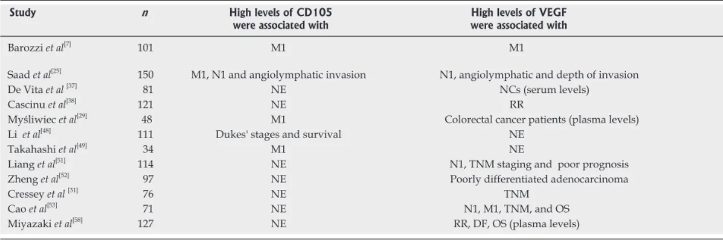
BV ultimately achieved FDA approval in 2004 as a first-line treatment for mCRC in combination with che-motherapy, based on its statistically and clinically meaning-ful beneits on progression-free survival and OS and has since garnered additional approval[79].BV is the most used VEGF inhibitor with clear proof of eficacy in CRC, how -ever, optimal use of this agent at various stages of the dis-ease is still under investigation. Additionally, there are nu-merous other angiogenic agents targeting VEGF and other pro-angiogenic systems in clinical development[80].These novel targeted agents inhibit the VEGF pathway by target-ing the VEGF ligand, its receptors or by blocktarget-ing down-stream signaling pathway components. Anti-angiogenic agents include antibodies, small molecule tyrosine kinase (TK) inhibitors, antisense oligonucleotides and aptamers[81]. Table 1 summarized the main results of CD105 and VEGF studies.
CONCLUSION
Despite major advances, in terms of knowledge and treatment of CRC in recent years, the single most well-documented prognostic marker of pathologic stage remains the gold standard for disease stage at diagnosis. Angiogenesis plays an important role in the growth and progressionof cancer but its role as a prognostic factor
is still controversial.Most studies report that endoglin and vascular endothelial growth factor family expres-sion are indicators of poor prognosis in CRC patients. Beyond these controversies, the ultimate clinical implica-tion of tumor angiogenesis is the development of anti-angiogenic therapy, targeting tumor vasculature.
REFERENCES
1 Svagzdys S, L�sauskait� V, Pavalkis D, N�dz�lski�n� I, Pranys D, Tam�lis A. Microv�ss�l d�nsity as n�w prognos� tic mark�r aft�r radioth�rapy in r�ctal canc�r. BMC Cancer
2009� 9� 95
2 Des Guetz G, Uzzan B, Nicolas P, Cuch�rat M, Mor�r� JF, B�namouzig R, Br�au JL, P�rr�t GY. Microv�ss�l d�nsity and VEGF �xpr�ssion ar� prognostic factors in color�ctal canc�r. M�ta�analysis of th� lit�ratur�. Br J Cancer 200�� 94� 1�23�1�32
3 Brenner H, Hoffm�ist�r M, Haug U. Should color�ctal can� c�r scr��ning start at th� sam� ag� in Europ�an countri�s? Contributions from d�scriptiv� �pid�miology. Br J Cancer
200�� 99� 532�535
4 Aljebreen AM. Clinico�pathological patt�rns of color�ctal canc�r in Saudi Arabia� young�r with an advanc�d stag� pr�s�ntation. Saudi J Gastroenterol 200�� 13� �4���
5 Center MM, J�mal A, Smith RA, Ward E. Worldwid� varia� tions in color�ctal canc�r. CA Canc�r J Clin 2009� 59� 3���3�� � Henry KA, Niu X, Bosco� FP. G�ographic dispariti�s in
color�ctal canc�r survival. Int J Health Geogr 2009� 8� 4� � Barozzi C, Ravaioli M, D'Errico A, Grazi GL, Poggioli G,
Cavrini G, Mazziotti A, Grigioni WF. R�l�vanc� of biologic mark�rs in color�ctal carcinoma� a comparativ� study of a broad pan�l. Cancer 2002� 94� �4���5�
� Zavoral M, Suchan�k S, Zavada F, Dus�k L, Muzik J, S�if�rt B, Fric P. Color�ctal canc�r scr��ning in Europ�. World J Gas-troenterol 2009� 15� 590��5915
9 Alexander DD, Wat�rbor J, Hugh�s T, Funkhous�r E, Grizzl� W, Mann� U. African�Am�rican and Caucasian dis� pariti�s in color�ctal canc�r mortality and survival by data sourc�� an �pid�miologic r�vi�w. Cancer Biomark 200�� 3� 301�313
10 Bosetti C, L�vi F, Rosato V, B�rtuccio P, Lucchini F, N�gri E, La V�cchia C. R�c�nt tr�nds in color�ctal canc�r mortality in Europ�. Int J Cancer 2011� 129� 1�0�191
2�� Jun� 10, 2011|Volum� 2|Issu� �|
WJCO|www.wjgn�t.com
Study n High levels of CD105 were associated with
High levels of VEGF were associated with
Barozzi et al[�]
101 M1 M1
Saad et al[25]
150 M1, N1 and angiolymphatic invasion N1, angiolymphatic and d�pth of invasion
D� Vita et al[3�]
�1 NE NCs �s�rum l�v�ls�
Cascinu et al[3�]
121 NE RR
Myśliwiec et al[29]
4� M1 Color�ctal canc�r pati�nts �plasma l�v�ls�
Li et al[4�]
111 Duk�s' stag�s and survival NE
Takahashi et al[49]
34 M1 NE
Liang et al[51]
114 NE N1, TNM staging and poor prognosis
Zh�ng et al[52]
9� NE Poorly diff�r�ntiat�d ad�nocarcinoma
Cr�ss�y et al [31]
�� NE TNM
Cao et al[53]
�1 NE N1, M1, TNM, and OS
Miyazaki et al[5�]
12� NE RR, DF, OS �plasma l�v�ls�
Table 1 The main results of CD105 and VEGF studies
11 Fairley TL, Cardin�z CJ, Martin J, All�y L, Fri�dman C, Ed� wards B, Jamison P. Color�ctal canc�r in U.S. adults young�r than 50 y�ars of ag�, 199��2001. Cancer 200�� 107� 1153�11�1 12 Brenner H, Alt�nhof�n L, Hoffm�ist�r M. S�x, ag�, and
birth cohort �ff�cts in color�ctal n�oplasms� a cohort analy� sis. Ann Intern Med 2010� 152� �9���03
13 Gurzu S, Jung J, Azamirei L, Mezei T, Cîmpean AM, Szen� tirmay Z. Th� angiog�n�sis in color�ctal carcinomas with and without lymph nod� m�tastas�s. Rom J Morphol Embryol
200�� 49� 149�152
14 Cascinu S, G�orgoulias V, K�rr D, Maughan T, Labianca R, Ychou M. Color�ctal canc�r in th� adjuvant s�tting� p�rsp�c� tiv�s on tr�atm�nt and th� rol� of prognostic factors. Ann Oncol 2003� 14 Suppl 2� ii25�ii29
15 Calvo HJ, Ort�ga GD, Pardo RJM, Lóp�z, MAJ, Cubo T. Biologia mol�cular d�l proc�sso m�tastásico d�l canc�r colo� r�ctal. Cirugia Española2000,68 � 5���5��
1� Zafar SY, Ab�rn�thy AP, Abbott DH, Grambow SC, Mar� c�llo JE, H�rndon JE 2nd, Row� KL, Kolimaga JT, Zullig LL, Patwardhan MB, Prov�nzal� DT. Comorbidity, ag�, rac� and stag� at diagnosis in color�ctal canc�r� a r�trosp�ctiv�, paral� l�l analysis of two h�alth syst�ms. BMC Cancer 200�� 8� 345 1� Ries LA, Wingo PA, Mill�r DS, How� HL, W�ir HK, Ros�n�
b�rg HM, V�rnon SW, Cronin K, Edwards BK. Th� annual r�port to th� nation on th� status of canc�r, 19�3�199�, with a sp�cial s�ction on color�ctal canc�r. Cancer 2000� 88� 239��2424
1� Townsend MC, B�auchamp D, Ev�rs MB, Mattox LK. Sabis� ton T�xtbook of Surg�ry, 1�th �d.� Saund�rs� Canada, 200� 19 Liu CL, Fan ST, Lo CM, Law WL, Ng IO, Wong J. H�patic
r�s�ction for color�ctal liv�r m�tastas�s� prosp�ctiv� study.
Hong Kong Med J 2002� 8� 329�333
20 Lambert LA, Colacchio TA, Barth RJ. Int�rval h�patic r�s�c� tion of color�ctal m�tastas�s improv�s pati�nt s�l�ction*
Curr Surg 2000� 57� 504
21 Abdalla EK, Vauth�y JN, Ellis LM, Ellis V, Pollock R, Broglio KR, H�ss K, Curl�y SA. R�curr�nc� and outcom�s following h�patic r�s�ction, radiofr�qu�ncy ablation, and combin�d r�s�ction/ablation for color�ctal liv�r m�tastas�s.
Ann Surg 2004� 239� �1��25� discussion �25��2� 22 de Haas RJ, Wich�rts DA, Flor�s E, Azoulay D, Castaing D,
Adam R. R1 r�s�ction by n�c�ssity for color�ctal liv�r m�tas� tas�s� is it still a contraindication to surg�ry? Ann Surg 200�� 248� �2���3�
23 Lang K, Korn JR, L�� DW, Lin�s LM, Earl� CC, M�nzin J. Factors associat�d with improv�d survival among old�r color�ctal canc�r pati�nts in th� US� a population�bas�d analysis. BMC Cancer 2009� 9� 22�
24 Brenner H, Hoffm�ist�r M, Arndt V, Haug U. G�nd�r dif� f�r�nc�s in color�ctal canc�r� implications for ag� at initia� tion of scr��ning. Br J Cancer 200�� 96� �2���31
25 Saad RS, Liu YL, Nathan G, C�l�br�zz� J, M�dich D, Silv�r� man JF. Endoglin �CD105� and vascular �ndoth�lial growth factor as prognostic mark�rs in color�ctal canc�r. Mod Pathol
2004� 17� 19��203
2� Gómez-Domínguez E, Trap�ro�Marugán M, d�l Pozo AJ, Cant�ro J, Gisb�rt JP, Maté J. Th� color�ctal carcinoma prog�
nosis factors. Signiicance of diagnosis delay. Rev Esp Enferm Dig 200�� 98� 322�329
2� Bilchik AJ, DiNom� M, Saha S, Turn�r RR, Wi�s� D, Mc� Cart�r M, Hoon DS, Morton DL. Prosp�ctiv� multic�nt�r tri� al of staging ad�quacy in colon canc�r� pr�liminary r�sults.
Arch Surg 200�� 141� 52��33� discussion 533�534.
2� Graziano F, Cascinu S. Prognostic mol�cular mark�rs for planning adjuvant ch�moth�rapy trials in Duk�s' B color�c� tal canc�r pati�nts� how much �vid�nc� is �nough? Ann On-col 2003� 14� 102��103�
29 Myśliwiec P, Pawlak K, Kukliński A, Kedra B. Combined p�riop�rativ� plasma �ndoglin and VEGF��a ass�ssm�nt in
color�ctal canc�r pati�nts. Folia Histochem Cytobiol 2009� 47� 231�23�
30 Pang RW, Poon RT. Clinical implications of angiog�n�sis in canc�rs. Vasc Health Risk Manag 200�� 2� 9��10�
31 Cressey R, Wattananupong O, L�rtpras�rtsuk� N, Vinit� k�tkumnu�n U. Alt�ration of prot�in �xpr�ssion patt�rn of vascular �ndoth�lial growth factor �VEGF� from solubl� to c�ll�associat�d isoform during tumourig�n�sis. BMC Cancer
2005� 5� 12�
32 Giatromanolaki A, Stathopoulos GP, Tsiompanou E, Papadimitriou C, G�orgoulias V, Gatt�r KC, Harris AL, Koukourakis MI. Combin�d rol� of tumor angiog�n�sis, bcl�2, and p53 �xpr�ssion in th� prognosis of pati�nts with color�ctal carcinoma. Cancer 1999� 86� 1421�1430
33 Kitadai Y. Angiog�n�sis and lymphangiog�n�sis of gastric canc�r. J Oncol 2010� 2010� 4���25
34 Kwon KA, Kim SH, Oh SY, L�� S, Han JY, Kim KH, Goh RY, Choi HJ, Park KJ, Roh MS, Kim HJ, Kwon HC, L�� JH.
Clinical signiicance of preoperative serum vascular endo� th�lial growth factor, int�rl�ukin��, and C�r�activ� prot�in l�v�l in color�ctal canc�r. BMC Cancer 2010� 10� 203
35 Tanigawa N, Amaya H, Matsumura M, Lu C, Kitaoka A, Matsuyama K, Muraoka R. Tumor angiog�n�sis and mod� of m�tastasis in pati�nts with color�ctal canc�r. Cancer Res
199�� 57� 1043�104�
3� Mosch B, R�iss�nw�b�r B, N�ub�r C, Pi�tzsch J. Eph r�c�p� tors and �phrin ligands� important play�rs in angiog�n�sis and tumor angiog�n�sis. J Oncol 2010� 2010� 1352�5
3� De Vita F, Orditura M, Li�to E, Infusino S, Morgillo F, Mar� tin�lli E, Cast�llano P, Romano C, Ciardi�llo F, Catalano G, Pignat�lli C, Galizia G. El�vat�d p�riop�rativ� s�rum vas� cular �ndoth�lial growth factor l�v�ls in pati�nts with colon carcinoma. Cancer 2004� 100� 2�0�2��
3� Cascinu S, Staccioli MP, Gasparini G, Giordani P, Catalano V, Ghis�lli R, Rossi C, Bald�lli AM, Graziano F, Saba V, Mu� r�tto P, Catalano G. Expr�ssion of vascular �ndoth�lial gro� wth factor can pr�dict �v�nt�fr�� survival in stag� II colon canc�r. Clin Cancer Res 2000� 6� 2�03�2�0�
39 Rodrigo JP, Cabanillas R, Chiara MD, García P�dr�ro J,
Astudillo A, Suárez Nieto C. [Prognostic signiicance of an� giog�n�sis in surgically tr�at�d supraglottic squamous c�ll carcinomas of th� larynx]. Acta Otorrinolaringol Esp 2009� 60� 2�2�2��
40 Gasparini G, B�vilacqua P, Bonoldi E, T�stolin A, Galassi A, V�rd�rio P, Boracchi P, Gugli�lmi RB, P�zz�lla F. Pr�dictiv� and prognostic mark�rs in a s�ri�s of pati�nts with h�ad and n�ck squamous c�ll invasiv� carcinoma tr�at�d with concurr�nt ch�moradiation th�rapy. Clin Cancer Res 1995� 11�13�5�13�3
41 Hollingsworth HC, Kohn EC, St�inb�rg SM, Roth�nb�rg ML, M�rino MJ. Tumor angiog�n�sis in advanc�d stag� ovarian carcinoma. Am J Pathol 1995� 147� 33�41
42 Takagi S, In�naga R, Oya R, Nakamura S, Ik�mura K. Blood v�ss�l d�nsity corr�lat�s with th� �ff�cts of targ�t�d intra� art�rial carboplatin infusion with concurr�nt radioth�rapy for squamous c�ll carcinomas of th� oral cavity and oropha� rynx. Br J Cancer 200�� 94� 15�0�15�5
43 Zhang SC, Miyamoto S, Kamijo T, Hayashi R, Has�b� T, Ishii G, Fukayama M, Ochiai A. Intratumor microv�ss�l d�nsity in biopsy sp�cim�ns pr�dicts local r�spons� of hy� popharyng�al canc�r to radioth�rapy. Jpn J Clin Oncol 2003� 33� �13��19
44 Hasan J, By�rs R, Jayson GC. Intra�tumoural microv�s� s�l d�nsity in human solid tumours. Br J Cancer 2002� 86� 15���15��
Th� risk of d�v�loping m�tastatic dis�as� in color�ctal can� c�r is r�lat�d to CD105�positiv� v�ss�l count. J Surg Oncol
200�� 93� 44��455
4� Yan G, Zhou XY, Cai SJ, Zhang GH, P�ng JJ, Du X. Lym� phangiog�nic and angiog�nic microv�ss�l d�nsity in human primary sporadic color�ctal carcinoma. World J Gastroenterol
200�� 14� 101�10�
4� Li C, Gardy R, S�on BK, Duff SE, Abdalla S, R�n�han A, O'Dwy�r ST, Haboubi N, Kumar S. Both high intratumoral microv�ss�l d�nsity d�t�rmin�d using CD105 antibody and �l�vat�d plasma l�v�ls of CD105 in color�ctal canc�r pati�nts corr�lat� with poor prognosis. Br J Cancer 2003� 88� 1424�1431 49 Takahashi N, Kawanishi�Tabata R, Haba A, Tabata M, Ha�
ruta Y, Tsai H, S�on BK. Association of s�rum �ndoglin with m�tastasis in pati�nts with color�ctal, br�ast, and oth�r solid tumors, and suppr�ssiv� �ff�ct of ch�moth�rapy on th� s�� rum �ndoglin. Clin Cancer Res 2001� 7� 524�532
50 Lee SJ, Kim JG, Sohn SK, Cha� YS, Moon JH, Kim SN, Ba� HI, Chung HY, Yu W. No association of vascular �ndoth�� lial growth factor�A �VEGF�A� and VEGF�C �xpr�ssion with survival in pati�nts with gastric canc�r. Cancer Res Treat
2009� 41� 21��223
51 Liang JF, Wang HK, Xiao H, Li N, Ch�ng CX, Zhao YZ, Ma YB, Gao JZ, Bai RB, Zh�ng HX. R�lationship and prognostic significanc� of SPARC and VEGF prot�in �xpr�ssion in colon canc�r. J Exp Clin Cancer Res 2010� 29� �1
52 Zheng S, Han MY, Xiao ZX, P�ng JP, Dong Q. Clinical
signiicance of vascular endothelial growth factor expression
and n�ovascularization in color�ctal carcinoma. World J Gastroenterol 2003� 9� 122��1230
53 Cao D, Hou M, Guan YS, Jiang M, Yang Y, Gou HF. Expr�ssion of HIF�1alpha and VEGF in color�ctal canc�r� association with clinical outcom�s and prognostic implications. BMC Cancer 2009� 9� 432
54 Paule B, Basti�n L, D�sland�s E, Cuss�not O, Podgorniak MP, Allory Y, Naïmi B, Porch�r R, d� La Taill� A, M�nashi S, Calvo F, Mourah S. Solubl� isoforms of vascular �ndoth�lial growth factor ar� pr�dictors of r�spons� to sunitinib in m�tastatic r�nal c�ll carcinomas. PLoS One 2010� 5� �10�15 55 Cook KM, Figg WD. Angiog�n�sis inhibitors� curr�nt
strat�gi�s and futur� prosp�cts. CA Cancer J Clin 2010� 60� 222�243
5� Duhoux FP, Machi�ls JP. Antivascular th�rapy for �pith�lial ovarian canc�r. J Oncol 2010� 2010� 3�254�
5� Hanrahan V, Curri� MJ, Gunningham SP, Morrin HR, Scott PA, Robinson BA, Fox SB. Th� angiog�nic switch for vascular �ndoth�lial growth factor �VEGF��A, VEGF�B, VEGF�C, and VEGF�D in th� ad�noma�carcinoma s�qu�nc� during color�ctal canc�r progr�ssion. J Pathol 2003� 200� 1�3�194
5� Miyazaki T, Okada N, Ishibashi K, Ogata K, Ohsawa T, Ishiguro T, Nakada H, Yokoyama M, Matsuki M, Kato H,
Kuwano H, Ishida H. Clinical signiicance of plasma level
of vascular �ndoth�lial growth factor�C in pati�nts with color�ctal canc�r. Jpn J Clin Oncol 200�� 38� �39��43
59 Zlobec I, St��l� R, Nigam N, Compton CC.A pr�dictiv� mod�l of r�ctal tumor r�spons� to pr�op�rativ� radioth�r�
apy using classiication and regression tree methods. Clin Cancer Res 2005� 15 � 5440�5443
�0 He M, Ch�ng Y, Li W, Liu Q, Liu J, Huang J, Fu X. Vascular �ndoth�lial growth factor C promot�s c�rvical canc�r m�tastasis via up�r�gulation and activation of RhoA/ ROCK�2/mo�sin cascad�. BMC Cancer 2010� 10� 1�0 �1 Thiele W, Sl��man JP. Tumor�induc�d lymphangiog�n�sis�
a targ�t for canc�r th�rapy? J Biotechnol 200�� 124� 224�241 �2 Stintzing S, H�in�mann V, Moosmann N, Hidd�mann W,
Jung A, Kirchn�r T. Th� tr�atm�nt of color�ctal carcinoma with monoclonal antibodi�s� th� importanc� of KRAS mutation analysis and EGFR status. Dtsch Arztebl Int 2009�
106� 202�20�
�3 Gille J. Antiangiog�nic canc�r th�rapi�s g�t th�ir act tog�th�r� curr�nt d�v�lopm�nts and futur� prosp�cts of growth factor� and growth factor r�c�ptor�targ�t�d approach�s. Exp Dermatol 200�� 15� 1�5�1��
�4 Sparano JA, Gray R, Giantonio B, O'Dwy�r P, Comis RL. Evaluating antiangiog�n�sis ag�nts in th� clinic� th� East�rn Coop�rativ� Oncology Group Portfolio of Clinical Trials.
Clin Cancer Res 2004� 10� 120��1211
�5 Gruenberger B, Tamandl D, Schu�ll�r J, Sch�ithau�r W, Zi�linski C, H�rbst F, Gru�nb�rg�r T. B�vacizumab, cap�citabin�, and oxaliplatin as n�oadjuvant th�rapy for pati�nts with pot�ntially curabl� m�tastatic color�ctal canc�r. J Clin Oncol 200�� 26� 1�30�1�35
�� Ma AT, Ma BB, L�i KI, Mo FK, Chan AT. Clinical pr�dic� tors of r�spons� to c�tuximab�ch�moth�rapy in m�tastatic color�ctal canc�r. Hong Kong Med J 2010� 16� 20��212 �� Nussenbaum F, H�rman IM. Tumor angiog�n�sis� insights
and innovations. J Oncol 2010� 2010� 132�41
�� Boehm S, Roth�rmundt C, H�ss D, Jo�rg�r M. Antiangio� g�nic drugs in oncology� a focus on drug saf�ty and th� �ld�rly � a mini�r�vi�w. Gerontology 2010� 56� 303�309 �9 Mahfud M, Br�it�nst�in S, El�Badry AM, Puhan M,
Rick�nbach�r A, Samaras P, P�ssaux P, Lop�z�B�n S, Ja�ck D, Figu�ras J, Alain�Clavi�n P. Impact of pr�op�rativ� b�vacizumab on complications aft�r r�s�ction of color�ctal liv�r m�tastas�s� cas��match�d control study. World J Surg
2010� 34� 92�100
�0 Tol J, Punt CJ. Monoclonal antibodi�s in th� tr�atm�nt of m�tastatic color�ctal canc�r� a r�vi�w. Clin Ther 2010� 32� 43��453
�1 Shih T, Lindl�y C. B�vacizumab� an angiog�n�sis inhibitor for th� tr�atm�nt of solid malignanci�s. Clin Ther 200�� 28� 1��9�1�02
�2 Rougier P, Mitry E. [Targ�t�d bioth�rapy� a r�volution in th� manag�m�nt of pati�nts with color�ctal canc�r?].
Gastroenterol Clin Biol 2009� 33� ��2���0
�3 Jubb AM, Hurwitz HI, Bai W, Holmgr�n EB, Tobin P, Gu�rr�ro AS, Kabbinavar F, Hold�n SN, Novotny WF, Frantz GD, Hillan KJ, Ko�pp�n H. Impact of vascular �ndoth�lial growth factor�A �xpr�ssion, thrombospondin�2 �xpr�ssion, and microv�ss�l d�nsity on th� tr�atm�nt �ff�ct of b�vacizumab in m�tastatic color�ctal canc�r. J Clin Oncol
200�� 24� 21��22�
�4 Tappenden P, Jon�s R, Paisl�y S, Carroll C. Syst�matic r�vi�w and �conomic �valuation of b�vacizumab and c�tuximab for th� tr�atm�nt of m�tastatic color�ctal canc�r. Health Technol Assess 200�� 11� 1�12�, iii�iv
�5 Naschberger E, Cron�r RS, M�rk�l S, Dimml�r A, Tripal P, Amann KU, Kr�mm�r E, Bru�ckl WM, Papadopoulos T, Hoh�nadl C, Hoh�nb�rg�r W, Stürzl M. Angiostatic immun� r�action in color�ctal carcinoma� Impact on survival and p�rsp�ctiv�s for antiangiog�nic th�rapy. Int J Cancer 200�� 123� 2120�2129
�� Hurwitz H, F�hr�nbach�r L, Novotny W, Cartwright T, Hainsworth J, H�im W, B�rlin J, Baron A, Griffing S, Holmgr�n E, F�rrara N, Fyf� G, Rog�rs B, Ross R, Kabbinavar F. B�vacizumab plus irinot�can, fluorouracil, and l�ucovorin for m�tastatic color�ctal canc�r. N Engl J Med
2004� 350� 2335�2342
�� Kabbinavar FF, Wallac� JF, Holmgr�n E, Yi J, C�lla D, Yost KJ, Hurwitz HI. H�alth�r�lat�d quality of lif� impact of
bevacizumab when combined with irinotecan, 5-luorouracil,
and l�ucovorin or 5�fluorouracil and l�ucovorin for m�tastatic color�ctal canc�r. Oncologist 200�� 13� 1021�1029 �� Ma WW, Hidalgo M. Exploiting nov�l mol�cular targ�ts
in gastroint�stinal canc�rs. World J Gastroenterol 200�� 13� 5�45�5�5�
�9 Yancopoulos GD. Clinical application of th�rapi�s targ�ting
2�9 Jun� 10, 2011|Volum� 2|Issu� �|
VEGF. Cell 2010� 143� 13�1�
�0 Hubbard J, Groth�y A. Antiangiog�n�sis ag�nts in color�c� tal canc�r. Curr Opin Oncol 2010� 22� 3�4�3�0
�1 Prat A, Casado E, Cortés J. N�w approach�s in angiog�nic targ�ting for color�ctal canc�r. World J Gastroenterol 200�� 13� 5�5��5���