Ana Filipa Pinto
Dissertation presented to obtain the Ph.D degree in Biochemistry
Instituto de Tecnologia Química e Biológica | Universidade Nova de Lisboa
Oeiras,
July, 2012
Reductive scavenging of reactive
oxygen species in prokaryotes
Rubrerythrin and Superoxide Reductase
O
2
+ 1e + 2H
+H
2
O
2
Reductive scavenging of
reactive oxygen species in
prokaryotes
Rubrerythrin and Superoxide Reductase
Ana Filipa Carapinha Pinto
Supervisors: Prof. Miguel S. Teixeira and Dr. Célia V. Romão
Dissertation presented to obtain the PhD degree in Biochemistry
Instituto de Tecnologia Química e Biológica
Universidade Nova de Lisboa
Quadro Comunitário de apoio (Bolsa de Doutoramento SFRH/ BD/ 41355/ 2007)
From left to right: Fernando Antunes, Cláudio Soares, Alessandro Giuffrè, Ana Filipa Pinto, João Vicente, Claudina Roudrigues-Pousada, Célia Romão and Miguel Teixeira
Second Edition, July 2012
Cover: 3D crystal structures of Superoxide reductase from Ignicoccus hospitalis (left) and
Desulforubrerythrin from Campylobacter jejuni (right).
Metalloproteins and Bioenergetics Unit
-Metalloenzymes and Molecular Bioenergetics Laboratory
Instituto de Tecnologia Química e Biológica Universidade Nova de Lisboa
Av. da República
Estação Agronómica Nacional 2780-157 Oeiras
Portugal
iii
This dissertation is the result of research work for four years at the Metalloenzymes and Molecular Bioenergetics Laboratory integrated in the Metalloproteins and Bioenergetics Unit at the Instituto de TecnologiaQuímica e Biológica from Universidade Nova de Lisboa, under the supervision of Prof. Miguel Teixeira and Célia Romão. The work here described concerns the study of two systems involved in the scavenging of Reactive Oxygen Species widely distributed in Prokaryotes: Rubrerythrins and Superoxide Reductases. A rubrerythrin-like protein from Campylobacter jejuni, Desulforubrerythrin, was studied by biochemical, spectroscopic and structural methods and a NADH-linked H2O2 reductase activity was measured. Superoxide detoxification in Ignicoccus hospitalis was studied by characterizing a non-canonical superoxide reductase by biophysical and structural methods.
This thesis is divided in four parts: Part I, organized in three chapters, comprising a general introduction to oxygen biochemistry (Chapter 1), to the two families of proteins concerning this thesis (Chapter 2) and to the pathogenic and hyperthermophilic organisms encoding for the proteins studied (Chapter 3). Part II concerns the experimental results obtained for desulforubrerythrin, a multidomain protein from Campylobacter jejuni, namely the biochemical and spectroscopic characterization of the protein (Chapter 4) and the analysis of its 3D crystal structure obtained by X-ray crystallography in two different redox states (Chapter 5). Part III (Chapters 6 and 7) focus on the study of a superoxide reductase enzyme from Ignicoccus hospitalis (wild-type and two site-directed mutants) and on the analysis of the 3D crystal structure obtained by X-ray crystallography.
V
I would like to acknowledge the following people for being indispensable for the work presented here:Prof. Miguel Teixeira - My supervisor, for giving the opportunity to work
in his lab and develop the work presented here, for the fruitful discussions and advices, for his knowledge and judgemental critics in every subject that definitely made my critical “eye” grow.
Dra. Célia Romão - My co-supervisor who has been there in the more “poltergeisty” moments and helped me go through. For all the discussions and advices and for always pushing me forward.
Dra. Diane Cabelli - For receiving me at the Brookhaven National
Laboratory, for her essential collaboration on the pulse radiolysis studies on superoxide reductases, for the discussions and advices.
Dra. Lígia Saraiva - For supervising and supporting all the molecular
biology work on superoxide reductases, performed in her lab and for the helpful advices.
Dr. Smilja Todorovic - For her resonance Raman studies on
rubrerythrins and superoxide reductases and for many helpful discussions and advices throughout the work presented here.
Prof. Peter Hilderbrandt - For his discussions and advices throughout
the four years of research work presented here.
Dr. João Rodrigues - For passing me on his heritage of the superoxide
reductases, his knowledge and helpful advices.
Dr. João Vicente - For being my “tutor” in the early stages of my
research work in the lab still as a university student. For his patience, knowledge and valuable advices.
Dr. Pedro Matias - For his strong collaboration in the X-ray structures
VI
crystallization and 3D structure determination.
Prof. Fernando Antunes - For allowing me visit his lab (FCUL) in order
to perform some assays on rubrerythrins activity and for helpful discussions as part of my thesis commission.
Prof. Harald Huber - For providing us with genomic DNA of
Nanoarchaeum equitans and Ignicoccus hospitalis.
Metalloproteins and Bioenergetics Unit - For all the fun, knowledge,
friendship, work discussions, gossip, not exactly in this order: the Metalloenzymes and Molecular Bioenergetics Laboratory Célia Romão, Sandra Santos, Liliana Pinto, Vera Gonçalves, Miguel Ribeiro, Cecília Miranda, Joana Carrilho and the Biological Energy Transduction Laboratory Manuela Pereira, Ana Paula Batista, Patrícia Refojo, Lara Paulo, Bruno Marreiros and Afonso Duarte. The previous members of this unit Andreia Veríssimo, Filipa Sousa, João Rodrigues, João Vicente and Fabrizio Testa.
Molecular Genetics of Microbial Resistance Laboratory - For the availability and help on the molecular biology work on superoxide reductases and for the good environment in the lab.
Family and Friends
A special acknowledgement to my friends and family for all the support.
FCT
VII
In this thesis a list of original publications is included: "Desulforubrerythrin from Campylobacter jejuni, a novel multidomain protein", Pinto AF, Todorovic S, Hildebrandt P,
Yamazaki M, Amano F, Igimi S, Romão CV, Teixeira M. 2011 J Biol Inorg Chem.;16(3):501-10
“Thermofluor-based optimization strategy for the stabilization
and crystallization of Campylobacter jejuni Desulforubrerythrin” Santos SP, Bandeiras TM, Pinto AF, Teixeira M, Romão CV.
2012 Protein Expr Purif.;81(2):193-200
"Reductive elimination of superoxide: Structure and mechanism of superoxide reductases", Pinto AF, Rodrigues JV, Teixeira M,
2010 Biochim Biophys Acta.; 1804(2):285-97
"Cloning, purification, crystallization and X-ray crystallographic analysis of Ignicoccus hospitalis neelaredoxin", Pinho FG, Romão CV, Pinto AF, Saraiva LM, Huber H, Matias PM,
Teixeira M, Bandeiras TM. 2010 Acta Crystallogr Sect F Struct Biol Cryst Commun.;66 (Pt 5):605-7
“Superoxide reductase from Nanoarchaeum equitans:expression, purification, crystallization and preliminary X-ray
crystallographic analysis”, Pinho FG, Pinto AF, Pinto LC, Huber
H, Romão CV, Teixeira M, Matias PM, Bandeiras TM. 2011 Acta Crystallogr Sect F Struct Biol Cryst Commun.;67(Pt 5):591-5
Three manuscripts are in preparation, based on studies presented in Chapters 5, 6 and 7:
“Three dimensional structure of the multidomain desulforubrerythrin from Campylobacter jejuni”, Pinto AF,
VIII
hospitalis” Pinto AF, Romão CV, Pinto LC, Huber H, Saraiva
LM, Cabelli D, Teixeira M, manuscript under preparation
“Insights on the superoxide reductase structures from the two
symbiotic organism: Ignicoccus hospitalis and Nanoarchaeum equitans reveal differences on the catalytic site”, Romão CV,
Pinto AF, Pinho FG, Barradas AR, Matias PM, Teixeira M,
IX
The purpose of this thesis is to contribute to a better understanding of systems involved in the scavenging of reactive oxygen species. The work focuses on an enzyme from the Rubrerythrin family that reduces hydrogen peroxide and one from the Superoxide Reductase family that reduces the superoxide anion. Both of these families are distributed widely across the three domains of life, Archaea, Bacteria and Eukarya, but are mainly found in anaerobic and microaerophilic prokaryotes. The target enzymes were a new multidomain rubrerythrin-like protein from the pathogenic bacterium Campylobacter jejuni, and a superoxide reductase from the hyperthermophilic archaeon Ignicoccus hospitalis. The objectives were to further understand these enzymes by establishing the nature and properties of their metal/catalytic centers and elucidate the reduction mechanisms of these two proteins at the molecular level, by biophysical methods and through the determination of their 3D structures by X-ray crystallography.In the genome of Campylobacter jejuni NCTC 11168 the gene cj0012c encodes a rubrerythrin-like protein. Using homology analysis, this protein has been proposed by Yamasaki and co-workers (Yamasaki, M., et al., FEMS Microbiol Lett, 2004. 235(1): p. 57-63) to have domains homologous to rubredoxin oxidoreductase and to rubrerythrin. The amino acid sequence revealed three iron binding domains: an N-terminal desulforedoxin-like domain, followed by a four-helix-bundle domain harbouring a non-sulfur diiron center, and a rubredoxin-like domain at the C-terminus.
X
reduction potentials of +240 mV (desulforedoxin-like center) and +185 mV (rubredoxin-like center). These centers are in the high-spin configuration in the as-isolated ferric form and are the main spectral contributors to its UV-visible spectrum. The protein further accommodates a diiron site with reduction potentials of +270 and +235 mV for the two sequential redox transitions Fe(III)-Fe(III)→(FeIII)-Fe(II) and Fe(III)-Fe(II)→Fe(II)-Fe(II). Resonance Raman spectroscopy combined with isotope labelling (D2O and H2O18) shows that the diiron
in the diferric state is probably bridged by a µ-oxo species. The protein is rapidly reoxidized by hydrogen peroxide but only slowly by oxygen. It has a significant NADH-linked hydrogen peroxide reductase activity of 3.9 ± 0.5 µmol H2O2 min-1mg-1, determined using two non-physiological
electron transfer proteins from Escherichia coli (a NADH oxidoreductase and a rubredoxin). The reduction of hydrogen peroxide has been the most reproducible activity observed in vitro for the rubrerythrin family. With its unique set of component domains, the protein is a novel example within the overall large diversity of domain organizations exhibited by this enzyme family. Given its component domains and homology to the rubrerythrin family, the protein was named by us as desulforubrerythrin (DRbr). To understand this unique domain architecture, the determination of its 3D structure by X-ray crystallography was carried out.
XI
structure was obtained in two different redox states. The arrangement of the rubredoxin and the diiron sites is such that electron transfer occurs from an external donor to the rubredoxin site, which then transfers the electron to the diiron center at c.a. 13 Å. In contrast, the desulforedoxin-like centers are located far away from the other metal sites (c.a. 34 Å between the Dx iron center and the diiron centers) and, despite the structure being now available, their function remains unclear. The 3D structure showed a diiron center in two different redox states with a redox-linked ligand and iron movement, a characteristic feature of the diiron site in rubrerythrins.Superoxide reductases (SOR) reduce superoxide to hydrogen peroxide. In many organisms, this system appears to be the only route for superoxide detoxification, as their genomes do not encode for other superoxide scavenging enzymes, the SODs.
The catalytic site is known to be composed of an iron bound to four histidines and a cysteine in a square pyramidal geometry. In the oxidized state a sixth ligand, a glutamate residue, is present in most SORs. However, several issues still remained to be explained, namely the number of catalytic intermediates of the reaction cycle and the role of specific amino acid residues close to the catalytic site.
A superoxide reductase from Ignicoccus hospitalis has been shown to lack the positively charged lysine residue (Lys 24, in Archaeoglobus fulgidus), conserved in the majority of SORs; this natural variation is also observed in other SORs from several Crenarchaeota. This residue is thought to have a relevant role for attracting the superoxide anion into the active site and stabilizing the first catalytic intermediate.
XII
found for other SORs. A pH induced wavelength shift at the visible band was also associated with the change of the Glu/H2O-SOR form to a
hydroxide bound species, with an associated pKa of 10.5 for the
wild-type protein, and 10.7 and 6.5 for the T24K and E23A mutants, respectively. The reduction potentials were determined to be E’0 (wt) =
+235, E’0 (E23A) = +283, E’0 (T24K) = +195 mV vs. SHE at pH 7. The
reduction potential was found to be pH independent for both the wt protein and the T24K mutant from pH 5 to 8.5. The E23A mutant is pH dependent in the pH range of 5 to 7.5 with an associated pKa of 6.3.
The reaction mechanism was studied by pulse radiolysis where the enzyme is exposed to known amounts of the superoxide anion and the electronic changes of the active site followed by visible absorption spectroscopy on a fast (microsecond to millisecond) timescale. The results show that the first intermediate detected is similar to those of the canonical enzymes and that the rate of its formation, 0.7 x 10⁹ M-1s-1 for the wt protein, is also within the range for the other enzymes; i.e., the lysine residue seems to be not essential for the mechanism of superoxide reduction. Similar studies on the two mutants revealed no significant differences on the rates for the formation of the first intermediate.
In the case of Ignicoccus hospitalis SOR, the data only reveals the formation of a single intermediate. In comparison with the results obtained for other SORs it was possible to establish that there are two types of apparent mechanisms, with one or two intermediates, independent of both the type of SOR, with one iron or two iron atoms, and its thermophilicity.
XIV
O objectivo desta tese de doutoramento é contribuir para uma melhor percepção dos sistemas envolvidos na desintoxicação das espécies reactivas de oxigénio. O trabalho desenvolvido centraliza-se no estudo de uma enzima pertencente à família das Rubreritrinas que reduz o peróxido de hidrogénio e no estudo de uma enzima que reduz o anião superóxido da família das Superóxido Reductases. Ambas as famílias estão extensamente distribuídas nos três domínios da vida: Archaea, Bacteria e Eucaria; contudo estas proteínas são mais abundantes em procariotas anaeróbios e microaerofilicos.
XV
visível, Ressonância Paramagnética Electrónica e Ressonância de Raman.Foram determinados experimentalmente os potenciais de redução para os dois centros de FeCys4, +240 mV (centro do tipo-desulforedoxina) e
+185 mV (centro do tipo-rubredoxina). Estes centros de ferro estão numa configuração de alto-spin na forma férrica e são os que mais contribuem espectralmente na região do UV-Visível. Para além destes dois centros, a proteína acomoda também um centro binuclear de ferro com potenciais de redução de +270 e +235 mV para as duas transições redox sequenciais Fe(III)-Fe(III) → Fe(III)-Fe(II) e Fe(III)-Fe(II) → Fe(II) -Fe(II). A espectroscopia de Ressonância de Raman acoplada à marcação isotópica (D2O e H2O18) revelou que os átomos de ferro do
centro binuclear no estado diférrico estão provavelmente ligados em ponte por uma espécie µ-oxo. A proteína é rapidamente reoxidada por peróxido de hidrogénio mas lentamente por oxigénio. A redução do peróxido de hidrogénio é a actividade enzimática in vitro mais reprodutível registada para a família das rubreritrinas. Tem uma actividade significativa de reductase de peróxido de hidrogénio associada à oxidação de NADH de 3.9 ± 0.5 µmol H2O2 min-1 mg-1,
XVI
cristalização da desulforubreritrina de Campylobacter jejuni. Verificou-se que esta proteína era estável a pH 6.2 e em concentrações de sal elevado, e por isso foi dialisada para a solução tampão selecionada (100 mM MES pH 6.2 e 500 mM NaCl). Cristais de elevada qualidade foram obtidos, permitindo a determinação da estrutura cristalina por difracção de raios-X da DRbr com uma resolução de 2.0 Å. Esta é a primeira estrutura cristalina de uma proteína do tipo rubreritrina, com esta arquitectura de domínios. A proteína cristalizou na forma tetramérica e tem uma configuração de domínios trocados. A estrutura cristalina 3D foi obtida em dois estados de redução diferentes. O arranjo estrutural do centro de ferro do domínio rubredoxina e do centro binuclear de ferro é tal que a transferência electrónica ocorre de um dador de electrões externo para o centro de ferro da rubredoxina, que por sua vez transfere os electrões para o centro binuclear que se encontra a uma distância de 13 Å aproximadamente. Em contraste, os centros de ferro dos domínios Dx estão muito distantes dos outros centros metálicos (34 Å, aproximadamente, entre o centro de ferro da Dx e o centro binuclear) e apesar de estruturalmente caracterizados, a sua função ainda não é conhecida. As estruturas 3D em diferentes estados de redução demonstram ainda que há um movimento de um átomo de ferro e de um ligando do mesmo no centro binuclear que é dependente do estado de redução, e é característico do centro binuclear em rubreritrinas.
As SOR reduzem superóxido a peróxido de hidrogénio. Em muitos organismos, este sistema aparece como o único mecanismo directo para a desintoxicação do superóxido, uma vez que os seus genomas não codificam para outras enzimas que eliminam o superóxido, como as superóxido dismutases (SOD).
XVII
Existem no entanto, várias questões a estudar, nomeadamente o número de intermediários catalíticos do ciclo da reacção e o papel de determinados resíduos de aminoácidos perto do centro catalítico. Para a superóxido reductase de Ignicoccus hospitalis verificou-se a ausência de um resíduo carregado positivamente, a lisina 24 (numeração da SOR de Archaeoglobus fulgidus), conservado na maioria das SORs; esta variação natural é também observada em outras SORs de vários organismos Crenarchaeota. Este resíduo é sugerido como tendo um papel relevante para atracção do anião superóxido para o centro activo e de estabilizar o primeiro intermediário catalítico.Neste trabalho, a proteína nativa e dois mutantes dirigidos foram expressos, purificados e caracterizados: o mutante E23A, onde o resíduo glutamato que actua como sexto ligando do centro de ferro na forma férrica da SOR foi mutado para uma alanina, e o mutante T24K, onde o resíduo de lisina foi reintroduzido na mesma posição em que é encontrado noutras SORs. As formas férrica das SORs de Ignicoccus hospitalis partilham as mesmas características espectrais encontradas em outras SORs. A variação do espectro eletrónico induzido pela variação do pH foi também observada, resultando na transformação da forma Glu/(H2O)-SOR para a forma da SOR ligada ao grupo hidróxido,
com um pKa associado de 10.5 para a proteína nativa, e 10.7 e 6.5
para os mutantes T24K e E23A, respectivamente. Os potenciais de redução foram também determinados: E’0 (nativa) = +235, E’0 (E23A) =
+283, E’0 (T24K) = +195 mV vs. SHE a pH 7. O potencial de redução
revelou-se independente dos valores de pH para a proteína nativa e o mutante T24K no intervalo de valores de pH de 5 a 8.5. O mutante E23A é dependente do pH no intervalo de valores de pH 5 a 7.5, com um pKa associado de 6.3.
XVIII
espectroscopia de absorção na região do visível numa rápida escala de tempo (microssegundos a milissegundos). Os resultados obtidos demonstraram que o primeiro intermediário detectado é semelhante ao obtido para enzimas canónicas, e a constante de velocidade da sua formação, 0.7 x 10⁹ M-1s-1 para a proteína nativa, está também dentro dos valores encontrados para outras enzimas; conclui-se assim que o resíduo de lisina ausente (lisina 24) parece não ser essencial no mecanismo de redução do superóxido. Estudos semelhantes para os dois mutantes não revelaram diferenças significativas, relativas à constante de velocidade de formação do primeiro intermediário.
Para a SOR de Ignicoccus hospitalis, os dados revelaram apenas a formação de uma espécie intermediária. Em comparação com os resultados obtidos para outras SORs foi possível estabelecer a existência de dois tipos de mecanismo aparente, com um ou dois intermediários, independentemente do tipo de SOR (com um ou dois ferros) e da termofilicidade do organismo.
A estrutura 3D obtida por cristalografia de raios-X, da SOR nativa de Ignicoccus hospitalis revelou que a proteína homotetramerica tem resíduos carregados positivamente, como por exemplo resíduos de arginina e lisina, localizados perto do centro activo. Esta análise sugere que estes resíduos poderão estar envolvidos na atracção do anião superóxido para o centro activo. Para o mutante E23A a estrutura 3D revelou que a ausência do resíduo de glutamato, e por isso semelhante a um intermediário catalítico na forma férrica, a cadeia lateral de uma lisina (lisina 21) está direccionada para o centro de ferro. O papel deste tipo de resíduo em atrair o substrato para o centro activo e de estabilizar uma espécie intermediária, é mantido nesta SOR.
XX
Å Angstrom (10-10 meters)
BCA Bicinchoninic acid
Bfr Bacterioferritin (hemoferritin)
bp base pairs
Da Dalton
Dfx Desulfoferrodoxin
DNA Deoxyribonucleic acid
Dps DNA protecting protein under starved conditions
Dx Desulforedoxin
e electron
EDTA Ethylenediaminetetraacetic acid E’0 Conditional Reduction potential
ε Molar absortivity
EPR Electron Paramagnetic Resonance
FMN Flavin monucleotide
FTIR Fourier transform infrared spectroscopy Fur Ferric uptake regulator
g EPR g-factor
H+ proton
HTH Helix-turn-Helix
IPTG Isopropylthiogalactoside
LB Luria Bertani
M Molar
MES 2-(N-morpholino) ethanesulphonic acid
Mm Molecular mass
MW Molecular weight
NAD+ Nicotinamide adenine dinucleotide
NADH Reduced nicotinamide adenine dinucleotide NADP+ Nicotinamide adenine dinucleotide phosphate
NADPH Reduced nicotinamide adenine dinucleotide phosphate
OD optical density
PAGE polyacrylamide gel electrophoresis
XXI
PMSF phenylmethylsulfonyl fluorideRbr Rubrerythrin
Rd Rubredoxin
ROS Reactive Oxygen Species
RR Resonance Raman
S Spin quantum number
SDS sodium dodecyl sulfate
SHE Standard hydrogen electrode
SOD Superoxide dismutase
SOR Superoxide reductase
TPTZ 2,4,6-tripyridyl-triazine
Tris Tris (hydroxymethyl)-aminomethane
UV Ultraviolet
Vis Visible
wt wild-type
Latin abbreviations
i.e. id est, that is to say
e.g. exempli gratia, for example
et al. et alia, and other people
c.a. circa, approximately
Aminoacid abbreviations
A Ala Alanine
C Cys Cysteine
D Asp Aspartate
E Glu Glutamate
F Phe Phenylalanine
G Gly Glycine
H His Histidine
I Ile Isoleucine
XXII
M Met Methionine
N Asn Asparagine
P Pro Proline
Q Gln Glutamine
R Arg Arginine
S Ser Serine
T Thr Threonine
V Val Valine
W Trp Tryptophan
XXIII
Foreword ... iii Acknowledgements ... V Thesis publications ... VII Summary ... IX Sumário ... XIV Abbreviations ... XX
Part I - General Introduction
1 – Oxygen ... 5 1. 1 - Oxygen Chemistry ... 7
1.1.1 - Oxygen Reduction ... 8 1.1.2 - Reactive Oxygen Species ... 9 1.1.2.1 - Superoxide ... 9 1.1.2.2 - Hydrogen peroxide ...11 1.1.2.3 - Hydroxyl Radical ...12
1.2 - Oxygen Toxicity ...13
1.2.1 - ROS generation in biological systems ...13 1.2.1.1 - Superoxide and Hydrogen peroxide ...14 1.2.1.2 - Fenton Reaction and Hydroxyl ...15 1.2.2 - Cellular responses to ROS ...16 1.2.2.1 - SoxR(S), two component regulatory system...17 1.2.2.2 - OxyR regulator ...19 1.2.2.3 - Fur and PerR regulators...20 1.2.3 - ROS and pathogens ...23
2 - ROS detoxification enzymes ...29 2.1 - Scavenging Systems: SOD and Peroxidases ...29 2.2 – Rubrerythrin ...35
XXIV
2.2.3.2 - Four-helix bundle domain...41 2.2.4 - Hydrogen peroxide reduction mechanism ...45 2.2.5 - Physiological electron donors ...46
2.3 - Superoxide Reductase ...47
2.3.1 - Amino acid sequence analysis ...49 2.3.2 - Physiological studies ...50 2.3.3 - Properties of the iron sites ...52 2.3.4 - Superoxide reduction mechanism ...54 2.3.5 - Role of specific aminoacid residues ...58 2.3.6 - Physiological electron donors – Reductive path ...58
3 - Microaerobes, anaerobes and hyperthermophiles: response to oxidative stress ...61 3.1 - Campylobacter jejuni ...61
3.2 - Hyperthermophiles: Ignicoccus hospitalis and Nanoarchaeum equitans ...64
Part II - Desulforubrerythrin: Experimental Results
Introduction ...73
4 - Desulforubrerythrin, a novel multidomain protein
4.1 - Experimental Procedures ...79 4.2 - Results and Discussion ...83 4.3 - Conclusions ...101
5 - Desulforubrerythrin 3D Structure
XXV
Introduction ...117
6 - Superoxide reduction in the crenarchaeon Ignicoccus hospitalis
6.1 - Experimental Procedures ...121 6.2 - Results and Discussion ...125 6.3 - Final Conclusion...136
1
Footnote ...141
7 - Function and Structure of SORs
7.1 - Experimental Procedures ...145 7.2 – Results and Discussion ...148
Part IV - General Discussion and Conclusion
8 - General Discussion and Conclusion:Reductive pathways for ROS Scavenging ...159 8.1 - Rubrerythrin: Structure and Function ...160
Aminoacid sequence ...161 Structure ...164 Hydrogen peroxide reduction mechanism ...166
8.2 - Superoxide Reductases: Structure and Function...169
Aminoacid sequence ...169 Structure ...172 Superoxide reduction mechanism ...173
8.3 - Conclusions/Final Remarks ...177
1
3
Chapter 1
1 - Oxygen ... 5 1. 1 - Oxygen Chemistry ... 7
1.1.1 - Oxygen Reduction ... 8 1.1.2 - Reactive Oxygen Species ... 9 1.1.2.1 - Superoxide ... 9 1.1.2.2 - Hydrogen Peroxide ...11 1.1.2.3 - Hydroxyl Radical ...12
1.2 - Oxygen Toxicity ...13
5
1 – Oxygen
Molecular oxygen or dioxygen (O2) discovery has been considered the
most important event in the history of chemistry, but at the same time it has been controversial. Three scientists in the 18th century made important contributions to the field and to the discovery of oxygen (1). The first report of discovery and isolation of O2 belongs to Joseph
Priestley in 1774 (Figure 1.1) when, by heating the red mineral mercuric oxide in a sealed glass chamber, a new gas was liberated and a candle burned furiously. Priestley also showed that this released element was responsible for a mouse to live longer and that this element was regenerated by a sprig of mint left in the same chamber where the mouse had died, in a process later called oxygenic photosynthesis (1). Due to the existing phlogiston theory (2), Priestley named this element as “dephlogisticated air” and described his methods and discoveries to a group of scientists in Paris, where a famous French Chemist was present, Antoine Lavoisier (Figure 1.1).
Figure 1.1 – The three discoverers of oxygen. From left to right: Carl Wilhem Scheel
(1742-1789) from The Popular science monthly in 1887, Joseph Priestley (1733-1804),
6
Lavoisier at the time of Priestley visit had been also involved in experiments with the red mercuric oxide and realized that upon heating, the metallic liquid was restored, with the release of a gas, which he assumed to be carbon monoxide (1). Knowing the experiments of Priestley allowed Lavoisier to discover that this gas was present in the atmospheric air. After nine months of Priestley publication Lavoisier published also his discovery, without any credit to Priestley work. In 1777 he named it oxygen, as part of all acids (oxy in Greek means acid and “gene” former). Lavoisier further established oxygen as an oxidant, in the combustion process and that it is essential for biological respiration (3). In spite of Priestley and Lavoisier contributions and discoveries, Carl Scheele, a Swedish chemist, should be the one to be given the first credits on O2 discovery (4) (Figure 1.1).Probably in 1771, Scheele made its first attempt by discovering a “fire air”, which supported combustion but he failed to publish his findings, probably by not understanding its breakthrough (1). Scheele was also exchanging correspondence with Lavoisier and by Spring of 1774, described him ways of preparing the “fire air”. Later on, Lavoisier denied knowing this letter sent by Scheele. Only by 1777 he was able to publish his findings but at that time Priestley and Lavoisier had already published their discoveries, that led to new interpretations of chemical theory (1). In the end of 18th century it was established that oxygen comprises 21% of Earth’s atmosphere and is fundamental for aerobic life and toxic in high concentrations, and also the essential oxidant for the combustion of organic molecules.
Regarding the deleterious effects of oxygen and its derivatives, the reactive oxygen species (ROS), they were not understood until 1954, when Gerschman et al. (5) discovered that oxygen poisoning and X-ray irradiation generated oxygen-derived free radicals.
7
Oxygen started to be introduced as a product of photosynthetic oxidation of water by prokaryotes (cyanobacteria). The transformation of Earth’s atmosphere to an oxidizing environment took about 750 million years, and during that adaption period, transition metals that were present in the anaerobic systems in the reduced forms were oxidized and precipitated as metal oxides, e.g., Fe2+ → Fe2O3, Fe3O4 and Mn2+→MnO2 (7). With an increasing oxidizing atmosphere revolutionary
consequences for the anaerobic life occurred and some species adapted and evolved to oxygen-utilizing aerobic life forms, while others were retreated to oxygen-free environments (8).
The decreased bioavailability of iron by its oxidation was concomitant with the oxidation of copper (I) to soluble copper (II) (6) ideally used as a metal redox active center in enzymes with higher reduction potentials (0 to 0.8 V vs. SHE) in comparison with the generally lower reduction potentials of iron–containing enzymes in anaerobic organisms (9). The high concentrations of atmospheric oxygen also turned possible the formation of ozone (O3) by solar radiation, generating a protective shield
against UV light, which allowed the establishment of terrestrial life (3). The duality of oxygen concerns in one hand its fundamental role in bioenergetics, as it allows extracting maximal energy from the reduced organic compounds associated with its reduction to water and, on the other hand, the reduction of oxygen, in a stepwise non-controlled way that leads to the formation of ROS, such as the superoxide anion or hydrogen peroxide.
1. 1 - Oxygen Chemistry
The oxygen molecule (O2) has a great oxidizing power, and its
four-electron reduction to water has a reduction potential of +0.815 V (vs. SHE, pH 7, 25ºC) (10). O2 is very stable and kinetically inert, due to its
electronic configuration. It has a triplet ground state, whose antibonding
molecular orbitals (π*y,z) have two unpaired electrons (total spin S =1),
8
Figure 1.2 – Simplified molecular orbital diagrams of molecular oxygen (O2), superoxide
(O2.-), peroxide (O22-) and singlet oxygen (1O2). Adapted from (11).
Due to the conservation of spin rule, the O2 molecule reacts only in a
process in which the total spin of the system does not change. This important restriction on oxidation by O2, is only overcome by spin
inversion, or when the reductant is also a paramagnetic species, e.g., a transition metal ion or a radical species (Figure 1.3) (3).
1.1.1 - Oxygen Reduction
The reduction of O2 can occur upon one electron reaction with a
paramagnetic species, radical species or by previous formation of singlet oxygen (Scheme 1.1).
Figure 1.3 – Oxygen one-electron reduction steps and respective reduction potentials,
adapted from (3). The reduction potentials are in Volts at pH 7 and 25ºC, vs SHE.
z
z
O2
2-O2
.-O2
1
O2
+2.31
-0.33
+0.89
+0.38
O2 O2.- H2O2 HO. + H2O 2H2O
9
Scheme 1.1 – Dioxygen reduction
a) One electron reaction
The one electron reduction of molecular oxygen, which has a low reduction potential (E’0 = -0.33 V) generates the superoxide radical, and
may occur only in the presence of reactants with a lower reduction potential (Figure 1.3), and with a spin state that may allow to overcome the kinetic barrier imposed by the spin restriction (12).
b)Quenching of carbon-centered radicals by O2
Molecular oxygen can react with carbon radicals (e.g., benzyl and alkyl groups) to generate peroxyl radicals that will then initiate a radical chain reaction, where secondary peroxyl radicals will be produced (3).
c) Formation of singlet molecular oxygen, 1O2
The singlet molecular oxygen is a very reactive species since the spin restriction is overcome (Figure 1.2). This singlet state can be generated by excitation of the ground triplet state upon photosensitization reactions (12).
1.1.2 - Reactive Oxygen Species
1.1.2.1 - Superoxide
The superoxide anion (O2.-) has a pKa of 4.8 and at low pH values forms
HOO., the perhydroxyl species.
O2.-→ H
2O2 ; HO. (ROS)
O2 1e
C-O-O.
(Peroxyl radicals)
C.(Carbon centered radical)
(Singlet oxygen)1O 2
10
The kinetics of protonation and deprotonation in water for superoxide was studied by pulse radiolysis (10) (Figure 1.4). Its disproportionation is a second-order and pH dependent spontaneous process (eq 1.1-1.2), (Figure 1.4) and has a maximum rate (k ~107 M-1s-1) at a pH that corresponds to the pKa for HOO./O2.-. The kinetics of disproportionation
of HOO. are described by the following equations ( 10):
HOO. + O
2.- → HOO.- + O2, k = 1.0 x 10⁸ M-1 s-1 (eq 1.1)
HOO. + HOO. → H2O2 + O2, k = 8.6 x 10⁵ M-1 s-1 (eq 1.2)
O2.- + O2.- + 2H+ → O2 + H2O2, k < 0.3 M-1 s-1 (eq 1.3)
At pH 7, the equilibrium in aqueous media favours the disproportionation of O2.- with K = 4 x 10²⁰ (13).
Superoxide anion in aqueous solutions can act as a reducing agent for multiple species, such as hemes in cytochromes or Cu2+ in copper
proteins, e.g. plastocyanins (14-15). As an oxidant, it can oxidize
Figure 1.4 – Second-order rate constant for the decay of superoxide vs. pH. Observed
11
ascorbate (k= 2.7 x10 5 M-1 s-1), generating H2O2 (12). The speciesHOO. can abstract protons from allylic carbons in fatty acids, being the initiator of a lipid peroxidation chain reaction, but at neutral pH these reactions are no longer relevant. So, the reactivity of the superoxide anion in biological (aqueous) solutions at neutral pH involves mainly the reaction with other radicals and metal ions/metalloproteins (see section 1.2).
1.1.2.2 - Hydrogen peroxide
Hydrogen peroxide (H2O2) is electronically a non-radical, having no
unpaired electrons, being its reactivity basically dependent on its low energy bonds (H-OOH, 376 kJ/mol; HO-OH, 213 kJ/mol) as compared with the bond energies of 493 kJ/mol for O2 and 393 kJ/mol for O2.- (3).
In water it behaves as a very weak acid with a pKa = 11.8, and may be
formed by dimerization of hydroxyl radicals (12),
2HO. → H
2O2, k = 5 x 10⁹ M-1s-1 (eq 1.4)
by proton-induced disproportionation of superoxide ions (3),
O2.- + O2.- + 2H+ → O2 + H2O2 (eq 1.5)
and by the consecutive reduction of O2 by 2 electrons (3)
O2 + 2H+ + 2e → H2O2 (eq 1.6)
H2O2 may participate in some known reactions that will generate the
most reactive oxygen species, hydroxyl radical (OH.), by the Haber-Weiss reaction (12):
12
However, in the presence of a metal ion, such as ferrous iron, the reactivity is much faster, through the Fenton reaction, where a reduced metal reduces H2O2, generating the hydroxyl radical (12):
Fe2+ + H2O2 → Fe3+ + OH. +OH-, k = 76 M-1s-1 (eq 1.8)
This rate is related to the ferrous iron in solution, whereas for Fe2+ -citrate this rate increases to 4.9 x 10³ M-1s-1 (12).
This process may be potentiated since the superoxide anion reacts with ferric iron, regenerating the ferrous ion that may continue the production of hydroxyl, until hydrogen peroxide is exhausted.
1.1.2.3 - Hydroxyl Radical
In aqueous solutions OH. species is the most powerful oxidant among the ROS, having a standard reduction potential E’0 (OH./H2O) = 2.31 V.
It ionizes at very high pH values:
OH.→ O- + H+, (pK
a = 11.9) (eq 1.9)
The dimerization of hydroxyl will generate H2O2 at near
diffusion-controlled rates, k = 5 x 10⁹ M-1s-1, although in vivo it is unlike to occur because the steady state concentration of OH. is nearly zero. The reactivity of this radical is so high, that will react with any component in its vicinity with high rate constants (~109 M-1s-1), diffusing only 5-10
molecular diameters from its site of formation (12).
Hydroxyl can be generated by biologically relevant reactions such as the Fenton reaction (eq 1.8), or UV-induced homolytic cleavage of the O-O bond of H2O2 (eq 1.10).
H-O-O-H → 2OH.
13
Hydroxyl radical reactions may occur by hydrogen abstraction, as in lipid peroxidation, or by hydrogen addition, generating guanine or thymine radicals in DNA molecules (16-17).1.2 - Oxygen Toxicity
1.2.1 - ROS generation in biological systems
Oxygen toxicity in biological systems is believed to derive mainly from superoxide formed from the one-electron reduction of oxygen by components of the mitochondrial electron transfer chain and consequently generating H2O2 and OH. (12, 18). Superoxide dismutase
14
The rate of superoxide production in the cell was proposed to be around 3 µMs-1, under standard aerobic growth conditions (
28), corresponding to no more than 1% of the total oxygen consumption by respiring aerobic cells. But due to the scavenging system of superoxide (SOD in E. coli), the steady state concentration of O2.- is only 10-10 M, two fold
lower than the necessary amount to inhibit cell growth (29).
Hydrogen peroxide is produced at a rate of 4 µMs-1 and steady state concentrations in the cell have been estimated to be about 10-7 M in the exponential cell growth state, the concentration limit for toxicity being c.a. 10-5 M (12, 30). The concentration of hydrogen peroxide inside the cell is also very dependent on its concentration in the culture medium, since, in contrast with superoxide, it is able to diffuse across the membrane.
1.2.1.1 - Superoxide and Hydrogen peroxide
Studies done by Boehme et al. in 1976 (31) revealed that high pressures of oxygen created a specific phenotype in E. coli, where cells lost the ability to grow without supplements of nicotinamide, branched-chain aminoacids and sulfurous aminoacids; the same phenotype was displayed by sod- mutants (19). The growth failure registered is usually due to the loss of activity of specific enzymes such as dihydroxyacid dehydratases, involved in branched chain aminoacid synthesis, and aconitase, a tricarboxylic acid (TCA) cycle enzyme, in the presence of O2.-, as shown in vitro and in sod- mutants (32-35). These enzymes
have a solvent-exposed [4Fe-4S] 2+/1+ cluster, which has been proposed
to be responsible for the high oxygen and superoxide sensitivity of these enzymes, which become inactive due to release of iron ions (36). For studying H2O2 reactivity, the use of E. coli catalase (kat) mutants,
an enzyme able to scavenge this species by dismutation, is the best approximation for evaluating the cell damages by H2O2. The amount of
1µM of H2O2 was found to inhibit the growth of an E. coli strain MG1655
15
cysteine residues generating sulfenic acid adducts or even sulfinic acid. at a rate of 106 M-1s-1 (11) as well as to the oxidation of many metalloproteins, by the Fenton reaction (eq. 1.8).
1.2.1.2 - Fenton Reaction and Hydroxyl
The iron available in the cell is to be used for heme synthesis, for iron incorporation into iron-dependent enzymes and ferritin, an iron storage protein, and is also suspected of being responsible for ROS generation (36).
DNA damage by Fenton reaction products (eq 1.8) as hydroxyl, includes oxidative lesions and mutagenesis. DNA being a negatively charged phosphate molecule may associate Fe2+ and the H2O2
-dependent Fenton reaction occurs at its surface, affecting the DNA bases. As a consequence, by hydrogen abstraction, it may lead to strand breakage and base release (17). At physiological pH the Fenton reaction, that depends upon the coordination environment of the iron atom, an open coordination site or a readily dissociable ligand such as water (38), has a rate ranging from 103- 104 M-1s-1 (12). The Fenton reaction was demonstrated in vivo using sod- mutants, which lead to an increase of superoxide concentration in the cell and as a consequence 100 fold higher mutations; studies performed with a fur (ferric uptake regulator) mutant, which leads to increased iron content showed a 2.5 fold higher mutation rate (39) (Figure 1.5).
The extent of DNA damage may be counterbalanced by the action of several transcriptional regulators that respond to several types of oxidative stress, such as, elevated concentrations of iron, H2O2 or
16
1.2.2 - Cellular responses to ROS
Modulations of protein activity or redox regulation are important mechanisms for controlling the oxidative stress and avoid harmful consequences to the cell. Transcription factors containing redox centers sense elevated levels of reactive species like superoxide and hydrogen peroxide, as well as iron concentration, activating the expression of a varied number of proteins. The description of these regulatory transcription factors in prokaryotes will focus on those responding to superoxide (SoxR(S)), hydrogen peroxide (OxyR and PerR) and iron (FuR).
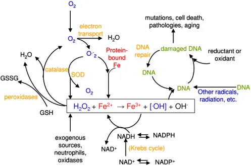
Figure 1.5 – Oxidative stress in the cell and DNA damage via Fenton reaction. H2O2 is
generated exogenously or by endogenous metabolism; O2.- is produced by one electron
reduction of oxygen by flavoproteins, metalloproteins; Superoxide dismutase (SOD) can scavenge O2.-, producing H2O2 which will react with the released Fe, in a Fenton reaction
producing OH., that will react with DNA, damaging and generating mutations and other
harmful processes. Scavenging systems like catalase and peroxidases can diminish the content of H2O2 in the cell. Adapted from (17).
H2O2+ Fe2+ →Fe3++ [.OH]+ OH
-O2 O2 O2 O .-2 SOD catalase peroxidases electron transport DNA repair (Krebs cycle) Protein-bound Fe damaged DNA DNA DNA DNA. Other radicals, radiation, etc.
H2O H2O
NADPH NADH NADP+ NAD+ NAD+ GSH exogenous sources, neutrophils, oxidases GSSG
mutations, cell death, pathologies, aging
reductant or oxidant
H2O2+ Fe2+ →Fe3++ [.OH]+ OH
-O2 O2 O2 O .-2 SOD catalase peroxidases electron transport DNA repair (Krebs cycle) Protein-bound Fe damaged DNA DNA DNA DNA. Other radicals, radiation, etc.
H2O H2O
NADPH NADH NADP+ NAD+ NAD+ GSH exogenous sources, neutrophils, oxidases GSSG
mutations, cell death, pathologies, aging
17
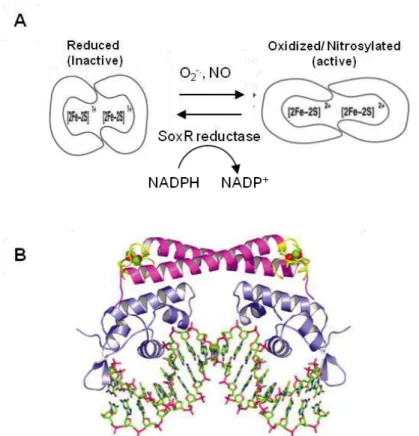
1.2.2.1 - SoxR(S), two component regulatory systemThe SoxR(S) system responds to superoxide or nitric oxide (NO) stress and is activated in two transcriptional steps (40). The discovery of this system occurred when Hassan and Fridovich (41) exposed E. coli to redox-cycling antibiotics and verified that the expression of MnSOD (manganese containing superoxide dismutase) was strongly induced. Some years later, two proteins, SoxR, a sensor protein that responds to oxidative stress and SoxS, a transcriptional activator that positively regulates a wide number of genes, were found out to be responsible for the high MnSOD expression in E. coli (42). The activation of this regulon is governed by the two proteins SoxR and SoxS that will trigger consecutive stages of transcription.
SoxR is a 17kDa protein containing a helix-turn-helix motif (HTH; DNA binding motif) in the N-terminal domain. It is a homodimer and each monomer contains one redox-active [2Fe-2S]2+/1+ cluster (
43). O2.- and
18
The reduction potential of SoxR [2Fe-2S]2+/1+ center, -285 mV (46), suggests that the reduction of this protein to its inactive, predominant redox state, is linked to the NAD(P)H/NAD(P)+
concentration ratio (E’0 =
-340 mV) (Figure 1.6). Since the majority of expressed genes induced by SoxS, will replenish the NAD(P)H pool in the cell, this two regulatory system, SoxR(S) is then autoregulated (48).
Figure 1.6 – A) SoxR activation and deactivation model (From (46)). B) Overall structure
19
1.2.2.2 - OxyR regulatorThe bacterial oxyR regulator responds to peroxide stress and is governed by OxyR. This protein has a molecular mass of 34kDa and belongs to the LysR family of transcription factors (50), with a HTH motif in the N-terminal region. OxyR forms a tetramer in solution and its active form occurs upon oxidation by hydrogen peroxide, which will trigger the formation of a disulfide bond, between Cys199 and Cys208 (E. coli numbering) (43). This active state, suffers a conformational shift that allows its binding to the promoter region of several genes, through direct contact with RNA polymerase (Figure 1.7).
In E. coli, OxyR activates the expression of several genes involved in H2O2 stress, such as katG (catalase), ahpCF (alkyl hydroperoxide
reductase), oxyS (transcription regulator that belongs to the SoxR(S) regulon), dps (DNA binding protein from starved cells), fur (ferric uptake regulator, see 1.2.2.3), gorA (glutathione reductase) and grxA
Figure 1.7 – Structure of the OxyR regulatory domain from E. coli. OxyR monomers in the
reduced (pdb 1I69) (A) and oxidized (pdb 1I6A) (B) forms are shown with the redox-active cysteines Cys-199 and Cys-208 (From (49)).
20
(glutaredoxin). The deactivation of OxyR is dependent on disulfide reducing systems such as glutathione together with glutaredoxin and thioredoxin reductase with thioredoxin. In vivo studies showed that glutathione and glutaredoxin are the major systems responsible for reducing the disulfide bond, which means that this protein is also autoregulated. The low reduction potential of the redox Cys center (-185 mV) means that OxyR is in the dithiol form in the cell and is the predominating form (46).
1.2.2.3 - Fur and PerR regulators
Fur-like proteins (Ferric uptake regulator) coordinate the regulation of iron assimilation. Fur acts as a positive transcriptional repressor; it represses transcription upon interaction with Fe2+ (Figure 1.8). Fur protein is a homodimer composed by two 17kDa subunits. Each subunit can be divided in two domains, an N-terminal DNA-binding domain with a regulatory binding site and a C-terminal domain rich in histidine residues which has a structural binding site and is involved in the dimerization (51-52). A divalent cation (ferrous iron in vivo) binds to the regulatory binding site allowing the protein to bind to the promoter region of iron-regulated genes, repressing gene transcription. The regulatory binding site can bind other divalent metal ions instead of Fe(II), such as Mn(II), Co(II), Zn(II) and Ni(II). The structural metal binding site has a very high affinity to Zn (II) (52-53) (Figure 1.8). The dimer active form binds the -35 and -10 promoter region of Fur-repressed genes. Fur-binding sites were originally found to form a 19-bp palindromic consensus sequence known as the ‘Fur box’.
21
sodB (FeSOD) (54-56).The regulation of Fur is part of an important link between redox stress management and iron homeostasis. In E. coli the fur gene is part of an operon with fdA (flavodoxin), which is able to maintain the ferrous ion pool in the cell, helping the activation of the Fur protein (58). This operon is induced by the SoxR(S) system and the fur gene has its own promoter, which is induced by OxyR (58).
Fur-like proteins able to sense signals other than iron have been found in gram positive and gram negative bacteria, such as Zur (Zinc uptake regulator), Mur (Manganese uptake regulator), Nur (Nickel uptake regulator), PerR (Peroxide regulator) and Irr (Iron responsive regulator) (51). In PerR, peroxide-regulon repressor, firstly characterized in
Figure 1.8 – A) Model of Fur-mediated gene repression (From (57)); B) The 3D structure
of Zn bound Fur from Vibrio cholerae (pdb 2W57; from (52)). Zinc ions are represented in spheres (yellow (Zn1) and orange (Zn2) from one monomer and green (Zn1 and Zn2) from the other monomer).
High Fe Low Fe
A
22
Bacillus (B.) subtilis, the iron-binding site has new functions such as metal-based sensor for peroxides (59). This regulator is the major one of the peroxide stress response in B. subtilis, but several other genomes were found to encode for a PerR-like protein, such as Campylobacter (C.) jejuni (60). This protein contains a structural Zn2+ site and can be activated to bind DNA by either Fe2+ or Mn2+ as co-repressors, bound to the regulatory binding site (Figure 1.9). In the active form with Fe2+/ Mn2+ bound to protein, H2O2 will oxidize the iron
atom, as in a Fenton reaction, generating a hydroxyl or ferryl species. This reactive species will oxidize one of the metal-coordinating histidines, facilitating the dissociation of Fe3+ and blocking the access to
Fe2+ ions (
51). As a consequence, these results in lack of DNA-binding activity, allowing the induction of the genes whose transcription was blocked. These genes will encode for, e.g., Dps like protein (MrgA), catalase (KatA), alkyl hydroperoxide reductase (AhpCF), enzymes of heme biosynthesis (HemAXCDBL) and Zinc uptake ATPase (ZosA) (61).
Figure 1.9 - Representation of the PerR-Zn-Mn crystal structure (62) (pdb 3F8N). Ribbon
structure in green, the structural Zn2+ ions are in black and the Mn2+ regulatory ions are in
23
1.2.3 - ROS and pathogensSome organisms, including anaerobes or microaerobes are pathogenic, invading and colonizing a host. To counteract this invasion the host uses many strategies, including the generation of ROS to kill the pathogenic organisms. The immune system of the host comprises specialized cells, e.g., macrophages and neutrophils, that are able to internalize the pathogen (phagocytosis) and by an intense oxidative burst, will produce ROS and destroy the invading organism (64). Cells like macrophages and neutrophiles use NADPH oxidase (Nox), a multicomponent enzyme, to generate superoxide and to initiate the ROS production. Nox enzymes constitute a family of transmembrane proteins comprising seven isoforms Nox 1-5 and Duox 1-2 (65). The structure of all these isoforms consists of six transmembrane domains and a cytosolic C-terminus (Figure 1.10). The III and IV transmembrane domains bind two prosthetic heme groups and the C-terminus contains two domains that bind the cofactor flavin adenine nucleotide (FAD) and the electron donor, NADPH (65).
Nox use these cofactors to transfer electrons from the cytosolic donor, NADPH to FAD and then sequentially to each heme group and finally to the molecular oxygen on the opposite side of the membrane to generate O2.- (65) (eq 1.11).
Figure 1.10 – Proposed structure of the core region of NADPH oxidase (NOX) enzymes.
24
NADPH + 2O2 → NADP+ + 2O2–. + H+ (eq 1.11)
NADPH is provided by the induced pentose phosphate pathway and the superoxide is generated to the extracellular space. The engulfed pathogens are wrapped in a plasma membrane vesicle that will be exposed to high fluxes of superoxide. In acidic pH the superoxide will be as the more reactive and membrane-permeable HOO. species or can also generate H2O2, which can cross the membrane easily.
The generation of OH. is also possible inside the phagocytic vacuole, since neutrophiles generate both superoxide and hypochlorous acid (HOCl) (eq 1.12).
HOCl + O2.- → O2 + OH. + Cl- (eq 1.12)
Hypochlorous acid generation is catalyzed by myeloperoxidase, an enzyme that in neutrophiles, is secreted into the vacuole. It is a heme-containing enzyme with a non-specific peroxidase activity, oxidizing a varied number of substrates (66). The oxidation of highly concentrated Cl- anions in the vacuole, allows the generation of the hypochlorous
acid that besides reacting with O2.-, can oxidize many biological
samples (66).
Another reactive species generated in macrophages and neutrophiles is nitric oxide (NO). This species is highly reactive and can react with O2.-,
forming peroxynitrite (ONOO-) that can oxidize iron-sulfur proteins, among many others. In macrophages, the enzyme inducible nitric oxide synthase (iNOS) is responsible for catalyzing the generation of NO, using NADPH and oxygen (67).
27
Chapter 2
2 - ROS detoxification enzymes ...29 2.1 - Scavenging Systems: SOD and Peroxidases ...29 2.2 - Rubrerythrin ...35
2.2.1 - Aminoacid sequence analysis ...35 2.2.3 - Structural domains ...38 2.2.3.1 - Rubredoxin domain ...39 2.2.3.2 - Four-helix bundle domain...41 2.2.4 - Hydrogen peroxide reduction mechanism ...45 2.2.5 - Physiological electron donors ...46
2.3 - Superoxide Reductase ...47
28
This Chapter includes material published in
“Reductive elimination of superoxide: Structure and mechanism of
superoxide reductases”, Pinto AF, Rodrigues JV, Teixeira M., 2010
29
2 - ROS detoxification enzymes
Oxidative stress in aerobic and anaerobic (facultative and strict) organisms has different impacts and responses, but shares some pathways for scavenging and reacting with ROS.
In aerobes the ROS production has its main origin in the aerobic electron transfer chain associated with the respiratory chain, which links NADH oxidation to O2 reduction to H2O, and is composed of several
complexes containing flavins, quinones and iron-sulfur centers which are prone to oxidation by O2, producing O2.- and H2O2 (see Section
1.2.1). Exogenous sources of ROS occur upon colonization of a host, whose immune system produces O2.- and H2O2, as mentioned in
Chapter 1. For these reasons, organisms have to regulate the steady-state concentration of these species using scavenging systems consisting of enzymes and antioxidant compounds. The goal is to maintain a balance of ROS formation and elimination and its concentration below toxic levels.
The most ubiquitous enzymatic systems involved in the scavenging of ROS among aerobic organisms, but also in anaerobic and facultative organisms, are superoxide dismutases (SOD) and peroxidases, of which catalase is a particular case.
2.1 - Scavenging Systems: SOD and Peroxidases
Superoxide Dismutase (SOD)
Superoxide dismutases constitute a family of enzymes able to catalyze the disproportionation of superoxide by redox-cycling of the metal in the active site (68). The disproportionation of superoxide is accomplished by SODs in two-steps (eq 2.1 and 2.2), which are both first-order in respect to O2.- and with kinetic rate constants in the range of 109 M-1 s-1
30
M(n+1)+ + O2.- + H+→ Mn+(H+) + O2 (eq 2.1)
Mn+(H+) + O
2.- + H+ → M(n+1)+ + H2O2 (eq 2.2)
2O2.- + 2H+ → O2 + H2O2 (eq 2.3)
The dismutation of superoxide follows a ping pong mechanism, where two O2.- will bind in two steps, the first coupled to uptake of a proton
which will facilitate substrate reduction and binding for the second O2
.-substrate molecule (69-72).
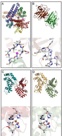
According to the metal cofactor each SOD harbors, they can be classified into three groups: Cu and Zn-containing superoxide dismutase (Cu,Zn-SOD), SODs specific for Fe, Mn or for either of the two metals (Fe-SOD, Mn-SOD or Fe/Mn-SOD) and the SODs that use Ni (Ni-SOD) (68). Although sharing the same function the overall structure differs for the three types of SODs (Figure 2.1).
Fe-SOD and Mn-SOD aminoacid sequences are extremely conserved namely on what concerns the residues of the active site and some nearby residues. The active site contains one Fe/Mn ion coordinated in a trigonal bipyramid geometry by three histidines, an aspartate and a molecule of OH-/H2O (69). Fe-SODs are considered the most primitive
SODs due to the Fe prevalence in a reducing environment as in the primitive atmosphere. Consistent with the diminished bioavailability of Feand appearance of O2, the MnSOD and CuZnSOD evolved (71).
31
The Ni-SODs enzymes were only recently found in Streptomyces bacteria and in cyanobacteria. The active site has a Ni ion bound in a square planar geometry to two thiolates from two cysteines and two backbone nitrogens from histidine and cysteine residues (73).In E. coli Fe-SOD is synthesized to almost 20 µM and O2.- is maintained
at 0.1 nM (28), which shows that the catalytic efficiency of SODs and the abundance of protein in the cytosol leads to a low level of O2.-,
non-hazardous to cell metabolites.
Figure 2.1 - A comparison of the enzyme structures and active sites for the four SODs, (A)
Streptomyces coelicolor NiSOD
(pdb: 1T6U), (B) human CuZnSOD (pdb: 1PU0), (C) E. coli FeSOD (pdb: 1ISA) and (D)
32
Peroxidases
Hydrogen peroxide is mainly scavenged by two types of enzymes. Peroxidases scavenge H2O2 by using it to oxidize other substrates (eq
2.4) and catalases directly catalyse the dismutation of H2O2 to O2 and
H2O (eq 2.5):
RH2 + H2O2 → R + 2H2O (eq 2.4)
H2O2 + H2O2 → O2 + 2H2O (eq 2.5)
Peroxidases are a large family of enzymes that have a wide range of substrates/electron donors. There are heme and non-heme peroxidases and in this last group the most important enzymes are thiol-specific peroxidases, glutathione peroxidases (Gpx) and peroxiredoxins (Prx) (74). Glutathione peroxidase encompasses a family of multiple isozymes, which catalyze the reduction of H2O2 or organic
hydroperoxides to water or corresponding alcohols using reduced glutathione. Some of these isozymes have selenium-dependent glutathione peroxidase activity (75). Peroxiredoxins are a ubiquitous family of antioxidant enzymes. Alkyl hydroperoxide reductase (AhpC) is a 2 Cys-peroxiredoxin and is the most studied enzyme from Prxs and is identified in a variety of prokaryotes (76). It is able to reduce a large number of substrates from H2O2 to organic compounds (76).
Catalases are among the most studied of enzymes. Three classes of proteins with no aminoacid sequence and structure similarity but with catalase activity have been defined (77). The class that is most
widespread in nature and which has been most extensively characterized is composed of mono functional, tetrameric heme-catalases subdivided on large (> 75 kDa) or small (< 60 kDa) subunits enzymes (77). The second, less widespread class is composed of
33
includes the non-heme or Mn-containing catalases, which have only been found in prokaryotes (77).
The mechanism of H2O2 dismutation by heme-catalases occurs in two
stages. The first substrate molecule is reduced to water, oxidizing catalase to an oxyferryl porphyrin cation species, named compound I. A second H2O2 molecule can complete the catalytic cycle reducing
compound I back to Fe3+ along with the generation of water and O2 (eq
2.6; E for enzyme and Por for porphyrin) (78).
E(Por-Fe3+) +H
2O2 → Compound I (Por+-Fe4+=O) +H2O
Compound I (Por+-Fe4+=O) + H
2O2 → E(Por-Fe3+) + H2O + O2 (eq 2.6)
Catalases do not follow Michaelis-Menten kinetics except at very low substrate concentrations, and different enzymes are affected differently at higher substrate concentrations. Turnover rates range from 54 000 s-1 to 833 000 s-1, which are considered the highest turnover rates for an
enzymatic system (78).
McCord and Fridovich in 1971 (79) proposed a correlation between oxygen tolerance and the content of SOD and catalase enzymes, so that anaerobic organisms unable to cope with oxygen would not need to express these scavenging enzymes. This hypothesis led to misleading assumptions, that in anaerobes the presence of O2 would impair growth