RESEARCH ARTICLE
Identification of Conserved MEL-28/ELYS
Domains with Essential Roles in Nuclear
Assembly and Chromosome Segregation
Georgina Gómez-Saldivar1☯, Anita Fernandez2☯
*, Yasuhiro Hirano3, Michael Mauro2, Allison Lai2, Cristina Ayuso1, Tokuko Haraguchi3,4, Yasushi Hiraoka3,4, Fabio Piano5,6,
Peter Askjaer1*
1Andalusian Center for Developmental Biology (CABD), CSIC/Junta de Andalucia/Universidad Pablo de Olavide, Seville, Spain,2Biology Department, Fairfield University, Fairfield, Connecticut, United States of America,3Graduate School of Frontier Biosciences, Osaka University, Suita, Japan,4Advanced ICT Research Institute Kobe, National Institute of Information and Communications Technology, Kobe, Japan,
5Department of Biology and Center for Genomics and Systems Biology, New York University, New York, New York, United States of America,6New York University, Abu Dhabi, United Arab Emirates
☯These authors contributed equally to this work. *[email protected](AF);[email protected](PA)
Abstract
Nucleoporins are the constituents of nuclear pore complexes (NPCs) and are essential regulators of nucleocytoplasmic transport, gene expression and genome stability. The nucleoporin MEL-28/ELYS plays a critical role in post-mitotic NPC reassembly through recruitment of the NUP107-160 subcomplex, and is required for correct segregation of mitotic chromosomes. Here we present a systematic functional and structural analysis of MEL-28 inC.elegansearly development and human ELYS in cultured cells. We have identi-fied functional domains responsible for nuclear envelope and kinetochore localization, chromatin binding, mitotic spindle matrix association and chromosome segregation. Sur-prisingly, we found that perturbations to MEL-28’s conserved AT-hook domain do not affect MEL-28 localization although they disrupt MEL-28 function and delay cell cycle progression in a DNA damage checkpoint-dependent manner. Our analyses also uncover a novel mei-otic role of MEL-28. Together, these results show that MEL-28 has conserved structural domains that are essential for its fundamental roles in NPC assembly and chromosome segregation.
Author Summary
Most animal cells have a nucleus that contains the genetic material: the chromosomes. The nucleus is enclosed by the nuclear envelope, which provides a physical barrier between the chromosomes and the surrounding cytoplasm, and enables precisely controlled trans-port of proteins into and out of the nucleus. Transtrans-port occurs through nuclear pore com-plexes, which consist of multiple copies of ~30 different proteins called nucleoporins. Although the composition of nuclear pore complexes is known, the mechanisms of their
a11111
OPEN ACCESS
Citation:Gómez-Saldivar G, Fernandez A, Hirano Y, Mauro M, Lai A, Ayuso C, et al. (2016) Identification of Conserved MEL-28/ELYS Domains with Essential Roles in Nuclear Assembly and Chromosome Segregation. PLoS Genet 12(6): e1006131. doi:10.1371/journal.pgen.1006131
Editor:Orna Cohen-Fix, National Institute of Diabetes and Digestive and Kidney Diseases, UNITED STATES
Received:March 13, 2016
Accepted:May 26, 2016
Published:June 24, 2016
Copyright:© 2016 Gómez-Saldivar et al. This is an open access article distributed under the terms of the
Creative Commons Attribution License, which permits unrestricted use, distribution, and reproduction in any medium, provided the original author and source are credited.
Data Availability Statement:All relevant data are within the paper and its Supporting Information files.
Funding:We gratefully acknowledge funding from the Spanish Ministry of Economy and
assembly and function are still unclear. We have analyzed the nucleoporin MEL-28/ELYS through a systematic dissection of functional domains both in the nematode
Caenorhabdi-tis elegansand in human cells. Interestingly, MEL-28/ELYS localizes not only to nuclear
pore complexes, but is also associated with chromosomal structures known as kineto-chores during cell division. Our studies have revealed that even small perturbations in MEL-28/ELYS can have dramatic consequences on nuclear pore complex assembly as well as on separation of chromosomes during cell division. Surprisingly, inhibition of MEL-28/ ELYS causes cell-cycle delay, suggesting activation of a cellular surveillance system for chromosomal damages. Finally, we conclude that the structural domains of MEL-28/ELYS are conserved from nematodes to humans.
Introduction
Metazoans have an open mitosis, in which the nuclear envelope (NE) disassembles during pro-phase to allow chromosome segregation and then reassembles around condensing chromosomes at anaphase [1]. During this process, the nuclear pore complexes (NPCs) are disassembled then rapidly reconstructed. ELYS, a large AT-hook domain protein, is essential for the late-mitosis rebuilding of the NPC [2]. ELYS is the first NPC component to associate with chromatin at the end of mitosis [3,4] and this association is required for the recruitment of the NUP107-160 sub-complex of the NPC, which in turn recruits vesicles containing the membrane-bound nucleo-porins POM121 and NDC1 [4]. Thus ELYS binding to chromatin represents the first step in the post-mitotic building of the pore, and all other steps in its manufacture are dependent on this ELYS/chromatin interaction.
ELYS was originally identified in a cDNA subtraction screen seeking genes expressed at high levels in the mouse embryonic sac [5]. Mouseelysknockouts die in the preimplantation stage because of cell death within the inner cell mass [6]. ELYS function is essential in all meta-zoa and is particularly important in rapidly dividing cells [7,8]. InC.elegans, the orthologous MEL-28 protein dynamically localizes to the nucleoplasm and NPC at interphase and then at the kinetochore and spindle at metaphase [9,10]. Consistent with its localization pattern, embryos that lackmel-28function have severe defects with NE function, mitotic spindle assem-bly and chromosome segregation and are unviable.
The ELYS/chromatin interaction has been studied extensivelyin vitrousingXenopuscell extracts. ELYS binds to chromatin during interphase but not at metaphase [11], when it instead associates with the spindle and kinetochore [12]. Chromatin immobilization assays have shown that the most C-terminal fragment of ELYS, corresponding to amino acids (aa.) 2281–
2408, is sufficient for chromatin binding. This region includes the AT hook, a motif that binds to AT-rich DNA. However the aa. 2281–2408 fragment with a mutated AT hook and a C-ter-minal fragment that excludes the AT hook (aa. 2359–2408) also bound to chromatin [4]. A nucleosome binding assay showed that a large C-terminal fragment that includes the AT hook (aa. 2281–2408) was sufficient to bind to nucleosomes, whereas a piece that includes just the AT hook (aa. 2281–2358) or just the region C-terminal to the AT hook (aa. 2359–2408) could not bind to nucleosomes [13]. Additionally, incubation ofXenopusextracts with the C-termi-nal 208-aa. fragment of ELYS prevented native ELYS from binding to sperm chromatin and also prevented the recruitment of other nucleoporins to the nuclear rim, phenocopying theelys
loss-of-function phenotype [11]. However, introducing a C-terminal fragment with a mutated AT hook does not disrupt nuclear pore assembly and is less effective at outcompeting the endogenous ELYS from binding to chromatin [4]. Thesein vitroexperiments suggest that both
The funders had no role in study design, data collection and analysis, decision to publish, or preparation of the manuscript.
the AT hook and other domains of the C terminus are important for the ELYS/chromatin interaction and the subsequent rebuilding of the NPC.
The ELYS/chromatin association has also been studied using mousein vitrofertilization. During fertilization in mice, sperm chromatin is rebuiltde novousing histones present in the oocyte. Experiments usingin vitrofertilized mouse oocytes depleted of histones showed that ELYS does not localize to the NE of the sperm pronucleus in the absence of histones, which in turn prevents the recruitment of other nucleoporins [14]. ELYS can be artificially targeted to the NE in the absence of histones by fusing it with a domain from an inner NE protein. This chimeric ELYS protein not only localizes to the NE but also recruits the other nucleoporins. This suggests that ELYS binding to chromatin is required for its localization to the nuclear rim, which in turn allows the remainder of the nuclear pore to be built.
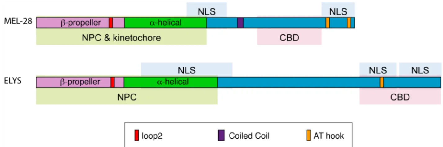
The overall architecture of MEL-28/ELYS is similar throughout the metazoa (see schematic representations in Figs2Cand7B). All metazoan MEL-28/ELYS homologs include an N-ter-minalβ-propeller domain, a centralα-helical domain, and a C-terminal domain that includes at least one AT hook. Crystal structure determination of the N-terminal domain of mammalian ELYS showed that it forms a seven bladedβ-propeller structure with an extra loop decorating each of the propeller blades [15]. In human cells, the N-terminal 1018 amino acids of ELYS (which includes theβ-propeller domain and the centralα-helical domain but not the C-termi-nal AT hook) is sufficient to localize the protein to NPCs [15]. Mutational disruption of the conserved loop on blade 6 of theβ-propeller domain (“loop2”) prevents the 1–1018 aa. frag-ment from localizing to the nuclear rim.
Despite the interest in defining the functional domains of MEL-28/ELYS, until now there have been no studies in which the phenotypic consequences of disrupting specific domains have been studied in developing animals. In this work, we have dissected the MEL-28 protein and studied its localization and function in liveC.elegansembryos. We have identified regions of MEL-28 required for its roles in meiosis as well as in chromatin binding and post-mitotic nuclear pore construction. Our parallel studies in HeLa cells show that the domains required for proper localization inC.elegansare conserved in human ELYS, suggesting that conclusions from functional analyses of MEL-28 inC.elegansare broadly applicable to vertebrate ELYS.
Results
MEL-28 is required for meiotic chromosome segregation
We previously reported thatC.elegansMEL-28 is broadly expressed [10]. However, a promoter study of 127 genes inC.elegansembryos suggested that MEL-28 is highly enriched in the intes-tinal E lineage ~200 min after fertilization [16]. We therefore revisited MEL-28 expression to analyze it in greater detail. Immunofluorescence analysis detected similar levels of MEL-28 in nuclei of all embryonic cells (S1A Fig) and all postembryonic tissues (S1B Fig). Next, using CRISPR-Cas9 technology [17], we generated a GFP knock-inmel-28allele to analyze the expression of endogenous MEL-28 by live microscopy. Similar to the observations with anti-bodies against MEL-28, GFP::MEL-28 localized to the NE in all cell types during embryonic and larval development and in adults (S1C Fig). Thus, we conclude that MEL-28 is ubiqui-tously expressed throughoutC.elegansdevelopment.
MEL-28 strongly accumulated on condensed oocyte chromosomes (S1C Fig; [9,18]). More-over, we noted during our initial studies ofmel-28mutant or RNAi-treated embryos that for-mation and migration of the maternal pronucleus was often more severely affected than the paternal pronucleus [9,10]. Based on these observations we speculated that MEL-28 might have important functions in meiosis.C.elegansoocytes are arranged in a linear fashion in the proximal part of the gonad, where each oocyte is numbered relative to the spermatheca (-1, -2,
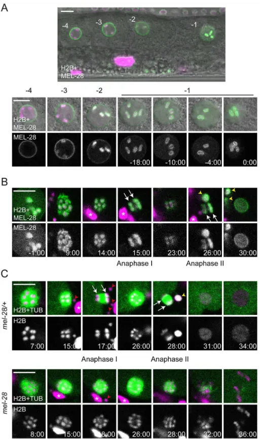
-3, etc.) [19]. The -1 oocyte completes maturation including germinal vesicle breakdown immediately before ovulation and fertilization triggers rapid progression through meiosis I and II. To examine these processes we performed livein uterorecordings of animals expressing GFP::MEL-28 and mCherry::HisH2B. In the -4 oocyte, MEL-28 localized to the NE and was absent from condensed chromosomes (Fig 1A). In the -3 and -2 oocytes MEL-28 gradually moved away from the NE and accumulated uniformly on meiotic chromosomes. Later, in the -1 oocyte MEL-28 redistributed to cover the surface of meiotic chromosomes (Fig 1A;S1 Video), in some cases completely enclosing the chromosomes and in other cases similar to the
“cup-shaped”localization of kinetochore proteins, such as KNL-1 and KNL-3 [20]. The associ-ation of MEL-28 with chromosomes persisted throughout meiosis I and II until pronuclear for-mation ~30 minutes after germinal vesicle breakdown (Fig 1B;S1 Video). The localization pattern of MEL-28 suggested a possible role during segregation of meiotic chromosomes, simi-lar to the situation in mitosis [9,10]. We therefore analyzedmel-28(t1684)embryos expressing GFP::β-tubulin and mCherry::HisH2B.mel-28(t1684)encodes a premature termination codon at aa. 766 and behaves like a strong loss-of-function of MEL-28, presumably due to nonsense-mediated mRNA decay [10]. Maternal contribution enables homozygousmel-28(t1684) her-maphrodites to develop until adulthood but they produce only unviable embryos (hereafter referred to asmel-28embryos, whereas embryos produced by heterozygous siblings are referred to as control ormel-28/+embryos) with severe NE assembly defects [10]. Strikingly,
inmel-28embryos chromosomes failed to segregate in anaphase I (n = 5/6 embryos) and
anaphase II (n = 4/6) and, consequently,mel-28embryos had either no (n = 4/6) or a single (n = 2/6) polar body, whereas control embryos had two polar bodies (n = 6/6;Fig 1C;S2 Video). In addition, chromosomes inmel-28embryos were not organized in a pronucleus but appeared scattered in the cytoplasm (Fig 1C; 36:00). To our knowledge, this is the first report describing the involvement of 28/ELYS in meiosis, expanding previously described MEL-28 functions and establishing an important role in chromosome segregation during both meio-sis and mitomeio-sis.
The MEL-28 N-terminus is required for NPC association
To characterize which regions of MEL-28 are required for its different functions, we examined full-length and truncated versions of MEL-28 fused to GFP and tracked their localization in
liveC.elegansembryos. While most transgenes are expressed (S2 Fig;S4 Fig), some exhibit
localization patterns distinct from length MEL-28 (see below). During interphase full-length MEL-28 was mainly localized to the NE but was also found in the nucleoplasm (Fig 2A;
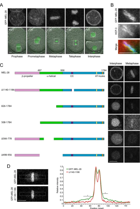
S3 Video;S9 Video). In prophase and prometaphase, MEL-28 left the NE before complete NE breakdown and associated to the condensing chromosomes. By metaphase, MEL-28 appeared as two lines parallel to the metaphase plate, resembling the characteristic pattern of holocentric kinetochore proteins, and less abundantly to the area of the mitotic spindle (Fig 2A–2D). Dur-ing anaphase, MEL-28 associated to decondensDur-ing chromosomes, and re-localized to reform-ing NE in telophase (Fig 2A;S3 Video).
Fig 1. MEL-28 is essential for female meiosis.(A) GFP::MEL-28 (green in merged images) was expressed in oocytes and accumulated at kinetochores of meiotic chromosomes (visualized with mCherry::HisH2B; magenta in merge). Shown are the four most proximal oocytes where position -1 is immediately next to the spermatheca. The -1 oocyte was observed every two minutes until germinal vesicle breakdown. (B) GFP:: MEL-28 associated with chromosomes throughout meiosis I and II and accumulated at the NE at pronuclear
Recently, Bilokapic and Schwartz found that the N-terminal half of ELYS containing theβ -propeller andα-helical domains localized to the NE in HeLa cells [15]. However, the relevance of these domains has not been analyzed in the context of full-length MEL-28/ELYS. We first deleted theβ-propeller and most of theα-helical domain (GFP::MEL-28826-1784) and found that both NE localization during interphase and kinetochore localization in mitosis were abro-gated (Fig 2C). Instead, the truncated protein was found in the nucleoplasm and weakly associ-ated with chromosomes during interphase and metaphase, respectively (note that kinetochore localization appears as two parallel lines whereas a single line reflects more uniform chromo-some association). Similar mis-localization was observed on deletion of aa. 1–507 (GFP::MEL-28508-1784) or aa. 498–956 (GFP::MEL-28Δ498–956), whereas deletion of aa. 566–778 (GFP:: MEL-28Δ566–778) also abolished the weak association to mitotic chromosomes (Fig 2C).
Together, these results demonstrate that both theβ-propeller and theα-helical domain are required for targeting MEL-28 to NPCs and to kinetochores. All four N-terminally truncated MEL-28 proteins accumulated in the nucleus in interphase, suggesting that the C-terminal unstructured domain of MEL-28 contains one or more nuclear localization signals (NLS’s; see below).
Finally, we assessed whether the truncations in theβ-propeller andα-helical domains inter-fered with MEL-28 function. As expected from the severe mis-localization, ectopic expression of any of the four MEL-28 truncations failed to restore viability ofmel-28embryos (Table 1), suggesting that the localization of MEL-28 to NPCs and kinetochores is essential to MEL-28 function. We conclude from these experiments that the N terminus of MEL-28 is required for proper MEL-28 localization and functions. Whereas its importance for NPC localization is concordant with data on ELYS our experiments revealed a novel role in kinetochore association.
MEL-28 loop2 is required during meiosis and mitosis
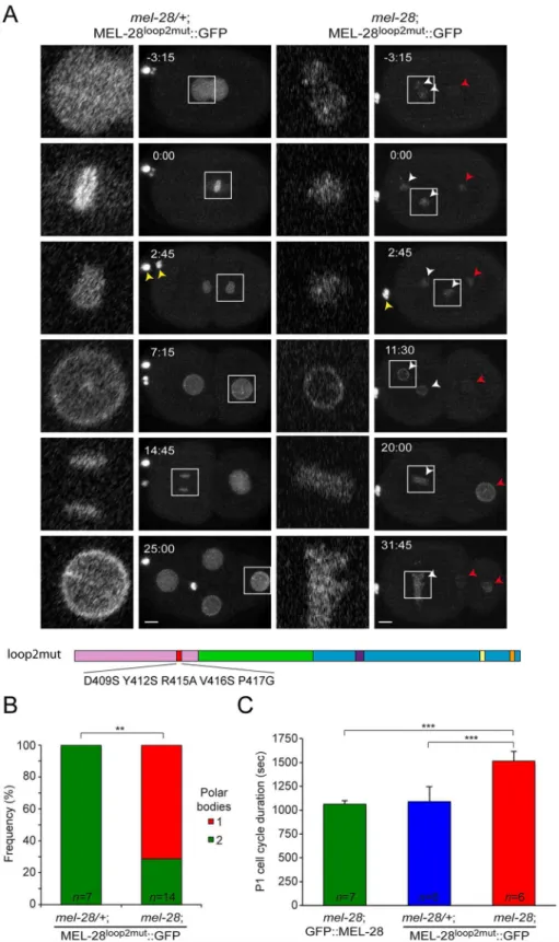
Bilokapic and Schwartz identified through protein crystallization and sequence alignments two conserved loops (loop1 and loop2) on the surface of theβ-propeller of ELYS [15]. When they substituted 5 aa. within loop2 the structural fold of theβ-propeller was maintained but NPC localization of the N-terminal half of ELYS (aa. 1–1018) fused to GFP was abrogated in HeLa cells. To test the relevance of loop2 in the context of full-length protein we introduced the equivalent aa. substitutions in MEL-28 (D409S/Y412S/R415A/V416S/P417G; MEL-28loop2mut;
Fig 3A). Inmel-28/+embryos MEL-28loop2mut::GFP localized normally during interphase and mitosis (Fig 3A, left panels; compare with wild type GFP::MEL-28 inFig 2A;S4 Video;S3 Fig), suggesting that loop2 residues are not essential for association of full-length MEL-28 with NPCs or kinetochores. However, MEL-28loop2mut::GFP was not able to substitute for endoge-nous MEL-28:mel-28embryos expressing MEL-28loop2mut::GFP were unviable (Table 1) and had frequent meiosis defects as evidenced by failure in polar body extrusion and presence of multiple female pronuclei (Fig 3A, right panels;S4 Video;Fig 3B). Moreover, pronuclei were abnormally small, contained less MEL-28loop2mut::GFP and did not position properly. In 83%
ofmel-28; MEL-28loop2mut::GFP embryos (n = 10/12) female and male pronuclei did not meet
before the first mitotic division. Instead, only the male pronucleus was positioned between the
formation. (C) Chromosomes (magenta) and meiotic spindles (green) were observedin utero. Anaphase I and II were characterized by abundant microtubules between segregating chromosomes in controlmel-28/+
animals (top) whereas chromosomes failed to segregate in homozygousmel-28mutants (bottom). White arrows point to segregating chromosomes. Red arrowheads and white asterisks mark sperm and somatic nuclei, respectively, outside the fertilized oocyte; yellow arrowheads indicate polar bodies. Time is indicated relative to germinal vesicle breakdown (min:sec). Scale bars, 5μm.
Fig 2. MEL-28 N-terminal domains are required for NPC and kinetochore localization.(A) Still images from time-lapse recording of embryo carrying a GFP insertion into the endogenousmel-28locus. Time is indicated relative to anaphase onset (min:sec). (B) Metaphase plate of early embryo expressing GFP::MEL-28 (green in merge) analyzed by immunofluorescence with a specific antibody against HCP-3/CENP-A (red in merge) and Hoechst (blue in merge) to visualize chromosomes. MEL-28 localized to kinetochores, which appear as lines on
centrosomes, whereas female pronuclei exhibited shorter migration and remained in the ante-rior of the embryo. During mitosis chromosomes failed to congress to the metaphase plate (Fig 3A; 0:00) and severe segregation defects were observed (Fig 3A; 20:00–31:45). We also noticed alterations in cell cycle timing, in particular for the posterior P1 blastomere at the two-cell stage. Inmel-28; GFP::MEL-28 andmel-28/+; MEL-28loop2mut::GFP embryos the cell cycle of P1 lasted ~1075 sec, whereas it lasted ~1513 sec (41% delay) inmel-28embryos expressing MEL-28loop2mut::GFP (Fig 3C). Other frequent defects included cleavage furrow regression (37%; n = 6/16) and abnormal positioning of cells within the eggshell (53%; n = 8/15).
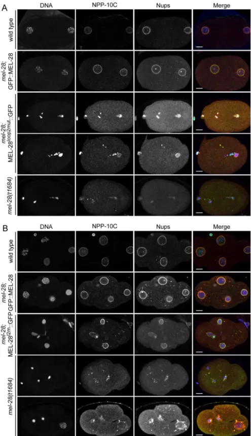
To analyze if the conserved loop2 is required for MEL-28’s role in NPC assembly we per-formed immunofluorescence onmel-28; MEL-28loop2mut::GFP embryos and compared them with wild type,mel-28, andmel-28; GFP::MEL-28 embryos. One-cell and four-cell stage embryos were analyzed for meiotic and mitotic defects, respectively, using mAb414 to visualize multiple Nups and specific antibodies against NPP-10C/NUP96, which is a component of the NUP107 complex [21]. Uniform peripheral signal was observed at pronuclei of wild type and
mel-28; GFP::MEL-28 one-cell stage embryos, whereas fragmented pronuclei with inconsistent
Nup signal was detected inmel-28; MEL-28loop2mut::GFP andmel-28embryos (Fig 4A). Analy-sis of four-cell stagemel-28; MEL-28loop2mut::GFP embryos confirmed the defects in chromo-some segregation observed by live imaging and revealed that although nuclei with peripheral
both sides of the chromosomes. (C) Cropped images from embryos expressing different MEL-28 truncations fused to GFP. Except GFP::MEL-28 and GFP::MEL-28Δ1140–1186embryos, all embryos also expressed un-tagged
endogenous MEL-28. Purple boxes in MEL-28 cartoons indicate a putative coiled-coil domain (aa. 1127–1160) whereas yellow (aa. 1630–1642) and orange (aa. 1746–1758) boxes indicate AT-hook sequences: their homology to the consensus AT-hook sequence is low and high, respectively. (D) Cropped images from metaphase embryos expressing GFP::MEL-28 or GFP::MEL-28Δ1140–1186. Images were processed identically to facilitate visualization
of full-length GFP::MEL-28 associated with the mitotic spindle. Signal intensities in boxed areas were quantified in raw images, normalized and plotted (n = 5, GFP::MEL-28; n = 2, GFP::MEL-28Δ1140–1186).
*p<0.05 by unpaired two-tailed t-test. Scale bars, 5μm.
doi:10.1371/journal.pgen.1006131.g002
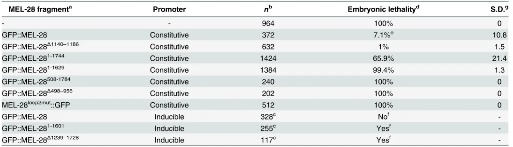
Table 1. Rescue efficiencies by MEL-28 fragments.
MEL-28 fragmenta Promoter nb Embryonic lethalityd S.D.g
- - 964 100% 0
GFP::MEL-28 Constitutive 372 7.1%e 10.8
GFP::MEL-28Δ1140–1186 Constitutive 632 1% 1.5
GFP::MEL-281-1744 Constitutive 1424 65.9% 21.4
GFP::MEL-281-1629 Constitutive 1384 99.4% 1.3
GFP::MEL-28508-1784 Constitutive 240 100% 0
GFP::MEL-28Δ498–956 Constitutive 202 100% 0
MEL-28loop2mut::GFP Constitutive 512 100% 0
GFP::MEL-28 Inducible 328c Nof
-GFP::MEL-281-1601 Inducible 255c Yesf
-GFP::MEL-28Δ1239–1728 Inducible 117c Yesf
-aGFP::MEL-28 fusions were expressed inmel-28(t1684)mutants. b,cThe number of embryosbor wormscanalyzed is indicated. dPercentage of unhatched embryos.
eThis strain was also homozygous forunc-32(e189), which may have in
fluenced the incomplete rescue.
fQualitative analysis was done for fragments under control of an inducible promoter due to heterogeneity of the expression. gStandard deviation.
Fig 3. MEL-28 loop2 is required during meiosis and mitosis.(A) Still images from time-lapse recordings of control (left) andmel-28(right) embryos expressing MEL-28loop2mut::GFP. Note the presence of two polar bodies in the left embryo but only a single polar body in the right embryo (yellow arrowheads). Concordantly, two oocyte-derived pronuclei were observed in the right embryo (white arrowheads). Red arrowheads indicate sperm-derived chromosomes. Whole-embryo images are max projections; inserts are single
Nup localization are formed, these are smaller than in wild type andmel-28; GFP::MEL-28 embryos (Fig 4B). The NE phenotypes inmel-28; MEL-28loop2mut::GFP embryos were less severe when compared tomel-28embryos. As previously reported, nuclear reformation and NPC assembly was strongly inhibited inmel-28embryos although a few cells had larger nuclei with irregular NE-structure (Fig 4B; bottommel-28embryo).
From these data we conclude that MEL-28’s loop2 is essential for correct chromosome seg-regation both in meiosis and mitosis but not strictly required for post mitotic NPC assembly, nor for incorporation into the NE.
Identification of MEL-28 nuclear localization and chromatin association
domains
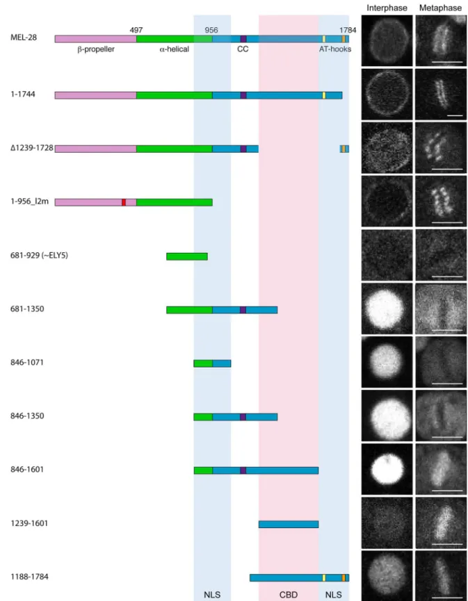
The observation that perturbations in MEL-28’s N-terminal half do not prevent nuclear accu-mulation of MEL-28 prompted us to analyze the C-terminus for functional domains. We first expressed GFP::MEL-281-1744, which lacks 40 aa. from the C-terminal end including one of the two AT-hook motifs. This short truncation did not interfere with MEL-28 localization in inter-phase nor during mitosis (Fig 5;S5 Video). However, expression of GFP::MEL-281-1744rescued lethality in only ~35% ofmel-28embryos (Table 1), indicating that the C-terminal AT hook of MEL-28 contributed significantly to MEL-28 activity. Next, we deleted aa. 1239–1728, includ-ing the other AT-hook motif. This reduced slightly the NE accumulation at interphase (Fig 5; GFP::MEL-28Δ1239–1728;S6 Video). Importantly, expression of GFP::MEL-28Δ1239–1728was not able to rescue the embryonic lethality ofmel-28embryos (Table 1), which suggests that there are domains within this region required for MEL-28 function. Despite several attempts, we were unable to express a MEL-28 aa. 1–956 fragment consisting of wild typeβ-propeller andα -helical domains (S4 Fig). In contrast, a similar fragment, but with the five aa. substitutions in loop2 described above was efficiently expressed (MEL-281-956_l2m::GFP;S7 Video). MEL-281-956_l2m::GFP localized to the cytoplasm and NE, but its relative NE accumulation compared to kinetochore localization was dramatically reduced (S3 Fig). As expected, expression of MEL-281-956_l2m::GFP did not rescue the embryonic lethality ofmel-28embryos (Table 1). Taken together with the results presented inFig 2, we conclude that although the N-terminalβ -pro-peller andα-helical domains are the main determinants for NPC and kinetochore localization, the C-terminal portion of MEL-28 also contributes significantly.
A divergent ~300 aa. MEL -28/ELYS homolog termed ELY5 was recently identified in sev-eral fungi [22,23]. Although our experiments presented above would suggest that the part of MEL-28 equivalent to ELY5 (identified as aa. 696–927 by [24]) does not contain the domains required for NPC localization we nevertheless expressed a fragment containing aa. 681–929 fused to GFP. As expected, this fragment did not localize to the NE or to kinetochores but showed instead diffuse cytoplasmic signal throughout the cell cycle (Fig 5; GFP::MEL-28681-929;
S2B Fig).
We next expressed a series of overlapping fragments from aa. 681 to the C-terminal end. All fragments that contained aa. 846–1071 accumulated efficiently in the nucleus (Fig 5; GFP:: MEL-28681-1350, GFP::MEL-28846-1071, GFP::MEL-28846-1350, and GFP::MEL-28846-1601;S4A Fig; GFP::MEL-28846-1167;S8 Video). A shorter fragment consisting of aa. 846–956 behaved similarly to free GFP (S4A Fig; GFP::MEL-28846-956). Nuclear accumulation was also detected for GFP::MEL-281188-1784, but not for GFP::MEL-281161-1601or GFP::MEL-281239-1601(Fig 5;
confocal sections. Scale bars, 5μm. (B) Frequency of embryos with a single or two polar bodies.**p<0.01 by Fisher exact test. (C) Timing from P0 division to P1 division is significantly delayed inmel-28embryos expressing MEL-28loop2mut::GFP.
***p<0.001 by unpaired two-tailed t-test.
Fig 4. Mutation of MEL-28 loop2 impairs chromosome segregation.One-cell stage (A) and 4-cell stage (B) embryos frommel-28mutants expressing either GFP::MEL-28 or MEL-28loop2mut::GFP were compared
with wild type andmel-28embryos by immunofluorescence. Embryos were analyzed with Hoechst (blue in merge), a specific antibody against NPP-10C/NUP96 (green in merge) and mAb414 recognizing multiple nups (red in merge). Scale bars, 5μm.
doi:10.1371/journal.pgen.1006131.g004
Fig 5. Identification of MEL-28 chromatin binding domain and nuclear localization signals.Cropped images from embryos expressing different MEL-28 truncations fused to GFP. Except GFP::MEL-281-1744and GFP::MEL-281188-1784, fusion
proteins were expressed from thehsp-16.41promoter in gastrulating embryos. Excluding GFP::MEL-281-1744embryos, all
S4A Fig). These observations are consistent with MEL-28 having at least two NLS’s mapping to the regions 846–1071 and 1601–1784. Moreover, using the NLS prediction software“cNLS Mapper”[25] we identified several putative mono- and bipartite NLSs in these regions: two in the central region (aa. 942–970 and 1033–1062 with scores 5.9 and 5.2, respectively) and three in the C-terminal region (aa. 1606–1636, 1682–1709 and 1741–1773 with scores 5.7, 7.4 and 5.3, respectively). Analysis of these C-terminal fragments also revealed that aa. 1239–1601 con-fer strong chromatin binding during mitosis (Fig 5).
The AT-hook domain is dispensable for MEL-28 localization, but
essential for its functions
Comparing the behavior of GFP::MEL-281239-1601and GFP::MEL-281188-1784indicated that MEL-28’s two AT hooks are not required for chromatin association, at least during mitosis (Fig 5). Moreover,in vitrobinding experiments found no difference in chromatin affinity between recombinant peptides that contained either the C-terminal 128 aa ofXenopusELYS including the single ELYS AT hook or a variant with mutated AT hook although the former was more efficient in competition assays [4]. In agreement with the competition assay, it was independently demonstrated that the same 128-aa. peptide efficiently binds nucleosome beads but not when the AT hook is mutated [13]. However, both studies concluded that the 128-aa. peptide contains residues outside the AT hook important for chromatin and nucleosome inter-action. We attempted to address this in further detail, but we were unable to detect expression of a construct encoding the C-terminal 161 aa. of MEL-28 fused to GFP (S4B Fig; GFP::MEL-281624-1784). A shorter 48-aa. fragment containing a single AT hook localized similarly to free GFP (S4A FigGFP::MEL-281740-1784). As a complementary approach, we examined the conse-quences of deleting the AT hooks from full-length MEL-28. We first comparedmel-28/+
embryos expressing GFP::MEL-281-1629(GFP::MEL-28ΔAT) withmel-28embryos expressing
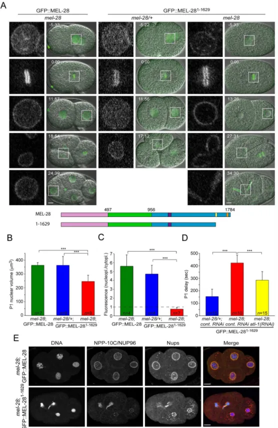
full-length MEL-28 fused to GFP. Time-lapse confocal microscopy demonstrated that the mel-28/+; GFP::MEL-281-1629embryos developed normally and the fluorescent protein localized similarly to GFP::MEL-28 (Fig 6A; compare left and middle panels;S9andS10Videos). In the absence of endogenous MEL-28, GFP::MEL-281-1629still accumulated at the periphery of inter-phase nuclei and to kinetochores of mitotic chromosomes (Fig 6A; right panels;S10 Video). This was in contrast to the severe phenotypes observed in MEL-28loop2mut::GFP embryos (Fig 3A) and suggested that MEL-28’s function in post-mitotic nuclear assembly is not strictly dependent on the AT hook domain. However,mel-28; GFP::MEL-281-1629embryos were unvi-able (Table 1) and displayed several defects. Most prominently, daughter nuclei were often (n = 5/7) trapped at the cleavage furrow during cytokinesis of the anterior AB blastomere of two-cell stage embryos (Fig 6A, right panels; 27:31–34:30). More direct evidence for chromo-some segregation failure was obtained by immunofluorescence analysis of four-cell stage embryos, which also demonstrated that NPP-10C/NUP96 and other Nups accumulated at the NE ofmel-28; GFP::MEL-281-1629embryos, albeit in an irregular pattern (Fig 6E). In addition, nuclear growth was significantly reduced in GFP::MEL-281-1629embryos (Fig 6A, third row;
Fig 6B), consistent with defects in NPC-mediated nucleocytoplasmic transport [26]. While nuclei frommel-28; GFP::MEL-28 andmel-28/+; GFP::MEL-281-1629grew to the same size (363.8 ± 19μm3and 363.3 ± 63μm3; respectively), the maximum volume of P1 nuclei was
reduced by 32% inmel-28; GFP::MEL-281-1629embryos (346.6 ± 44μm3). We also noticed that
the nucleoplasmic pool of GFP::MEL-281-1629was strongly diminished inmel-28embryos compared to GFP::MEL-28 inmel-28embryos and GFP::MEL-281-1629inmel-28/+embryos (Fig 6A and 6C). Whereas the ratio between nucleoplasmic and cytoplasmic GFP signal was similar betweenmel-28; GFP::MEL-28 andmel-28/+; GFP::MEL-281-1629embryos (5.60 ± 1.29
and 4.72 ± 0.99; respectively), the ratio was 87% lower inmel-28; GFP::MEL-281-1629embryos (0.76 ± 0.18). These data are compatible with a model in which GFP::MEL-281-1629has reduced affinity for interphase chromatin and therefore accumulates at NPCs: inmel-28/+embryos interaction of GFP::MEL-281-1629with endogenous MEL-28 accumulates the former in the nucleoplasm, potentially interacting with chromatin.
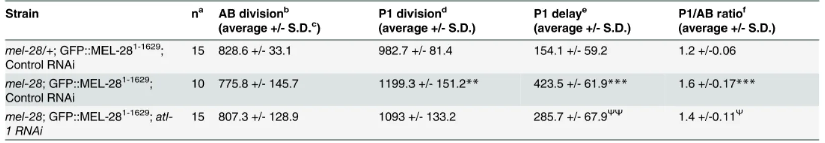
During time-lapse recordings of 2-cell stagemel-28embryos, we realized that division of the P1 blastomere was much delayed relatively to the AB division. In wild-type embryos the P1 cell division is delayed by ~2.5 min compared to AB division. This P1 delay is dependent on check-point proteins and is thought to have evolved to protect the germ-line lineage from aneuploidy. Thus, inhibition of DNA replication or induction of DNA damage is typically associated with extended P1 delay. When we compared embryos expressing GFP::MEL-281-1629an increase in P1 delays by 176% was observed inmel-28versus mel-28/+embryos (423.5 ± 61.9 sec versus 154.1 ± 59.2 sec;Table 2;Fig 6D). The presence of chromatin bridges inmel-28; GFP::MEL-281-1629embryos (Fig 6E) suggested that chromosomes might be entangled, potentially as con-sequence of stalled replication and/or double-stranded DNA breaks. To address if the DNA damage checkpoint indeed is involved in the extended P1 delay inmel-28; GFP::MEL-281-1629 embryos, we depleted ATL-1, theC.eleganshomolog of ATR by RNAi [27]. This mitigated the P1 delay (285.7 ± 67.9 sec), which suggested that removal of the AT-hook domain from MEL-28 activates DNA damage and thereby an exaggerated delay of P1 cell division. However, depletion of ATL-1 did not fully rescue P1 cell-cycle timing, which suggests that other check-points are also activated inmel-28; GFP::MEL-281-1629embryos. In conclusion, although
expressing GFP::MEL-281-1629. Measurements were performed on fully-grown P1 nuclei. (D) Asynchrony between division of AB and P1 blastomeres was significantly delayed inmel-28embryos expressing GFP:: MEL-281-1629; this delay was partially reduced by depletion of ATL-1. (E) Four-cell stage embryos from
mel-28mutants expressing either GFP::MEL-28 or GFP::MEL-281-1629were analyzed with Hoechst (blue in
merge), a specific antibody against NPP-10C/NUP96 (green in merge) and mAb414 recognizing multiple nups (red in merge). Scale bars, 5μm.***p<0.001 by unpaired two-tailed t-test.
doi:10.1371/journal.pgen.1006131.g006
Table 2. Loss of MEL-28’s AT Hooks causes checkpoint-dependent cell division delays.
Strain na AB divisionb
(average +/- S.D.c) P1 division d
(average +/- S.D.)
P1 delaye (average +/- S.D.)
P1/AB ratiof (average +/- S.D.)
mel-28/+; GFP::MEL-281-1629;
Control RNAi
15 828.6 +/- 33.1 982.7 +/- 81.4 154.1 +/- 59.2 1.2 +/-0.06
mel-28; GFP::MEL-281-1629;
Control RNAi
10 775.8 +/- 145.7 1199.3 +/- 151.2** 423.5 +/- 61.9*** 1.6 +/-0.17***
mel-28; GFP::MEL-281-1629;
atl-1 RNAi
15 807.3 +/- 128.9 1093 +/- 133.2 285.7 +/- 67.9ΨΨ
1.4 +/-0.11Ψ
Significant differences by two-tailed t-test:
**different from controlmel-28/+ embryos, p<0.01; ***different from controlmel-28/+ embryos, p<0.001;
Ψ
different frommel-28control RNAi embryos p<0.01;
ΨΨ
different frommel-28control RNAi embryos p<0.001.
aNumber of embryos analyzed via real-time DIC microscopy.
bTime in seconds between P0 cytokinesis onset and AB cytokinesis onset. cStandard deviation.
dTime in seconds between P0 cytokinesis onset and P1 cytokinesis onset. eTime in seconds between AB cytokinesis onset and P1 cytokinesis onset. fratio of time to P1 over time to AB cytokinesis onset.
doi:10.1371/journal.pgen.1006131.t002
GFP::MEL-281-1629localizes properly to the NE and kinetochores, depletion of MEL-28’s AT-hook domain causes reduced nuclear growth, mis-segregation of chromosomes and activates the ATR DNA damage checkpoint.
MEL-28/ELYS localization domains are evolutionary conserved
To explore the degree of conservation of localization domains we expressed human full-length ELYS (ELYS1-2275) and 14 ELYS truncations fused to GFP in HeLa cells. As reported, ELYS1-2275 was enriched at the NE in interphase and in a pattern coincident with kinetochores in meta-phase (Fig 7A;S5 Fig; punctate localization on metaphase chromosomes was observed in single confocal sections as well as in maximum intensity projections). Two fragments containing the entireβ-propeller andα-helical domains (ELYS1-1101and ELYS1-1700) still accumulated at the NE but had increased cytoplasmic signal, suggesting that, like for MEL-28, sequences outside theβ-propeller andα-helical domains contribute to efficient NPC targeting (Fig 7A;S6 Fig). In contrast, all truncations from the N-terminal end abolished NE signal, including a deletion of ELYS aa. 1–178 (ELYS179-2275), indicating that theβ-propeller is critically required for incorpo-ration of ELYS into the NE. A short N-terminal fragment, ELYS1-329, was also not detected at the NE, which implies that although the first 178 aa. of ELYS are needed for NPC localization, they are not sufficient.
Two internal fragments, ELYS600-1101and ELYS600-1700, were nuclear in interphase whereas ELYS1430-1700was mostly cytoplasmic (Fig 7). This suggests that both MEL-28 (Fig 5; MEL-28846-1071) and ELYS have at least one NLS at equivalent locations within the central region of the protein. Nuclear accumulation was also observed for two non-overlapping C-terminal frag-ments, ELYS1851-2034and ELYS2034-2275. In agreement with earlier predictions [5], this suggests the presence of NLS’s in the AT-hook-containing last 425 aa. of ELYS, similar to our mapping of a potential NLS to the AT-hook domain of MEL-28 (Fig 5; MEL-281188-1784) and would rep-resent another functional conservation between ELYS and MEL-28. We also noted that the shortest C-terminal ELYS fragments were enriched in nucleoli, whereas longer fragments (e.g. ELYS179-2275, ELYS476-2275, and ELYS600-2275) were excluded from these compartments (Fig 7).
Interestingly, all 14 ELYS truncations localized differently from full-length ELYS during metaphase. The three N-terminal fragments (ELYS1-329, ELYS1-1101, and ELYS1-1700) and the three internal fragments (ELYS600-1101, ELYS600-1700, and ELYS1430-1700) were not detected on mitotic chromosomes (Fig 7). In contrast, truncations from the N-terminal end increased the abundance of ELYS on chromosomes aligned on the metaphase plate. Importantly, the pattern was more diffuse on the chromosomes compared to the punctate pattern of full-length ELYS (S5 Fig). This was particularly prominent for ELYS1700-2275and ELYS1851-2275, but was also observed for the longer ELYS476-2275, ELYS600-2275, and ELYS1430-2275fragments. These results suggest that the C-terminus of ELYS has affinity for chromatin but that the ability to interact with chromosomes is reduced in the context of full-length ELYS, which specifically localizes to kinetochores. Thus, we conclude that association with mitotic chromosomes is also conserved
fromC.elegansto humans. Because of the similarity between MEL-28 and ELYS in terms of
structural organization despite low primary sequence homology, we propose that the func-tional assignments for MEL-28 domains presented in this work are likely to be relevant in more complex animals, including humans.
Discussion
C.elegansMEL-28 and human ELYS have divergent amino acid sequences, with at best 23%
Fig 7. Mapping of ELYS localization domains.(A) Transiently transfected HeLa cells expressing full-length human ELYS (1–2275) or ELYS fragments fused to GFP (green in merge) were fixed and counterstained to visualize DNA (blue in merge). Maximum intensity projection of z-sections spanning the metaphase plate is shown for ELYS; other images represent single confocal sections. Scale bars, 10μm. (B) Schematic representation of ELYS and analyzed fragments. Orange (aa. 1980–1989) boxes indicate AT-hook
Previous work demonstrated that MEL-28/ELYS is essential for mitotic chromosome segre-gation inC.elegansand vertebrates [9,10,28]. Here we show that MEL-28 is also required for meiotic chromosome segregation inC.elegansoogenesis. InC.elegans, chromosome segrega-tion during female meiosis is kinetochore-independent, and instead depends on microtubule growth in the region between separating chromosomes and lateral microtubule attachments to the separating chromosomes [20,29]. It may be that these lateral attachments to chromosomes are less stable in the absence of MEL-28, leading to failure of chromosome segregation. Alter-natively there could be defects to the architecture of the meiotic spindle when MEL-28 is dis-rupted, as has been shown for the mitotic spindle inmel-28RNAi-treated embryos [9,21]. It is important to note that the cell cycle proceeds inmel-28embryos despite the penetrant failure in meiotic chromosome segregation, which suggests thatmel-28does not affect the anaphase-promoting complex [30,31].
In bothC.elegansand HeLa cells, full-length MEL-28/ELYS localizes to the nucleoplasm and NPCs at interphase and to the kinetochore at mitosis [9,10,12,28]. Here we observed that
inC.elegans, localization to NPCs and the kinetochore is dependent on both the N-terminalβ
-propeller domain and the centralα-helical domain, corresponding to the N-terminal 956 aa. residues. Mammalian ELYS NPC localization also requires theβ-propeller andα-helical domains [15] and here we have shown that these domains are also necessary for the localiza-tion of ELYS to kinetochores at metaphase. Similar to previous studies of human ELYS [15], we have found that the conserved loop decorating blade 6 (“loop2”) is structurally conserved amongst the vertebrate and invertebrate MEL-28/ELYS homologs. When loop2 was disrupted by five substitution mutations in mouse ELYS, this prevented a 1018-aa. N-terminal ELYS frag-ment (corresponding to theβ-propeller andα-helical domains) from localizing properly to the NE [15]. We found disruption of loop2 within an equivalent N-terminal fragment of MEL-28 (aa.1-956) caused a reduction of localization at the NPC and nucleoplasm, with a correspond-ing increase in cytoplasmic fluorescence. Interestcorrespond-ingly, the full-length MEL-28 fusion with the loop2 defect had the wild-type localization pattern, suggesting that domains in the C terminus contribute to nuclear rim localization. Even so, mutations of loop2 severely disrupted MEL-28 function and caused cell cycle delay, nuclear expansion defects, problems with chromosome segregation during mitosis and meiosis, and ultimately embryonic inviability. However, NPC components were recruited to the reforming nuclei relatively efficiently. This suggests that the chromosomal functions of MEL-28 are more sensitive to defects to loop2 than the nuclear pore functions of MEL-28.
In vitroanalyses studying the C-terminal domain of ELYS usingXenopusextracts have
sug-gested that there are at least two domains, including the AT hook, required for chromatin bind-ing [4,11,13]. Our results studying the C terminus of human ELYS are consistent with this. We identified at least two domains needed for metaphase chromatin localization. The C-termi-nal end of ELYS corresponding to aa. 1851–2275 bound to metaphase chromatin. However the aa. 1851–2034 fragment (which includes the AT hook) and a smaller aa. 2034–2275 C-terminal fragment were both excluded from metaphase chromatin, suggesting that both the AT hook and the domain C-terminal to the AT hook are required for metaphase chromatin binding.
TheC.elegansMEL-28 data also suggest that both the AT hooks and other C-terminal
domains are involved in chromatin binding.C.elegans mel-28(t1684)embryos expressing GFP::MEL-281-1629had reduced fluorescence in the nucleoplasm at interphase, consistent with an inefficient chromatin binding. These embryos also showed defects in recruitment of NPC
sequences. Truncations containing ELYS aa. residues 600–1101, 1851–2024 and/or residues 2034–2275 (blue shading) were efficiently imported.
components that would be expected if MEL-28 could not effectively bind to chromatin [3,4]. We studied multiple C-terminal fragments of MEL-28 (that also lacked the N-terminalβ -pro-peller and the centralα-helical domains). Such fragments that include aa. 1239–1601 localized to the metaphase chromatin, but fragments lacking this domain were excluded from metaphase chromatin. This suggests that aa. 1239–1601, just N-terminal to the AT hooks in MEL-28, comprise a chromatin-binding domain. Notably, MEL-28 fragments with an intact N terminus (including theβ-propeller domain and the centralα-helical domain) localized to the kineto-chore regardless of the presence of aa. 1239–1601, showing that metaphase kinetochore localization does not require this domain. With human ELYS, in contrast, fragments were completely excluded from the chromatin and kinetochores unless they contained the C termi-nal domain including aa. 1851–2275.
In contrast to the behavior of C-terminal MEL-28 and ELYS fragments, full-lengthC. ele-gansand human proteins were enriched at kinetochores with no apparent affinity for other parts of the metaphase chromosomes. Moreover, disruption of kinetochores blocks recruit-ment of MEL-28 to mitotic chromosomes [9]. However, several observations indicate that full-length MEL-28 and ELYS also interact with chromatin. Firstly, ELYS bound to chromatin in interphaseXenopusegg extracts [4,11,13]. Secondly, DamID experiments inC.elegansadults showed specific interaction of MEL-28 throughout all chromosomes [32]. As a possible expla-nation for the different behavior at interphase and mitosis we speculate that MEL-28 and ELYS might undergo conformational changes in mitosis that lower their affinity for chromatin. Upon deletion of N-terminal regions, the chromatin association domain(s) in the C-terminus of MEL-28 and ELYS become more accessible and confer binding to metaphase chromosomes. Such a“shielding”mechanism is concordant with the gradual increase in association to meta-phase chromosomes as more residues are deleted from the N-terminus of ELYS. Alternatively, or in combination with conformational changes of MEL-28 and ELYS, condensed mitotic chromosomes might provide a less favorable binding site for MEL-28/ELYS.
MEL-28 is efficiently targeted to the NPC and the kinetochore even without AT hooks. However, theΔAT-hooks version of MEL-28 clearly lacks MEL-28 function;mel-28(t1684)
embryos expressing MEL-281-1629were defective in NPC assembly and nearly all died before
Fig 8. Overview of MEL-28 and ELYS localization domains.The N-terminal halves of MEL-28 and ELYS are sufficient to localize to NPCs (green shading) although less efficiently than full-length proteins. In the case of MEL-28, the N-terminus is also sufficient to localize to kinetochores. Both proteins contain central and C-terminal domains that are imported into nuclei (blue shading) and C-terminal domains that confer binding to chromatin (pink shading). A conserved loop2 motif in the N-terminalβ-propeller is important for NPC localization in the context of truncated proteins. Both the loop2 motif and the AT-hook domain of MEL-28 are essential for embryonic viability.
doi:10.1371/journal.pgen.1006131.g008
hatching. This shows that having MEL-28 placed at the NE is not sufficient for efficient recruit-ment of the remaining components of the NPC but that this depends on the AT-hook domain. In addition, these embryos show chromatin bridges and activate a checkpoint associated with DNA breakage. Previous work has suggested a role for MEL-28 in chromosome congression and segregation [9,10], and our observations suggest that these functions require the AT hooks.
The second, or most C-terminal, of the two predicted AT hooks clustered at the C terminus is a canonical AT hook whereas the penultimate is less well conserved [10]. Interestingly, the MEL-28 fusion missing its last AT hook retained some MEL-28 function, asmel-28(t1684) ani-mals expressing this fusion showed partial penetrance embryonic lethality, with over one third of the embryos surviving (Table 1). Since removal of both AT hooks causes 99% embryonic lethality, either the penultimate AT hook or the short domain between the AT hooks must con-tribute to MEL-28 function. In either case, mostmel-28(t1684)embryos expressing the version lacking the last AT hook are unviable, so the last AT hook is clearly needed for full MEL-28 function.
In conclusion, human ELYS andC.elegansMEL-28 have similar functional domains. Both orthologs depend on an intactβ-propeller domain and centralα-helical domains for NPC and kinetochore organization. Theβ-propeller domain contains several loops, and our work has demonstrated that loop2, a region that contributes to ELYS localization in mammals [15], is also critical for MEL-28 function. Both MEL-28 and ELYS also have several putative NLS’s tra-versing the central and C terminal regions of the protein and a C-terminal chromatin-binding domain. One major difference between MEL-28 and ELYS is that chromatin and kinetochore binding is strictly dependent on the C-terminal chromatin-binding domain in ELYS. In con-trast, MEL-28 fragments lacking the C terminus are still delivered to the kinetochore as long as the N terminus is intact although in a more irregular manner. It is possible that MEL-28 kinet-ochore localization is more robust to perturbation because of the unique holocentric structure of the kinetochore inC.elegans.
Materials and Methods
Plasmid constructions
DNA fragments to express MEL-28 full length and truncations were generated by PCR amplifi-cation (KAPA HiFi; KAPA Biosystems, Wilmington, USA) or restriction enzyme digestion and inserted into appropriate cloning vectors. In all cases,mel-28introns were maintained. Plasmid details are listed inS1 Table.
Nematode strains and transgenesis
The wild type strain used was theC.elegansBristol strain N2. Transgenic strains were gener-ated by any of three different methods: MosSCI [33], CRISPR-Cas9 [17] or microparticle bombardment [34]. GE2633 (mel-28(t1684)) was obtained from theCaenorhabditisGenetic Centers. Other strains are listed inS3 Table. Strains were cultured at 15–25°C using standard
C.elegansmethods [35].
C
.
elegans
embryonic lethality rescue experiments
Rescue experiments were performed according to the promoter used to express the different MEL-28 fragments. For constitutive promoters homozygous L4 larvae were placed on individ-ual plates to develop and lay eggs for 24 h at 20°C. Then, the adults were removed and the number of eggs was determined. Twenty four hours later embryonic lethality was calculated by counting unhatched embryos. For constructs with thehsp-16.41heat shock inducible pro-moter, young gravid adults were incubated for 1 h at 32°C and allowed to recover and lay eggs for 24 h at 20°C. The adults were then removed and rescue of embryonic lethality was deter-mined by the presence of viable offspring after 24 h at 20°C.
C
.
elegans
RNAi
We carried out RNAi as described [36] with minor adaptations. In total, 10–15 synchronized L4 hermaphrodites were placed on NGM plates (+ 1 mM IPTG + 100μg/ml ampicillin) seeded
withE.coliproducing double-stranded RNA (alt-1RNAi clone sjj_T06E4.3 from [37]) and incubated for 20-24h at 20°C before analysis of cell cycle timing by live DIC microscopy.
Cell culture
HeLa cells were a gift from Dr. Hiroshi Kimura (see [38] for the cell origin). WI-38 cells were purchased from ATCC (Manassas, VA, USA). These cells were maintained in DME medium containing 10% fetal bovine serum (FBS) at 37°C in a humidified 5% CO2. K562 cells were
obtained from the Riken Cell Bank (Tsukuba, Japan) and maintained in RPMI1640 medium containing 10% FBS. HeLa cells were grown in a glass-bottom culture dish (MatTech, USA). GFP fusion plasmids (1μg) were transfected into the cells with Lipofectamine 2000
(Invitro-gen) according to manufacturer’s protocol. After 24 hours transfection, the cells were fixed with 4% formaldehyde for 10 min, permeabilized with 0.1% Triton X-100 in PBS for 5 min. For immunostaining, the cells were blocked by blocking buffer (PBS containing 10% Blocking One (Nacalai tesque, Japan) and 0.1% Triton X-100), and then probed with CENP-A anti-body (generous gift from Dr. Tatsuo Fukagawa (Osaka University), [39]), followed by Alexa Fluor 568-conjugated anti-mouse IgG secondary antibody (1:500, Lifetechnologies, USA). The cells were stained with 40,6-diamidino-2-phenylindole (DAPI) at 100 ng/ml for 10 min at room
temperature. After washing 3-times with 0.1% Triton X-100 in PBS, the cells were mounted on ProLong Diamond antifade mountant (Molecular Probes, Carlsbad, CA). The cells were observed by confocal microscopy (LSM510META and LSM780; Zeiss; operated by built-in software) equipped with a C-Apo 40x NA 1.2 water immersion lens.
C
.
elegans
immunofluorescence
C.elegansembryos and larvae were collected and processed by freeze cracking and methanol
fixation as described [40]. The following primary antibodies were used: mouse monoclonal antibody (mAb) 414 (Covance, Princeton, NJ, USA,1:250), mouse monoclonal antibody MH27 (1:50; [41], provided by the Developmental Studies Hybridoma Bank), rabbit polyclonal
α-HCP-3 antiserum MH3N (1:200; generous gift from Dr. Mark Roth [42]), rabbit polyclonal
α-NPP10-C/NUP96 antiserum GBLC (1:300; [21]), rabbit polyclonalα-MEL-28 antiserum BUD3 (1:200–250; [10]). Secondary antibodies were Alexa Fluor 546-conjugated goat anti-mouse antibodies (Invitrogen, 1:1000), Alexa Fluor 488- and Alexa Fluor 633-conjugated goat anti-rabbit antibodies (Invitrogen, 1:1000). For DNA staining, Hoechst 33258 (Hoechst) was used at 5μg/ml. Confocal images forS1A Figwere obtained with a Nikon A1R microscope
through a Plan Apo VC 60x/1.4 objective (Nikon, Tokyo, Japan) using a pinhole of 1 airy unit. All other immunofluorescence images were acquired with a confocal Leica SPE microscope equipped with an ACS APO 636/ 1.3 objective (Leica, Wetzlar, Germany) using a pinhole of 1 airy unit.
Live imaging
C.eleganssamples were mounted between a coverslip and a 2% agarose pad; embryos were
released by dissecting young adult hermaphrodites and mounted in 3μL M9 buffer, whereas
larvae and adults were mounted in 3μL 10 mM levamisole HCl (Sigma-Aldrich, St. Louis, MI,
USA). Forin uteroimaging of oocytes and newly fertilized embryos, young adult hermaphro-dites were anesthetized in 20μL 5 mM ethyl 3-aminobenzoate methanesulfonate (aka Tricaine;
Sigma-Aldrich), 0.5 mM levamisole HCl, 0.5x M9 for 15–20 minutes prior to mounting in 3μL
of the same buffer on 2% agarose pads. Vaseline was added between the slide and the coverslip to avoid compression of the animals and melted VALAP (1:1:1 mixture of Vaseline, lanolin, and paraffin) was used to seal the cover slip. Confocal epifluorescence and DIC images were recorded at 22–24°C with a Nikon A1R microscope through a Plan Apo VC 60x/1.4 objective (Nikon, Tokyo, Japan) using a pinhole of 1.2–1.4 airy unit.
Image processing and analysis
For preparation of Fig panels images were processed with FIJI (fiji.sc/Fiji) and Adobe Photo-shop CS5 or CS6 (Adobe, San Jose, CA, USA). Identical adjustment of brightness and contrast was applied to all comparable panels within each Fig without changing gamma. Quantification of fluorescence signal at the NE, cytoplasm and nucleoplasm was performed on raw 12 bit images. Fluorescence intensity was normalized by background subtraction; forC.elegans, images of wild type embryos acquired with identical microscope settings were used, with exception ofS2B Fig
Statistical analysis
Statistical analysis was performed with Origin 8.0 (OriginLab, Northampton, MA, USA), Microsoft Excel (Microsoft, Redmond, WA, USA) and online Graphpad tools (http:// graphpad.com).
Supporting Information
S1 Fig. MEL-28 is ubiquitously expressed.(A) Embryos were fixed and analyzed with
Arrow points to a mature oocyte with MEL-28 localization to condensed chromosomes. Scale bars, 10μm.
(TIF)
S2 Fig. Analysis of MEL-28 expression levels.(A) Compared to a strain that expresses GFP::
MEL-28 from the endogenousmel-28locus after CRISPR/Cas9-mediated GFP knock-in (top panel), expression of GFP::MEL-28 full-length and mutant proteins from transgenes inserted by microparticle bombardment or MosSCI is either similar or lower, thus arguing against the possibility of artifacts induced by overexpression. Confocal images were acquired with identical settings (laser power = 7% and PMT high voltage = 150) except Pmex-5::GFP::MEL-28 (yellow
asterisk; laser power = 9%) andmel-28; Ppie-1::GFP::MEL-28Δ498–956(red asterisk; laser
power = 8%). (B) Comparison of GFP::MEL-28 fragments expressed from heat shock-induced single copy transgenes containing thehsp-16.41promoter. Older embryos are shown because induction is inefficient in young embryos. Confocal images were taken with identical settings (laser power = 5% and PMT high voltage = 150). (C) A GFP::MEL-281740-1784fragment expressed under control of thepie-1promoter is visible in early embryos and localizes diffusely throughout the cell (laser power = 8% and PMT high voltage = 160). Wild type embryos not expressing GFP were observed with identical microscope settings and included as controls in A-C. Scale bars, 10μm.
(TIF)
S3 Fig. Impaired nuclear import and NPC localization of MEL-281-956_loop2mut.(A)
Confo-cal images of embryos expressing GFP::MEL-28 or MEL-281-956_loop2m::GFP. Both embryos also expressed endogenous untagged MEL-28. Scale bars, 5μm. (B) In interphase, the ratio of
nucleoplasmic versus cytoplasmic GFP signal was ~4.4-fold higher for full-length MEL-28 compared to MEL-281-956_loop2m(3.41 ± 1.28 versus 0.77 ± 0.07). Mutation of MEL-28 loop2 (MEL-28loop2m::GFP) in the context of full-length protein did not reduce nuclear enrichment (3.97 ± 0.44), suggesting that the impaired import of MEL-281-956_loop2m::GFP was mainly due to deletion of the C-terminal domain. (C) Accumulation of MEL-281-956_loop2m::GFP at the NE (relative to kinetochore localization) was also specifically reduced (0.94 ± 0.09, 0.94 ± 0.1, and 0.14 ± 0.09, respectively).p<0.001 by unpaired two-tailed t-test.
(TIF)
S4 Fig. Analysis of additional MEL-28 fragments.(A) Cropped images from embryos
express-ing different MEL-28 truncations fused to GFP. Except GFP::MEL-28 all embryos also expressed untagged endogenous MEL-28. (B) MEL-28 truncations for which several transgenic lines were obtained but without showing GFP expression, potentially reflecting reduced mRNA or protein stability.
(TIF)
S5 Fig. Full-length ELYS, but not ELYS fragments, strongly accumulates at kinetochores at
mitosis.Cells expressing full-length or truncated GFP-ELYS (green in merge) were analyzed
by immunofluorescence with a specific antibody against kinetochore protein CENP-A (red in merge) and DAPI (blue in merge). Single confocal sections (A) and maximum projection images (B) of metaphase cells are shown. Full-length ELYS co-localizes extensively with CENP-A whereas several C-terminal fragments are diffusely associated with metaphase chro-mosomes. Scale bars, 10μm.
(TIF)
S6 Fig. The C-terminal domain of ELYS is required for efficient targeting to the nuclear
envelope.Fluorescence intensity of the NE and cytoplasm was determined for HeLa cells
transiently expressing GFP fused to full-length ELYS (ELYS1-2275), ELYS1-1101, or ELYS1-1700. The ratio of NE versus cytoplasmic fluorescence was reduced by 70–71% for the two truncated ELYS proteins.p<0.001 by unpaired two-tailed t-test.
(TIF)
S1 Video. MaturingC.elegansoocytes expressing GFP::MEL-28 (green) and mCherry::
HIS-58 (magenta) observed by confocal microscopy.Time is indicated relative to germinal
vesicle breakdown. The video is a merge of two separate recordings: Frames -30 min to 2 min correspond toFig 1Awhereas frames 4 min to 44 min correspond toFig 1B. Playback speed is 360-720x.
(AVI)
S2 Video. FertilizedC.elegansoocytes expressing GFP::TBB-2 (green) and
mCherry::HIS-58 (magenta) observed by confocal microscopy.Time is indicated relative to germinal vesicle
breakdown. Corresponds to heterozygousmel-28/+(top) and homozygousmel-28(bottom) mutants inFig 1C. Playback speed is 360x.
(AVI)
S3 Video. Recording of earlyC.elegansembryos to evaluate MEL-28 coiled-coil domain.In
the top, control embryo expressing GFP::MEL-28 and in the bottom, embryo expressing GFP:: MEL-28Δ1140–1186both observed by confocal microscopy. Corresponds toFig 2C. Playback speed is 60x. Frames were taken every 3 seconds for 24 minutes.
(AVI)
S4 Video. Recording of earlyC.elegansembryos to evaluate MEL-28 loop2 region.
Hetero-zygous (top) and homoHetero-zygous (bottom)mel-28embryos expressing MEL-28loop2mut::GFP
observed by confocal microscopy.Corresponds toFig 3A. Playback speed is 60x. Stacks of 7
focal planes were acquired every 15 seconds for 35 minutes; videos represent maximum projec-tion images.
(AVI)
S5 Video. Recording of earlyC.elegansembryo to evaluate last MEL-28 AT-hook motif.
Embryo expressing GFP::MEL-281-1744observed by confocal microscopy.Corresponds to
Fig 5. Playback speed is 60x. Frames were taken every 4.6 seconds for 34 minutes. (AVI)
S6 Video. Recording of gastrulatingC.elegansembryos to evaluate MEL-28 chromatin
binding domain.In the top, control embryo expressing GFP::MEL-28 and in the bottom,
embryo expressing GFP::MEL-28Δ1239–1728observed by confocal microscopy. Red and yellow arrowheads indicate examples of dividing cells. GFP::MEL-28 is associated with kinetochores and chromatin at metaphase and anaphase, whereas GFP::MEL-28Δ1239–1728is absent from anaphase chromosomes. Corresponds toFig 5. Playback speed is 60x. Frames were taken every 6.6 and 4.6 seconds, respectively, for 7.5 minutes.
(AVI)
S7 Video. Recording of earlyC.elegansembryos to evaluate N-terminal vs. full-length
MEL-28.Embryos expressing full-length MEL-28loop2mut::GFP (top) or MEL-281-956_l2m::GFP
(bottom) observed by confocal microscopy. Corresponds toFig 5. Playback speed is 60x. Frames were taken every 6.6 and 4.6 seconds, respectively, for 3 minutes.
(AVI)
S8 Video. Recording of gastrulatingC.elegansembryos to evaluate minimal MEL-28
(bottom) observed by confocal microscopy. Corresponds toFig 5. Playback speed is 60x. Frames were taken every 4.6 seconds for 6 minutes.
(AVI)
S9 Video. EarlyC.elegansembryo expressing GFP::MEL-28 observed by confocal
micros-copy.Corresponds toFig 6A. Playback speed is 60x. Frames were taken every 3 seconds for 32
minutes. (AVI)
S10 Video. Recording of earlyC.elegansembryos to evaluate MEL-28 AT-hook domain.
Heterozygous (top) and homozygous (bottom)mel-28embryos expressing GFP::MEL-281-1629 observed by confocal microscopy. Corresponds toFig 6A. Playback speed is 60x. Frames were taken every 3 seconds for 32 minutes.
(AVI)
S1 Table. Plasmids used in this study.
(XLSX)
S2 Table. Primers used in this study.
(XLSX)
S3 Table. Strains used in this study.
(XLSX)
Acknowledgments
We wish to thank Dr. Katherina García García, Rocio Carranza Pazo and Cristina Martín Monteagudo for technical assistance. We also thank Drs. Tatsuo Fukagawa (Osaka University) and Mark Roth (Fred Hutchinson Cancer Research Center) for providing anti-CENP-A and anti-HCP-3 antibodies, respectively. Some strains were provided by the CGC, which is funded by the NIH office of Research Infrastructure programs (P40 OD010440).
Author Contributions
Conceived and designed the experiments: GGS AF YHiran MM AL TH YHirao FP PA. Per-formed the experiments: GGS AF YHiran MM AL CA PA. Analyzed the data: GGS AF YHiran MM AL CA TH YHirao FP PA. Wrote the paper: GGS AF YHiran PA.
References
1. Schellhaus AK, De Magistris P, Antonin W. Nuclear Reformation at the End of Mitosis. J Mol Biol. 2015. doi:10.1016/j.jmb.2015.09.016PMID:26423234.
2. Doucet CM, Talamas JA, Hetzer MW. Cell cycle-dependent differences in nuclear pore complex assembly in metazoa. Cell. 2010; 141(6):1030–41. Epub 2010/06/17. S0092-8674(10)00490-3 [pii] doi:
10.1016/j.cell.2010.04.036PMID:20550937.
3. Franz C, Walczak R, Yavuz S, Santarella R, Gentzel M, Askjaer P, et al. MEL-28/ELYS is required for the recruitment of nucleoporins to chromatin and postmitotic nuclear pore complex assembly. EMBO Rep. 2007; 8(2):165–72. PMID:17235358.
4. Rasala BA, Ramos C, Harel A, Forbes DJ. Capture of AT-rich Chromatin by ELYS Recruits POM121 and NDC1 to Initiate Nuclear Pore Assembly. Mol Biol Cell. 2008; 19(9):3982–96. Epub 2008/07/04. E08-01-0012 [pii] doi:10.1091/mbc.E08-01-0012PMID:18596237.
5. Kimura N, Takizawa M, Okita K, Natori O, Igarashi K, Ueno M, et al. Identification of a novel transcrip-tion factor, ELYS, expressed predominantly in mouse foetal haematopoietic tissues. Genes Cells. 2002; 7(4):435–46. Epub 2002/04/16. 529 [pii]. PMID:11952839.
6. Okita K, Kiyonari H, Nobuhisa I, Kimura N, Aizawa S, Taga T. Targeted disruption of the mouse ELYS gene results in embryonic death at peri-implantation development. Genes Cells. 2004; 9(11):1083–91. Epub 2004/10/28. GTC791 [pii] doi:10.1111/j.1365-2443.2004.00791.xPMID:15507119.
7. Davuluri G, Gong W, Yusuff S, Lorent K, Muthumani M, Dolan AC, et al. Mutation of the zebrafish nucleoporin elys sensitizes tissue progenitors to replication stress. PLoS Genet. 2008; 4(10): e1000240. doi:10.1371/journal.pgen.1000240PMID:18974873; PubMed Central PMCID: PMCPMC2570612.
8. Gao N, Davuluri G, Gong W, Seiler C, Lorent K, Furth EE, et al. The nuclear pore complex protein Elys is required for genome stability in mouse intestinal epithelial progenitor cells. Gastroenterology. 2011; 140(5):1547–55 e10. Epub 2011/02/15. doi:10.1053/j.gastro.2011.01.048PMID:21315719; PubMed Central PMCID: PMC3282118.
9. Fernandez AG, Piano F. MEL-28 is downstream of the Ran cycle and is required for nuclear-envelope function and chromatin maintenance. Curr Biol. 2006; 16(17):1757–63. PMID:16950115.
10. Galy V, Askjaer P, Franz C, Lopez-Iglesias C, Mattaj IW. MEL-28, a novel nuclear-envelope and kineto-chore protein essential for zygotic nuclear-envelope assembly in C. elegans. Curr Biol. 2006; 16 (17):1748–56. PMID:16950114.
11. Gillespie PJ, Khoudoli GA, Stewart G, Swedlow JR, Blow JJ. ELYS/MEL-28 chromatin association coordinates nuclear pore complex assembly and replication licensing. Curr Biol. 2007; 17(19):1657– 62. Epub 2007/09/11. S0960-9822(07)01858-1 [pii] doi:10.1016/j.cub.2007.08.041PMID:17825564.
12. Yokoyama H, Koch B, Walczak R, Ciray-Duygu F, Gonzalez-Sanchez JC, Devos DP, et al. The nucleo-porin MEL-28 promotes RanGTP-dependent gamma-tubulin recruitment and microtubule nucleation in mitotic spindle formation. Nature communications. 2014; 5:3270. doi:10.1038/ncomms4270PMID:
24509916.
13. Zierhut C, Jenness C, Kimura H, Funabiki H. Nucleosomal regulation of chromatin composition and nuclear assembly revealed by histone depletion. Nat Struct Mol Biol. 2014; 21(7):617–25. doi:10.1038/ nsmb.2845PMID:24952593; PubMed Central PMCID: PMCPMC4082469.
14. Inoue A, Zhang Y. Nucleosome assembly is required for nuclear pore complex assembly in mouse zygotes. Nat Struct Mol Biol. 2014; 21(7):609–16. doi:10.1038/nsmb.2839PMID:24908396.
15. Bilokapic S, Schwartz TU. Structural and functional studies of the 252 kDa nucleoporin ELYS reveal distinct roles for its three tethered domains. Structure. 2013; 21(4):572–80. doi:10.1016/j.str.2013.02. 006PMID:23499022; PubMed Central PMCID: PMCPMC4077343.
16. Murray JI, Boyle TJ, Preston E, Vafeados D, Mericle B, Weisdepp P, et al. Multidimensional regulation of gene expression in the C. elegans embryo. Genome Res. 2012; 22(7):1282–94. doi:10.1101/gr. 131920.111PMID:22508763; PubMed Central PMCID: PMCPMC3396369.
17. Dobrzynska A, Askjaer P, Gruenbaum Y. Lamin-Binding Proteins in Caenorhabditis elegans. Methods in enzymology. 2016; 569:455–83. doi:10.1016/bs.mie.2015.08.036PMID:26778571.
18. Ertl I, Porta-de-la-Riva M, Gomez-Orte E, Rubio K, Aristizabal-Corrales D, Cornes E, et al. Functional Interplay of Two Paralogs Encoding SWI/SNF Chromatin-Remodeling Accessory Subunits During Cae-norhabditis elegans Development. Genetics. 2016. doi:10.1534/genetics.115.183533PMID:
26739451.
19. Greenstein D. Control of oocyte meiotic maturation and fertilization. WormBook. 2005:1–12. doi:10. 1895/wormbook.1.53.1PMID:18050412.
20. Dumont J, Oegema K, Desai A. A kinetochore-independent mechanism drives anaphase chromosome separation during acentrosomal meiosis. Nat Cell Biol. 2010; 12(9):894–901. doi:10.1038/ncb2093
PMID:20729837; PubMed Central PMCID: PMCPMC3052858.
21. Galy V, Mattaj IW, Askjaer P. Caenorhabditis elegans nucleoporins Nup93 and Nup205 determine the limit of nuclear pore complex size exclusion in vivo. Mol Biol Cell. 2003; 14(12):5104–15. PMID:
12937276.
22. Asakawa H, Kojidani T, Mori C, Osakada H, Sato M, Ding DQ, et al. Virtual breakdown of the nuclear envelope in fission yeast meiosis. Curr Biol. 2010; 20(21):1919–25. doi:10.1016/j.cub.2010.09.070
PMID:20970342.
23. Liu HL, De Souza CP, Osmani AH, Osmani SA. The three fungal transmembrane nuclear pore complex proteins of Aspergillus nidulans are dispensable in the presence of an intact An-Nup84-120 complex. Mol Biol Cell. 2009; 20(2):616–30. Epub 2008/11/21. E08-06-0628 [pii] doi:10.1091/mbc.E08-06-0628
PMID:19019988.
24. Bilokapic S, Schwartz TU. Molecular basis for Nup37 and ELY5/ELYS recruitment to the nuclear pore complex. Proc Natl Acad Sci U S A. 2012; 109(38):15241–6. doi:10.1073/pnas.1205151109PMID: