Presence of two mitochondrial genomes in the mytilid
Perumytilus
purpuratus
: Phylogenetic evidence for doubly uniparental inheritance
Jaime Vargas
1, Montse Pérez
2, Jorge Toro
3and Marcela P. Astorga
11
Instituto de Acuicultura, Universidad Austral de Chile, Puerto Montt, Chile.
2Instituto Español de Oceanografía. Centro Oceanográfico de Vigo, Vigo, Spain.
3
Instituto de Ciencias Marinas y Limnológicas. Universidad Austral de Chile, Valdivia, Chile.
Abstract
This study presents evidence, using sequences of ribosomal 16S and COI mtDNA, for the presence of two mitochon-drial genomes inPerumytilus purpuratus. This may be considered evidence of doubly uniparental mtDNA inheri-tance. The presence of the two types of mitochondrial genomes differentiates females from males. The F genome was found in the somatic and gonadal tissues of females and in the somatic tissues of males; the M genome was found in the gonads and mantle of males only. For the mitochondrial 16S region, ten haplotypes were found for the F genome (nucleotide diversity 0.004), and 7 haplotypes for the M genome (nucleotide diversity 0.001), with a distance Dxy of 0.125 and divergence Kxy of 60.33%. For the COI gene 17 haplotypes were found for the F genome (nucleo-tide diversity 0.009), and 10 haplotypes for the M genome (nucleo(nucleo-tide diversity 0.010), with a genetic distance Dxy of 0.184 and divergence Kxy of 99.97%. Our results report the presence of two well-differentiated, sex-specific types of mitochondrial genome (one present in the male gonad, the other in the female gonad), implying the presence of DUI inP. purpuratus. These results indicate that care must be taken in phylogenetic comparisons using mtDNA se-quences ofP. purpuratus without considering the sex of the individuals.
Keywords: Mytilidae, 16S, COI,Perumytilus, DUI.
Received: December 29, 2014; Accepted: January 22, 2015.
Introduction
In most animal species, mitochondrial DNA (mtDNA) is inherited maternally (Aviseet al., 1987; Birky, 1995). Nevertheless, a different inheritance mode has been described in bivalves, known as doubly uniparental inheri-tance or DUI, (Skibinskiet al., 1994; Zouroset al., 1994a; Zouros, 2000, 2013; Passamonti and Ghiselli, 2009; Breton
et al., 2007). This type of mitochondrial inheritance has been found in seven families of bivalves (Theologidiset al., 2008), and in five of the 33 genera of the Mytilidae family (Teske et al., 2012), including Mytilus, Geukensia,
Musculista and Brachidontes (Theologidis et al., 2008). Among Mytilidae, the DUI mechanism has been well stud-ied inMytilus (Hoeh et al., 1997; Quesada et al., 1999; Zbawickaet al., 2003). This inheritance is characterized by the presence of two highly divergent mtDNAs, known as F (Female) and M (Male) mitochondrial genomes (Fisher and Skibinski, 1990; Hoehet al., 1991; Skibinskiet al., 1994; Zouroset al., 1994b).
In DUI species, the F genome is transmitted by fe-males to their male and female offspring, whereas the M
ge-nome is transmitted by males, and generally only to male offspring. Consequently, females are homoplasmic for the F genome and males contain both genomes (F and M), al-though their spermatozoa may be homoplasmic for the M genome (Venetiset al., 2006; Ghiselliet al., 2011). In fe-males, the M genome is generally lost after successive cell divisions, although it can be detected in small quantities in somatic tissues in adult females (Stewartet al., 1995; Gar-rido-Ramoset al., 1998; Dalziel and Stewart, 2002; Ghi-selliet al., 2011; Zouros, 2013). The distribution of mito-chondria inherited from sperm shows two different patterns in embryos. In the case of male embryos, mitochondria are aggregated in a single blastomere, which is the precursor of the male germ lineage, while in female embryos, mitochon-dria inherited from males are dispersed and disaggregated (Caoet al., 2004; Obata and Komaru, 2005; Cogswell et al., 2006; Milaniet al., 2011, 2012), meaning that usually only the F genome is present in their somatic tissues. It has also been proposed that the mechanism is due to the pres-ence of a factor known as Z, controlled by a nuclear locus with two alleles,Zandz(Kenchington et al., 2002). Fe-males with theZallele produce eggs that allow the retention of sperm mitochondria and their aggregation in the germ line of embryos that will become male. Females with thezz Send correspondence to Marcela Astorga. Instituto de Acuicultura,
Universidad Austral de Chile, Postal Office #1327, Puerto Montt, Chile. E-mail: marcelaastorga@uach.cl.
genotype produce eggs without the Z factor; as a result, the mitochondria of the fertilizing spermatozoon are dispersed or lost, and the embryos will all be female (see reviews by Kenchingtonet al., 2002, 2009; Passamonti and Ghiselli, 2009; Zouros, 2013).
Theologidis et al. (2008) cite 36 bivalve species known at that date to have DUI, all of North Atlantic origin; however Boyle and Etter (2013) reported DUI in a cosmo-politan species recorded in the South Pacific. Nevertheless there are few studies which report this process in species distributed in the southern hemisphere. The aim of this study was therefore to determine whether the mytilid
Perumytilus purpuratus(Mytilidae), an endemic species of the southern cone of South America, displays DUI. This species is of ecological importance, shaping community structure and acting as a bioengineer in the rocky intertidal; its geographical distribution ranges from the Pacific (Ecua-dor to Chile) to the Western Atlantic, as far north as La Lobería, Argentina (Guiñez and Castilla, 1999; Lancellotti and Vasquez, 2000; Prado and Castilla, 2006; Acevedoet al., 2010; Caro et al., 2011). The species belongs to a monospecific genus, phylogenetically close to the genus
Brachidontes(Aguirreet al., 2006; Trovantet al., 2013). In this study, fragments of the mitochondrial genes 16S and COI were sequenced from various female and male adult tissues ofP. purpuratus.The 16S and COI mtDNA were used as molecular markers representing the mitochondrial genome.
Materials and Methods
Sampling area
Adults of both sexes ofPerumytilus purpuratuswere collected from the rocky intertidal of Pelluco (41°12’S; 72°53’W; Puerto Montt, Chile) during the spring (Novem-ber and Decem(Novem-ber). Each individual was sexed by observa-tion of its gonads under a stereo microscope, and tissue samples were taken from the mantle and gonads of sexually mature adults. Samples were labeled and conserved in etha-nol (95%) at 4°C.
DNA extraction
Total DNA was extracted from 30 mg of mantle and gonad tissues from adults using the standard phenol method (Doyle and Doyle, 1987). The quality and quantity of DNA were assessed by electrophoresis in 1% agarose gels stained with SYBR safe (Invitrogen).
PCR amplification
The 16S region of mtDNA was amplified with the universal primer pair 16S-AR and 16S-BR (Palumbiet al., 1991). The cytochrome oxidase subunit I (COI) was ampli-fied using the primers COIaF and COIaR designed by Trovantet al.(2013). These amplifications were carried out in a final volume of 30mL of solution containing: 50 ng
DNA template for each individual adult, 2mL of 10X PCR Rxn Buffer, 0.2 mM of dNTPs, 2.5 mM of MgCl2, 0.2mM of each primer and 1 U ofTaqDNA polymerase. The am-plification protocol consisted of an initial denaturation at 95 °C for 9 min, followed by 35 cycles of 95 °C for 1 min, 40 °C for 1 min (for the 16S gene) or 45 °C for 1 min (for the COI gene) and 72 °C for 1 min, followed by a final ex-tension at 72 °C for 9 min. The amplified products were vi-sualized under UV light with SYBR safe dye in 1.5% agarose gels. The PCR products were purified with the Purelink PCR purification kit (Life Technologies) and sequenced using an automatic ABI Prism 377 sequencer (Applied Biosystems). Different annealing conditions were evaluated to facilitate amplification and obtain different mitochondrial genomes.
Sequence analysis
The sequences obtained were edited using the BLAST-2 and BIOEDIT 5.0.9 softwares (Hall, 1999), and multiple alignment was carried out with the CLUSTAL X program (Thompsonet al., 1994).
The number of polymorphic sites, number of haplo-types, haplotype diversity, nucleotide diversity and the av-erage number of different nucleotides were estimated using the DnaSP software version 5.53 (Librado and Rozas, 2009).
To detect differences in the number of mutations ac-cumulated between sample types, and so establish the ex-pansion history of each genome, an analysis of mutation frequency between sequence pairs (mismatch distribution) (Rogers and Harpending, 1992) was applied using the DnaSP software version 5.53 (Librado and Rozas, 2009).
The Neighbor Joining method (Saitou and Nei, 1987) implemented in MEGA 5 software (Tamuraet al., 2011) was used to represent the degree of similarity of sequences. For the 16S sequences, aBrachidontessequence retrieved from GenBank (accession n° DQ836016) was used as ex-ternal group.To determine the distance between female and male sequences, we calculated the number of base substitu-tions per site by averaging all sequence pairs between groups with standard error using the Maximum Composite Likelihood model and 1000 bootstrap replicates. The varia-tion rate among sites was modeled with a gamma distribu-tion (shape parameter = 1).
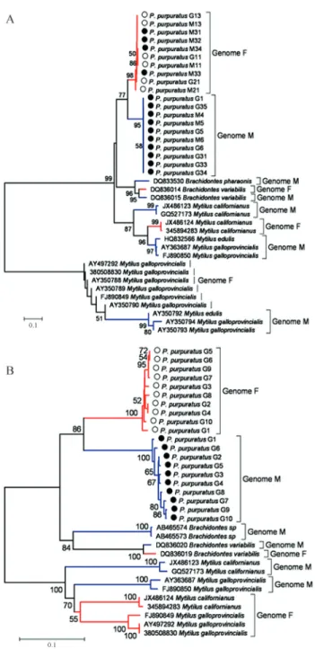
To evaluate the origin of DUI and the complex rela-tionships between the F and the M mitochondrial mole-cules, two phylogenies were constructed based on 16S and COI sequences obtained forP. purpuratusand sequences of F and M genomes fromBrachidontes variabilis,Mytilus edulis, M, californianusandM. galloprovincialis.
Results
A total of 105 sequences, from both tissues and both mitochondrial markers, were obtained from 35 samples of
P. purpuratus: 14 females and 21 males (details in Tables 1 and 2). Somatic and gonadal tissue for each individual was analyzed. A total of 65 sequences of 486 bp from the 16S region of mtDNA inP. purpuratuswere obtained, repre-senting the somatic tissues of 14 females and 20 males, and the gonadal tissues of 13 females and 18 males. For the COI gene, a total of 40 sequences of 540 bp were analyzed, cor-responding to the somatic tissues and the gonadal tissues of 10 females and 10 males. Only in two cases both genomes were amplified in a single individual and unfortunately these sequences were impossible to read (Phenograms in Figure S1). All the sequences of the 16S region were depos-ited in GenBank under accession numbers KF159809 to KF159878 and KF661909 to KF661918 and all the se-quences of COI gene under accession numbers KF661919 to KF661973 (sequence alignments are shown in Figu-re S2).
Two mitochondrial genomes were obtained in P. purpuratusmussels.In females only one type of genome was amplified (F genome) for both mitochondrial genes, in both somatic and gonadal tissues. In males, two kinds of genomes were found (F and M) for both mitochondrial genes. Specifically, in the gonadal tissues of males only the M genome was found, for both mitochondrial genes, while somatic tissue of males either the F or the M genome was found for 16S, and only the F genome was obtained with the COI primers used. In male mantles, 16S primers ampli-fied the M genome in 15 samples and the F genome in five others (Figure 1); the COI primers amplified only the F ge-nome in all the samples of male mantle tissues analyzed, while the M genome was amplified in all the male gonadal tissues.
A total of 65 sequences were analyzed for the 16S re-gion; 17 haplotypes were identified, showing 67 polymor-phic sites (S) with haplotype diversity h = 0.856; nucleotide diversity wasp or Pi = 0.064. For the F genome, 32 se-quences were obtained and 10 haplotypes were identified, with nine polymorphic sites; haplotype diversity was h = 0.833 and nucleotide diversity Pi = 0.004. For the M ge-nome, 33 sequences were obtained and seven haplotypes were identified, with seven polymorphic sites; haplotype diversity was h = 0.589 and nucleotide diversity Pi = 0.001 (Table 1). In total, nine haplotypes were obtained in female individuals and 10 in males; there are therefore two
haplo-Table 1- Indices of genetic variability based on mtDNA (16S) sequences F and M genomes, for females and males (gonadic and somatic tissues together) and total in the musselP. purpuratus.
Genome F Genome M Females Males Total
Number of sequences 32 33 27 38 65
Sequence length (bp) 486 486 486 486 486
S (polymorphic sites) 9 7 8 67 67
Number of haplotypes 10 7 9 10 17
Haplotype diversity±sd 0.83±0.04 0.59±0.1 0.84±0.04 0.69±0.08 0.86±0.03
Nucleotide diversity P 0.004 0.001 0.004 0.031 0.064
N° different nucleotides K 2.046 0.818 2.148 15.065 31.327
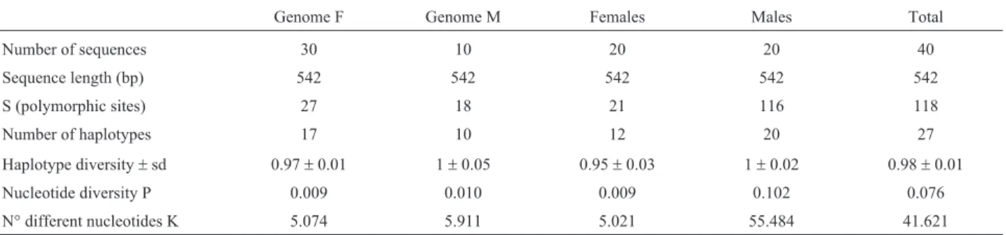
Table 2- Indices of genetic variability based on mtDNA (COI) sequences for F and M genome, females and males (gonadic and somatic tissues together) and total in the musselP. purpuratus.
Genome F Genome M Females Males Total
Number of sequences 30 10 20 20 40
Sequence length (bp) 542 542 542 542 542
S (polymorphic sites) 27 18 21 116 118
Number of haplotypes 17 10 12 20 27
Haplotype diversity±sd 0.97±0.01 1±0.05 0.95±0.03 1±0.02 0.98±0.01
Nucleotide diversity P 0.009 0.010 0.009 0.102 0.076
types shared between males and females, which are present in four out of five males that presented the F genome in mantle tissue.
A total of 40 sequences were analyzed for the COI gene; 27 haplotypes were identified with 118 polymorphic sites (S); haplotype diversity was h = 0.981 and nucleotide diversity Pi = 0.076. In the F genome, 30 sequences were obtained and 17 haplotypes were identified, with 27 poly-morphic sites; haplotype diversity was h = 0.966 and nucle-otide diversity Pi = 0.009. In the M genome, 10 sequences were obtained and 10 haplotypes were identified, with 18 polymorphic sites; haplotype diversity was h = 0.997 and nucleotide diversity Pi = 0.010 (Table 2). In total, 12 haplo-types were found in female individuals and 20 haplohaplo-types in males; there are therefore five haplotypes shared
be-tween males and females, which are present in five out of 10 males that presented the F genome in mantle tissue.
The F genome (found in females and males) showed higher genetic diversity than the M genome (found in males), for both mitochondrial genes (16S and COI) mainly in polymorphic loci, the number of haplotypes, haplotype diversity and different nucleotides (Table 1 for 16S region; Table 2 for COI gene).
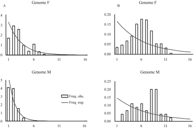
Distribution of pairwise differences between haplo-types (mismatch distributions) was used to estimate past population expansions by haplotypes. In this case, the F ge-nome ofP. purpuratusdisplayed a larger number of accu-mulated mutations than the M genome in its 16S sequences. However this pattern was less evident in the COI gene se-quences (Figures 2A and 2B, respectively).
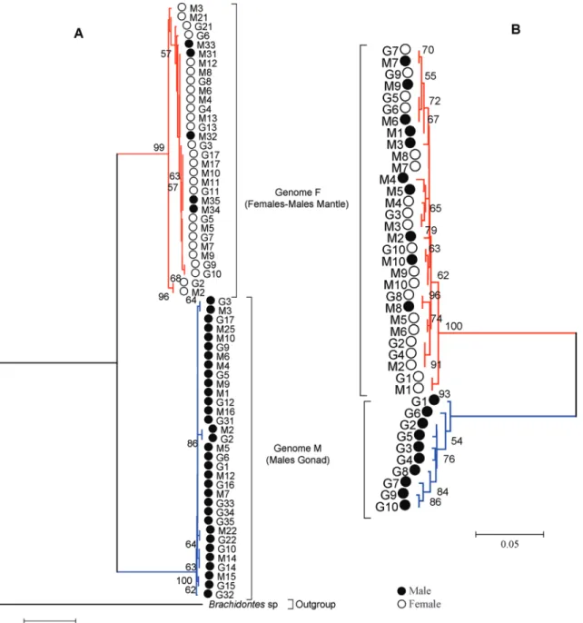
Based on a high bootstrap value for both genes ana-lyzed, the sequences were segregated into two well-defined and well-supported clades (Figure 3). One of the clades grouped female haplotypes (F) and somatic tissue of five males together, while the other clade grouped male haplo-types (M) alone, with no differences with regard to tissue type (somatic or gonadal). A significant genetic divergence was detected between F and M haplotypes: in the 16S re-gion sequences the genetic distance was Dxy = 0.125 and genetic differentiation Kxy was 60.33%, with high signifi-cance value (p < 0.001), estimated by permutations test. The corresponding values for the COI gene sequences were Dxy = 0.184 and genetic differentiation Kxy was 99.97% (p < 0.001).
Figure 1- Distribution of mitochondrial genomes by tissue-type and sex in the Chilean musselPerumytilus purpuratusevaluated with sequences of the 16S region. F: F-genome; M: M-genome.
Finally, in order to understand the origin of DUI, we analyzed both genomes (F and M haplotypes) from P. purpuratusin comparison with F and M haplotypes from other DUI species. The haplotypes were segregated by spe-cies of origin (taxon-joining pattern by Zouros, 2013), and a single clade was observed for each of theP. purpuratus
mitochondrial genes (Figure 4). Phylogenetic reconstruc-tion showed that species formareconstruc-tion predated the differentia-tion of the mitochondrial genomes (gender-joining pattern) inP. purpuratus.This was most obvious from the COI data (Figure 4 B).
Discussion
The phylogenetic reconstruction from both genes dis-tinguishes two clades: one specific to M haplotypes, ob-tained from males, present in 100% of the gonadal tissue samples for both genes and in somatic tissues for 16S; the other specific to F haplotypes, obtained from female so-matic and gonadal tissues and male soso-matic tissues. This conclusion is supported by high values of consistency and significant genetic divergence, displaying robust separa-tion between female (F) and male (M) haplotypes.
This indicates the presence of DUI inP. purpuratus. As expected for DUI species, males are heteroplasmic for both F and M genomes, and females are homoplasmic for the F genome only (Hoehet al., 1996, 1997, 2002; Quesada
et al., 1999; Zbawickaet al., 2003). Each sex presents only the sex-specific genome in its gonad. In general, differenti-ation at the mitochondrial genome level between F and M has been estimated at up to 20% in the Mytilidae family (Passamontiet al., 2003; Caoet al., 2004; Miziet al., 2005; Bretonet al., 2006; Theologidiset al., 2008), and as high as
50% in fresh water species of the Unionidae family (Doucet-Beaupréet al.,2010). InP. purpuratus,this value was found to be between 60% and 99%, depending on the mitochondrial gene. This is higher than has been observed for other species with DUI, and may result from the lower occurrence of the F genome detected in the male mantle with 16S primers, and non-detection of the M genome in the male mantle for the COI gene. Detection may be im-proved in the future by the design of specific genome prim-ers.
Terranovaet al.(2007) observed intraspecific vari-ability in Brachidontes variabilis for differentiation be-tween F and M haplotypes, and therefore in DUI ocurrence. This variability resulted from differences observed be-tween F and M haplotypes present in females and males re-spectively, similar toP. purpuratus. However this pattern was only observed in samples collected in the Indian Ocean, and was not apparent in samples collected in either the Pacific, the Red Sea or the Mediterranean (Terranovaet al., 2007). The authors argued that this may be due to the presence of three cryptic species with allopatric distribu-tion in each locadistribu-tion.
Moreover,P. purpuratusdisplayed greater variability in 16S F haplotypes than in M haplotypes, whether mea-sured by the number of accumulated mutations in its ge-nome over time (Figure 2) or by genetic variability indica-tors (Tables 1 and 2). Similar observations are reported in the mytilid Musculista senhousia (Passamonti, 2007), which may be explained by an older evolutionary history of the F genome than the M genome, with the latter appearing at a more recent date. However this deviation may also be due to the lower frequency with which the M genome is ob-served, meaning that its diversity may be underestimated. The pattern observed for the COI gene was more complex, since some indicators showed the same pattern, while oth-ers presented greater divoth-ersity in the M genome. The expla-nation suggested forM. senhousiais that this mechanism has evolved to protect mtDNA in females (e.g.antioxidant gene complexes) while selection could be relaxed in males (Passamonti, 2007; Zouros, 2013). In other mytilid species, however, it has been observed that M genomes are more variable than F genomes (Rawson and Hilbish, 1995; Stew-artet al., 1995; Zouros, 2013). Greater genetic diversity of the M genome may be due to several mechanisms, such as greater M mitochondrial replication rates during the early development of male embryos, differences in selection pressures, or the result of different effective sizes between genomes, as proposed by some authors (Skibinskiet al., 1994; Stewartet al., 1996; Schmidtet al., 1997; Hasegawa
et al., 1998; Ballard, 2000a,b).
The results obtained from the mismatch distribution indicate that the F genome has a longer history than the M genome, based on the greater number of accumulated muta-tions. This is more evident in the results for the 16S gene than the COI gene, as shown in the mismatch distribution
graphs (Figure 2). An empirical mismatch distribution that does not deviate from a unimodal distribution of pairwise differences among haplotypes and presents smooth distri-bution (Harpending, 1994) suggests recent expansion (Ro-gers and Harpending, 1992).
In addition to the evidence mentioned above, which indicates the presence of two mitochondrial genomes and the presence of DUI, the heteroplasmy (two genomes in the same individual) found in five males for the 16S region and 10 males for the COI gene confirms this mode of inheri-tance inP. purpuratus. The presence of M haplotypes in male tissues only (gonadal and somatic), and F haplotypes in both female tissue types and in male somatic tissues, confirms the existence of heteroplasmy in males of P. purpuratus, as expected for a DUI species (Mizi et al., 2005; Bretonet al., 2006; Venetiset al., 2006; Theologidis
et al., 2008; Cao et al., 2009; Passamonti and Ghiselli, 2009). However the M haplotype was more often amplified from gonadal tissues ofP. purpuratusmales with 16S and COI primers (100%) and less often from somatic tissues of males with 16S primers (75.0%). This has previously been observed by Passamonti and Scali (2001) and Ghiselliet al.
(2011) using cloning and Real-Time qPCR, respectively, and in the results reported by Terranovaet al.(2007), who detected evidence for DUI only with 16S-rDNA but not with COI. Previous studies have found evidence for such situations, where somatic tissues in males can be dominated by the F genome, with the occasional presence of small quantities of the M genome (Garrido-Ramoset al., 1998; Dalziel and Stewart, 2002; Ghiselliet al., 2011). This dif-ferent pattern may be tentatively related to the specific seg-regation mechanism of sperm mitochondria during male embryo development in DUI species (Cao et al., 2004; Obata and Komaru, 2005; Cogswellet al., 2006; Milaniet al., 2011, 2012), which drives sperm-derived mitochondria into the primordial mesodermal fate blastomeres (from which the adductor derives), whereas it allows only sto-chastic leakage into ectodermal fate blastomeres (from which the mantle originates). Our data for the M genome detected in the mantles of some males but not in others, may tentatively be explained by such stochastic events. In future work we propose to design new M- and F-specific primers to establish the degree of heteroplasmy with fre-quency estimators in this species.
Theologidis et al. (2007) demonstrated that in the original study by Saavedraet al.(1997), detection of the M genome in males ofMytilus galloprovincialiswas impossi-ble due to proimpossi-blems linked to mutations in primer annealing sites of the M genome. The fact that a greater number of both genomes was observed using 16S amplification than with COI primers may therefore result from a similar prob-lem to that encountered by Saavedraet al.(1997). For ex-ample, in our results, the M genome was amplified in males 1, 5, 6 and 9 using 16S primers, but not using COI primers (Figure 3). The next step required is to develop a research
strategy that will enable us to perform more detailed and in depth analyses of DUI inP. purpuratus.
Phylogenetic reconstruction carried out in order to understand the timing ofP. purpuratusspecies formation
vs.the separation between the two genomes and the origin of DUI showed a taxon-joining pattern. This may indicate that species formation predated genome differentiation in
P. purpuratus. Nonetheless, the loss of closely related spe-cies may in some way hide the presence of a gender-joining pattern, according to which DUI origin would be the first event. This differs from the pattern observed by Rawson and Hilbish (1995) and Zouros (2013) inMytilus, where a gender-joining pattern was found. The hypothesis proposed is that DUI in the genusMytilushad a single origin. In the case of the monospecific genusPerumytilus, the absence of closely related species makes it difficult to evaluate whe-ther the process of speciation was earlier or later than dif-ferentiation of the inheritance of mitochondrial genomes.
To conclude, the evidence presented here reveals the presence of two mitochondrial genomes that differentiate females (F haplotypes) from males (M and F haplotypes). These results not only confirmed the existence of two mito-chondrial genomes in this species, but also enabled us to de-tect the presence of sex-specific genomes in gonadal tissue of each type of sample (males and females). This indicates that caution must be taken when phylogenetic and phylo-geographic comparisons are done using mtDNA sequences ofPerumytilus purpuratuswithout considering the sex of the individuals.
Acknowledgments
Financial support was provided by the Fondo Nacio-nal de Ciencia y Tecnología FONDECYT (Project 1101007). The authors are grateful for the comments of the anonymous reviewers which helped them to improve this manuscript.
References
Acevedo J, Orellana FI and Guiñez R (2010) Experimental evalu-ation of the in situcopper toxicity on associated fauna of Perumytilus purpuratus(Bivalvia, Mytilidae), an ecosystem bioengineer. Rev Biol Mar Oceanogr 45:497-505.
Aguirre, ML, Perez, SI, and Sirch, YN (2006) Morphological variability of Brachidontes swainson (Bivalvia, Mytilidae) in the marine Quaternary of Argentina (SW Atlantic). Palaeogeogr Palaeoclimatol Palaeoecol 239:100-125. Avise JC, Arnold J, Ball RM, Bermingham E, Lamb T, Neigel JE,
Reeb CA and Saunders NC (1987) Intraspecific phylogeo-graphy: The mitochondrial DNA bridge between population genetics and systematics. Annu Rev Ecol Syst 489-522. Ballard JWO (2000a) Comparative genomics of mitochondrial
DNA inDrosophila simulans. J Mol Evol 51:64-75. Ballard JWO (2000b) Comparative genomics of mitochondrial
Birky CW (1995) Uniparental inheritance of mitochondrial and chloroplast genes: Mechanisms and evolution. Proc Natl Acad Sci 92:11331-11338.
Boyle E and Etter R (2013) Heteroplasmy in a deep-sea proto-branch bivalve suggests an ancient origin of doubly uni-parental inheritance of mitochondria in Bivalvia. Mar Biol 160:413-422.
Breton S, Burger G, Stewart DT and Blier PU (2006) Comparative analysis of gender-associated complete mitochondrial geno-mes in marine mussels (Mytilus spp.). Genetics 172:1107-1119.
Breton S, Beaupré HD, Stewart DT, Hoeh WR and Blier PU (2007) The unusual system of doubly uniparental inheri-tance of mtDNA: Isn’t one enough? Trends Genet. 23:465-474.
Cao LQ, Kenchington E and Zouros E (2004) Differential segre-gation patterns of sperm mitochondria in embryos of the blue mussel (Mytilus edulis). Genetics 166:883-894. Cao LQ, Ort BS, Mizi A, Pogson G, Kenchington E, Zouros E and
Rodakis GC (2009) The control region of maternally and pa-ternally inherited mitochondrial genomes of three species of the sea mussel genusMytilus. Genetics 181:1045-1056. Caro AU, Guiñez R, Ortiz V and Castilla JC (2011) Competition
between a native mussel and a non-indigenous invader for primary space on intertidal rocky shores in Chile. Mar Ecol Prog Ser 428:177-185.
Cogswell AT, Kenchington EL and Zouros E (2006) Segregation of sperm mitochondria in two- and four-cell embryos of the blue musselMytilus edulis: Implications for the mechanism of doubly uniparental inheritance of mitochondrial DNA. Genome 49:799-807.
Dalziel AC and Stewart DT (2002) Tissue-specific expression of male-transmitted mitochondrial DNA and its implications for rates of molecular evolution in Mytilus mussels (Bivalvia, Mytilidae). Genome 45:348-355.
Doucet-Beaupré H, Breton S, Chapman EG, Blier PU, Bogan AE, Stewart DT and Hoeh WR (2010) Mitochondrial phylo-genomics of the Bivalvia (Mollusca): Searching for the ori-gin and mitogenomic correlates of doubly uniparental inher-itance of mtDNA. BMC Evol Biol 10:e50.
Doyle JJ and Doyle JL (1987) A rapid DNA isolation procedure for small quantities of fresh leaf tissue. Phytochem Bull 19:11-15.
Fisher C and Skibinski DOF (1990) Sex-biased mitochondrial-DNA heteroplasmy in the marine musselMytilus. Proc R Soc B-Biol Sci 242:149-156.
Garrido-Ramos MA, Stewart DT, Sutherland BW and Zouros E (1998) The distribution of male-transmitted and female-transmitted mitochondrial DNA types in somatic tissues of blue mussels: Implications for the operation of doubly uni-parental inheritance of mitochondrial DNA. Genome 41:818-824.
Ghiselli F, Milani L and Passamonti M (2011) Strict sex-specific mtDNA segregation in the germ line of the DUI species Venerupis philippinarum (Bivalvia, Veneridae). Mol Biol Evol 28:949-961.
Guiñez R and Castilla JC (1999) A tridimensional self-thinning model for multilayered intertidal mussels. Am Nat 154:341-357.
Hall TA (1999) BioEdit: A user-friendly biological sequence alignment editor and analysis program for Windows 95/98/NT. Nucleic Acids Symp Ser 41:95-98.
Harpending HC (1994) Signature of ancient population growth in a low-resolution mitochondrial DNA mismatch distribution. Hum Biol 66:591-600.
Hasegawa M, Cao Y and Yang Z (1998) Preponderance of slightly deleterious polymorphism in mitochondrial DNA: Nonsynonymous/synonymous rate ratio is much higher within species than between species. Mol Biol Evol 15:1499-1505.
Hoeh WR, Blakley KH and Brown WM (1991) Heteroplasmy suggests limited biparental inheritance of Mytilus mitochon-drial DNA. Science 251:1488-1490.
Hoeh WR, Stewart DT, Sutherland BW and Zouros E (1996) Mul-tiple origins of gender-associated mitochondrial DNA lin-eages in bivalves (Mollusca, Bivalvia). Evolution 50:2276-2286.
Hoeh WR, Stewart DT, Saavedra C, Sutherland BW and Zouros E (1997) Phylogenetic evidence for role-reversals of gender-associated mitochondrial DNA inMytilus(Bivalvia, Myti-lidae). Mol Biol Evol 14:959-967.
Hoeh WR, Stewart DT and Guttman SI (2002) High fidelity of mi-tochondrial genome transmission under the doubly unipa-rental mode of inheritance in freshwater mussels (Bivalvia, Unionoidea). Evolution 56:2252-2261.
Hudson R, Boos D and Kaplan N (1992) A statistical test for de-tecting population subdivision. Mol Biol Evol 9:138-151. Kenchington E, MacDonald B, Cao LQ, Tsagkarakis D and
Zou-ros E (2002) Genetics of mother-dependent sex ratio in blue mussels (Mytilus spp.) and implications for doubly uni-parental inheritance of mitochondrial DNA. Genetics 161:1579-1588.
Kenchington EL, Hamilton L, Cogswell A and Zouros E (2009) Paternal mtDNA and maleness are co-inherited but not caus-ally linked in Mytilid mussels. PLoS ONE 4:e6976. Lancellotti DA and Vásquez JA (2000) Zoogeography of benthic
macroinvertebrates of the Chilean coast: Contribution for marine conservation. Rev Chil Hist Nat 73:99-129. Librado P and Rozas J (2009) DnaSP v5: A software for
compre-hensive analysis of DNA polymorphism data. Bioinfor-matics 25:1451-1452.
Milani L, Ghiselli F, Maurizii MG and Passamonti M (2011) Dou-bly uniparental inheritance of mitochondria as a model sys-tem for studying germ line formation. PLoS ONE 6:e28194. Milani L, Ghiselli F and Passamonti M (2012) Sex-linked
mito-chondrial behavior during early embryo development in Ruditapes philippinarum (Bivalvia Veneridae), a species with the Doubly Uniparental Inheritance (DUI) of mito-chondria. J Exp Zool B 318:182-189.
Mizi A, Zouros E, Moschonas N and Rodakis GC (2005) The complete maternal and paternal mitochondrial genomes of the Mediterranean musselMytilus galloprovincialis: Impli-cations for the doubly uniparental inheritance mode of mtDNA. Mol Biol Evol 22:952-967.
Nei M, (1987) Molecular Evolutionary Genetics. Columbia Uni-versity Press, New York, NY, 512 pp.
Palumbi S, Martin A, Romano S, McMillan WO, Stice L and Grabowski G (1991) The Simple Fool’s Guide to PCR, ver-sion 2.0. Department of Zoology and Kewalo Marine Labo-ratory, Honolulu, 47 pp.
Passamonti M (2007) An unusual case of gender-associated mito-chondrial DNA heteroplasmy: The mytilid Musculista senhousia (Mollusca Bivalvia). BMC Evol Biol 7(Suppl 2):S7.
Passamonti M and Ghiselli F (2009) Doubly uniparental inheri-tance: Two mitochondrial genomes, one precious model for organelle DNA inheritance and evolution. DNA Cell Biol 28:79-89.
Passamonti M and Scali V (2001) Gender-associated mitochon-drial DNA heteroplasmy in the venerid clam Tapes philippinarum(Mollusca Bivalvia). Curr Genet 39:117-124. Passamonti M, Boore JL and Scali V (2003) Molecular evolution and recombination in gender-associated mitochondrial DNAs of the Manila clamTapes philippinarum. Genetics 164:603-611.
Prado L and Castilla AC (2006) The bioengineerPerumytilus purpuratus (Mollusca, Bivalvia) in central Chile: Biodi-versity, habitat structural complexity and environmental heterogeneity. J Mar Biol Assoc UK 86:417-421.
Quesada H, Wenne R and Skibinski DOF (1999) Interspecies transfer of female mitochondrial DNA is coupled with role-reversals and departure from neutrality in the musselMytilus trossulus. Mol Biol Evol 16:655-665.
Rawson PD and Hilbish TJ (1995) Evolutionary relationships among the male and female mitochondrial-DNA lineages in theMytilus edulisspecies complex. Mol Biol Evol 12:893-901.
Rogers AR and Harpending H (1992) Population growth makes waves in the distribution of pairwise genetic differences. Mol Biol Evol 9:552-569.
Saavedra C, Reyero MI and Zouros E (1997) Male-dependent doubly uniparental inheritance of mitochondrial DNA and female-dependent sex-ratio in the mussel Mytilus galloprovincialis. Genetics 145:1073-1082.
Saitou N and Nei M (1987) The neighbor-joining method: A new method for reconstructing phylogenetic trees. Mol Biol Evol 4:406-425.
Schmidt TR, Jaradat SA, Goodman M, Lomax MI and Grossman LI (1997) Molecular evolution of cytochrome c oxidase: Rate variation among subunit VIa isoforms. Mol Biol Evol 14:595-601.
Skibinski DOF, Gallagher C and Beynon CM (1994) Sex-limited mitochondrial DNA transmission in the marine mussel Mytilus edulis. Genetics 138:801-809.
Stewart DT, Saavedra C, Stanwood RR, Ball AO and Zouros E (1995) Male and female mitochondrial DNA lineages in the blue mussel (Mytilus edulis) species group. Mol Biol Evol 12:735-747.
Stewart DT, Kenchington ER, Singh RK and Zouros E (1996) De-gree of selective constraint as an explanation of the different rates of evolution of gender-specific mitochondrial DNA lineages in the musselMytilus. Genetics 143:1349-1357. Tamura K, Peterson D, Peterson N, Stecher G, Nei M and Kumar
S (2011) MEGA5: Molecular Evolutionary Genetics Analy-sis using maximum likelihood, evolutionary distance, and
maximum parsimony methods. Mol Biol Evol 28:2731-2739.
Terranova MS, Lo Brutto S, Arculeo M and Mitton JB (2007) A mitochondrial phylogeography of Brachidontes variabilis (Bivalvia, Mytilidae) reveals three cryptic species. J Zool Syst Evol Res 45:289-298.
Teske PR, Papadopoulos I, Barker NP and McQuaid CD (2012) Mitochondrial DNA paradox: Sex-specific genetic structure in a marine mussel - despite maternal inheritance and pas-sive dispersal. BMC Genet 13:e45.
Theologidis L, Saavedra C and Zouros E (2007) No evidence for absence of paternal mtDNA in male progeny from pair mat-ings of the mussel Mytilus galloprovincialis. Genetics 176:1367-1369.
Theologidis I, Fodelianakis S, Gaspar MB and Zouros E (2008) Doubly uniparental inheritance (DUI) of mitochondrial DNA in Donax trunculus (Bivalvia, Donacidae) and the problem of its sporadic detection in Bivalvia. Evolution 62:959-970.
Thompson JD, Higgins DG and Gibson TJ (1994) CLUSTAL W: Improving the sensitivity of progressive multiple sequence alignment through sequence weighting, position-specific gap penalties and weight matrix choice. Nucleic Acids Res 22:4673-4680.
Trovant B, Ruzzante D, Basso N and Orensanz J.M. (2013). Dis-tinctness, phylogenetic relations and biogeography of inter-tidal mussels (Brachidontes, Mytilidae) from the south-western Atlantic. J Mar Biol Assoc UK 13:1469-7769. Venetis C, Theologidis I, Zouros E and Rodakis GC (2006) No
ev-idence for presence of maternal mitochondrial DNA in the sperm ofMytilus galloprovincialismales. Proc R Soc B Biol Sci 273:2483-2489.
Zbawicka M, Skibinski DOF and Wenne R (2003) Doubly uni-parental transmission of mitochondrial DNA length variants in the musselMytilus trossulus. Mar Biol 142:455-460. Zouros E (2000) The exceptional mitochondrial DNA system of
the mussel family Mytilidae. Genes Genet Syst 75:313-318. Zouros E (2013) Biparental inheritance through uniparental
trans-mission: The doubly uniparental inheritance (DUI) of mito-chondrial DNA. Evol Biol 40:1-31.
Zouros E, Ball AO, Saavedra C and Freeman KR (1994a) An un-usual type of mitochondrial DNA inheritance in the blue musselMytilus. Proc Natl Acad Sci USA 91:7463-7467. Zouros E, Ball AO, Saavedra C and Freeman KR (1994b)
Mito-chondrial DNA inheritance. Nature 368:818.
Supplementary Material
The following online material is available for this article: Figure S1 - Phenograms showing amplification of two genomes from one individual.
Figure S2 - Alignment of all sequences used for phylogen-etic reconstruction.
This material is available as part of the online article from http://www.scielo.br/gmb.
Associate Editor: Igor Schneider