Nuno Miguel Moura Espinha
Degree in Biochemistry
Bioprocess engineering of induced
pluripotent stem cells for application in
cell therapy and pre-clinical research
Dissertation to obtain Master Degree in Biotechnology
Supervisor: Dr. Maria Margarida de Carvalho Negrão Serra,
IBET/ITQB-UNL
Co-Supervisor: Dr. Ana Teresa de Carvalho Negrão Serra,
IBET/ITQB-UNL
Jury:
President: Prof. Dr. Pedro Miguel Ribeiro Viana Baptista
Examiner: Prof. Dr. Maria Alexandra Núncio de Carvalho Ramos Fernandes Supervisor: Dr. Maria Margarida de Carvalho Negrão Serra
I
Copyright
Bioprocess engineering of induced pluripotent stem cells for application in cell
therapy and pre-clinical research
Nuno Miguel Moura Espinha, FCT/UNL, UNL
A Faculdade de Ciências e Tecnologia e a Universidade Nova de Lisboa têm o direito, perpétuo
e sem limites geográficos, de arquivar e publicar esta dissertação através de exemplares
impressos reproduzidos em papel ou de forma digital, ou por qualquer outro meio conhecido
ou que venha a ser inventado, e de a divulgar através de repositórios científicos e de admitir a
sua cópia e distribuição com objetivos educacionais ou de investigação, não comerciais, desde
III
Acknowledgements
I would like to acknowledge all the people directly or indirectly involved in this thesis.
To Dr. Paula Alves, for giving me the opportunity to do my master thesis at the Animal Cell Technology Unit at ITQB/IBET, for the good working conditions offered and for being a strong example of leadership and professionalism.
To Dr. Margarida Serra, for her guidance, constant encouragement and support. I am grateful to her for inspiring me with her persistence, critical thinking, perfectionism and the motivation demonstrated throughout this whole work. Also, I would like to thank the chance of having attended the international conference “Stem Cells for Drug Screening and Regenerative Medicine” that positively contributed to my scientific formation.
To Dr. Ana Teresa Serra, for her kindness, constant good mood and, most of all, for her guidance during the development of the cell-based cardiotoxicity and cardioprotective assays presented in this work.
To Dr. Tomo Saric and his group for providing the Murine transgenic αPIG-iPS cell line, the starting point for this whole thesis, and for the support in RT-PCR and electrophysiology analysis. Also, I would like to thank Yuri Lages for his support during the perfusion bioreactors developed in this work.
To Marcos Sousa, for the constant availability, encouragement and all the advices with the environmentally controlled bioreactor processes. Also, to João Clemente, for his good mood and help during wave bioreactor cultures.
To Cláudia Correia, for having taught the majority of what I learned during this year and for always being there when it was most needed. A special thanks for her confidence, constant support, encouragement, scientific discussions and for being a good friend throughout the year.
To all the ACTU colleagues, for the good working environment, friendship and help during this year.
To my family, for all the support and understanding during all my academic formation. A special thanks to my mother for all the well prepared meals which I brought with me to work every day.
A special thanks to all my close friends and ITQB/IBET colleagues, for the hours of relaxation, and friendship. They were a huge support during this year.
V
Preface
This work was performed at the Animal Cell Technology Unit, IBET and ITQB-UNL,
within the scope of the project “CAREMI - Cardio Repair European Multidisciplinary Initiative” (HEALTH-F5-2010-242038), funded by the European Union (EU).
Part of this work has been included in poster communications and a submitted article.
Poster Communications
Correia C, Serra M, Sousa M, Espinha N, Brito C, Burkert K, Fatima A, Hescheler J, Carrondo MJT, Saric T, Alves PM (2013). “Novel Scalable Platforms for the Production of iPSC-derived Cardiomyocytes”. 1st International Meeting on Stem Cells for Regenerative Medicine and Drug Screening, Cantanhede, Portugal.
Correia C, Serra M, Sousa M, Espinha N, Brito C, Burkert K, Fatima A, Hescheler J, Carrondo MJT, Saric T, Alves PM (2013). “Novel Scalable Platforms for the Production of pure Cardiomyocytes derived from iPSC”. ESACT Meeting in Lille, France.
Correia C, Serra M, Sousa M, Espinha N, Brito C, Burkert K, Fatima A, Hescheler J, Carrondo MJT, Saric T, Alves PM (2013). “Improving the Production of Cardiomyocytes derived from iPSC for cardiac cell-based therapies”. Cardiac Biology - From Development to Regenerative Medicine Symposium. EMBL Heidelberg, Germany.
Correia C, Serra M, Sousa M, Espinha N, Brito C, Burkert K, Fatima A, Hescheler J, Carrondo MJT, Saric T, Alves PM (2013). “Novel Scalable Platforms for the Production of pure Cardiomyocytes derived from iPSC”. 8th International Meeting of the Portuguese Society for Stem Cells and Cell Therapies, Faro, Portugal.
Correia C, Serra M, Espinha N, Sousa M, Brito C, Burkert K, Fatima A, Hescheler J, Carrondo MJT, Saric T, Alves PM (2014). Towards Scalable Production and Cryopreservation of Functional iPSC-derived Cardiomyocytes. Scale-up and Manufacturing of Cell-based Theraphies III, San Diego, California, USA.
Submitted Article
Article submitted to Stem Cell Reviews and Reports (January, 2014)
VI
VII
Abstract
The production of cardiomyocytes (CMs) from induced pluripotent stem cells (iPSCs)
presents great potential for patient-specific regenerative therapies and cardiotoxicity drug
evaluation. The successful translation of iPSCs to these fields requires the development of
robust bioprocesses capable of producing CMs in high quality, quantity and purity.
Traditional protocols for CM differentiation of iPSCs lack control and robustness and are thus
inefficient. Furthermore, efficient cryopreservation and hypothermic storage strategies are a
demand, as cell banking and transport is a prerequisite for clinical and industrial
applications. The main aim of this thesis was the evaluation of different bioreactor systems
for the production and purification of miPSC-derived CMs. Novel strategies for CM
cryopreservation were tested. Also, CMs were used to study the cardioprotective effect of
antioxidant compounds.
The wave bioreactor was the most suitable system for CM differentiation, allowing
high differentiation yields (60 CMs/input of miPSC) and the production of clinically relevant
numbers of CMs (2.3x10⁹ CMs), simultaneously reducing bioprocess duration when
compared to stirred tank bioreactors. Produced CMs presented typical structural and
functional features. Moreover, CryoStorTMCS10 and FBS+10%DMSO (with ROCKi
pretreatment) revealed to be suitable solutions for cryopreservation of miPSC-derived CMs,
achieving high cell recoveries after thawing. In addition, HypoThermosol®-FRS enabled
hypothermic storage of CMs for up to 7 days. Finally, it was shown that CMs derived from
miPSCs present potential to be used in the development of cardioprotective assays.
This work demonstrates the establishment of a fully integrated bioprocess, capable of
producing high quality miPSC-derived CMs in environmentally controlled bioreactors and
ensuring efficient cryopreservation and storage of the produced cells. Hopefully, the
knowledge acquired with this work can be translated to human iPSCs, presenting a relevant
step forward towards the application of human CMs to clinical and industrial applications,
such as cardiac regeneration, disease modeling and cardiotoxicity and cardioprotective
cell-based assays.
Key Words: induced Pluripotent Stem Cells (iPSCs); cardiomyocyte differentiation; 3D
culture; environmentally controlled bioreactors; cryopreservation; cardioprotection
IX
Resumo
A produção de cardiomiócitos (CMs) derivados de células estaminais pluripotentes
induzidas (iPSCs) apresenta grande potencial para terapias regenerativas e avaliação de
drogas cardiotóxicas. A transferência de iPSCs para estas aplicações necessita do
desenvolvimento de bioprocessos robustos e capazes de produzir CMs de elevada qualidade,
quantidade e pureza. Protocolos tradicionais para diferenciação de iPSCs em CMs revelam
falta de controlo e robustez sendo ineficientes. Além disso, são necessárias estratégias
eficientes de criopreservação e armazenamento hipotérmico, sendo que a formação de
bancos de células e transporte é um pré-requisito para aplicações clinicas e industriais. O
principal objetivo desta tese foi a avaliação de diferentes sistemas de biorreatores para a
produção e purificação de CMs derivados de miPSCs. Foram avaliadas novas estratégias para
criopreservação de CMs. Ainda, CMs foram utilizados para estudar o efeito cardioprotector de
compostos antioxidantes.
O biorreator wave foi o sistema mais apropriado para a diferenciação de CMs,
permitindo elevados rendimentos de diferenciação (60 CMs/miPSCs) e a produção de
números clinicamente relevantes de CMs (2.3x10⁹ CMs), simultaneamente reduzindo a
duração do bioprocesso quando comparado a biorreatores de tanque agitado. Os CMs
produzidos apresentavam estrutura e funcionalidades típicas. Além disso, Cryostor TMCS10 e
FBS+10%DMSO (pre-tratado com ROCKi) revelaram ser soluções adequadas para a
criopreservação de CMs, atingindo recuperações celulares elevadas após descongelamento.
Ainda, HypoThermosol®FRS permitiu um armazenamento hipotérmico de CMs até 7 dias.
Por último, foi demonstrado que CMs derivados de miPSCs apresentam potencial para serem
usados no desenvolvimento de ensaios cardioprotectores.
Este trabalho demonstrou o estabelecimento de um bioprocesso integrado, capaz de
produzir elevadas quantidades de CMs derivados de miPSCs em biorreatores controlados e
assegurando criopreservação e armazenamento eficazes das células produzidas.
Esperançosamente, o conhecimento adquirido neste trabalho pode ser transferido para iPSCs
humanas, sendo um passo relevante para a utilização de CMs humanos para aplicações
clinicas ou industriais, tais como, regeneração cardíaca, modelos de doenças e ensaios
celulares de cardiotoxicidade e cardioprotecção.
Palavras-chave: células estaminais pluripotentes induzidas (iPSCs); diferenciação de
cardiomiócitos; cultura 3D; biorreatores; criopreservação; ensaios celulares de
XI
List of Abbreviations
AFP Alpha-fetoprotein
ALCAM Activated leukocyte cell adhesion molecule ANP Atrial natriuretic peptide
AP Alkaline phosphatase
AP Action potential
bFGF basic fibroblast growth factor
BMP Bone morphogenic protein
BR Bioreactor
BSA Bovine serum albumin
Cch Carbachol
CCD Charged–coupled device
cDNA complementary deoxyribonucleic acid
CMs Cardiomyocytes
CPA Cryoprotective agents
CS10 Cryostor CS10 solution
cTnT cardiac troponin T
Ct’s Cycle threshold
CVD Cardiovascular diseases
DMEM Dulbecco modified Eagle medium
DMSO Dimethyl sulfoxide
DO Dissolved oxygen
DKK1 Dickkopf homolog 1
EB Embryoid body
ECM Extracellular matrix
EDTA Ethylenediamine tetraacetic acid
eGFP enhanced green fluorescent protein
ESCs Embryonic stem cells
FACS Fluorescence-activated cell sorting
FBS Fetal bovine serum
FDA Fluorescein diacetate
FDA Food and Drug Administration
FGFs Fibroblast growth factors
FI Fold increase
GAPDH Glyceraldehyde-3-Phosphate Dehydrogenase
GFP Green fluorescent protein
GMP Good manufacturing practices
HCN4 Hyperpolarization activated cyclic nucleotide-gated potassium channel 4
HTS HypoThermosol solution
IgG Immunoglobulin G
IgM Immunoglobulin M
IMDM Iscove's modified Dulbecco's medium
iPSCs induced pluripotent stem cells
XII
LDH Lactate dehydrogenase
LDS Late development stage
LIF Leukemia inhibitory factor
LN2 Liquid nitrogen
MACS Magnetic-assisted cell separation
MDP Maximum diastolic potential
MEFs Murine embryonic fibroblasts
MEM- NEAA Minimum essential medium – non-essential amino acids
MI Myocardial infarction
miPSCs murine induced pluripotent stem cells Myl2 Myosin light chain, ventricular isoform Myl7 Myosin light chain, atrial isoform
NADH Nicotinamide adenine dinucleotide
Nkx2.5 Nk2 transcription factor related locus 5 Oct-4 Octamer-4 transcription factor
PBS Phosphate buffered saline
PBS Pneumatic Bioreactor System
Pen/Strep Penicillin-streptomycin
PFA Paraformaldehyde
PGI2 Prostaglandin I2
PI Propidium iodide
PSCs Pluripotent stem cells
RNA Ribonucleic acid
ROCKi Rho-associated kinase inhibitor
ROS Reactive oxygen species
RT-PCR Real time-polymerase chain reaction
RT-qPCR Real time-quantitative polymerase chain reaction SIRPA Signal-regulatory protein alpha
SSEA-1 Stage-specific embryonic antigen-1
T-Bra T Brachyury
TGF-β Transforming growth factor β
TMRM Tetramethylrhodamine methyl ester perchlorate
VCAM1 Vascular adhesion molecule 1
Vdd Velocity of diastolic depolarization VEGF Vascular endothelial growth factor
Vmax Maximum velocity of depolarization
Vvm Volume of gas flow per volume of culture medium per minute
WB Washing buffer
WNT Wingless/INT
α-MHC α-myosin heavy chain
2D Two-dimensional
XIII
Table of Contents
1. Introduction ... 1
1.1 Stem cell-based therapies for cardiac repair ... 1
1.1.1 Pluripotent stem cells: ESCs vs iPSCs... 2
1.2 Cardiomyocyte differentiation ... 5
1.2.1 Understanding cardiac differentiation ... 5
1.2.2 Embryoid body formation ... 7
1.2.3 Co-culture of PSCs with END2 cells... 8
1.2.4 Guided differentiation with growth factors and small molecules ... 9
1.2.5 Guided differentiation promoted by cyclic strains & oxygen ... 10
1.2.6 Scalable production of cardiomyocytes derived from PSCs ... 11
1.3 Purification of iPSC-derived cardiomyocytes ... 16
1.3.1 Genetic selection of cardiomyocytes ... 16
1.3.2 Non-genetic purification of cardiomyocytes ... 17
1.4 Characterization of cardiomyocytes ... 18
1.5 Cryopreservation and storage of PSC-derived cardiomyocytes ... 20
1.6 PSC-derived cardiomyocytes as promising cell models for cardiotoxicity assays ... 22
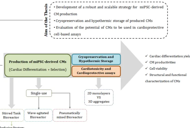
2. Aim of the thesis ... 25
3. Materials and Methods ... 27
3.1 miPSC culture on feeder layers ... 27
3.2 miPSC differentiation in fully controlled bioreactors ... 27
3.2.1 miPSC differentiation in stirred tank bioreactor ... 27
3.2.2 Disposable single-use bioreactors ... 29
3.3 Cryopreservation and hypothermic storage of CMs derived from miPSC ... 30
3.3.1 Cryopreservation of 2D monolayers ... 31
3.3.2 Cryopreservation of 3D aggregates ... 31
3.3.3 Hypothermic storage of CMs derived from miPSC ... 31
3.3.4 Assessment of cell recovery of CMs ... 31
3.4 Cardiotoxicity and cardioprotective assays ... 32
3.5 Cell viability evaluation ... 32
3.6 Evaluation of aggregate concentration and size ... 33
3.7 Culture yields determination ... 33
3.8 Characterization of miPSCs and miPSC-derived CMs ... 34
3.8.1 Structural characterization ... 34
XIV
3.9 Statistical analysis ... 36
4. Results and Discussion ... 37
4.1 Bioprocess engineering of miPSCs ... 37
4.1.1 Production of miPSC-derived CMs in fully controlled bioreactors ... 37
4.1.2 Development of a perfusion strategy in a stirred tank bioreactor ... 45
4.1.3 Structural and functional characterization of miPSC-derived CMs ... 48
4.2 Cryopreservation and hypothermic storage of miPSC-derived CMs ... 52
4.2.1 Cryopreservation of CMs as monolayers and aggregates ... 53
4.2.2 Hypothermic storage of CMs as monolayers and aggregates... 58
4.2.3 Characterization of cryopreserved and hypothermically stored CM monolayers ... 59
4.3 Evaluation of the potential of miPSC-derived CMs to be used in the development of cardioprotective cell-based assays ... 60
5. Conclusion ... 63
6. References ... 65
XV
List of Figures
Figure 1.1: Causes of death in Europe, 2011 (adapted from [2]) ... 1
Figure 1.2: Stem cell sources, pluripotency potential and generation of iPSCs ... 3
Figure 1.3: Schematic representation depicting sequential steps required for obtaining PSC-derived cardiomyocytes ... 6
Figure 1.4: Current methods for cardiac differentiation of human PSCs ... 7
Figure 1.5: 2D and 3D strategies for cultivation of PSCs (adapted from [15]) ... 12
Figure 1.6: Examples of different bioreactor designs used for stem cell bioprocessing ... 13
Figure 1.7: Basic characterization of PSC-derived cardiomyocytes ... 19
Figure 1.8: Main steps composing common cryopreservation procedure for mammalian cells ... 21
Figure 2.1: Thesis rational ... 25
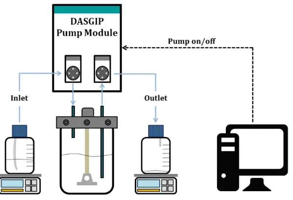
Figure 3.1: Perfusion apparatus used in continuous perfusion experiments ... 29
Figure 4.1: Characterization of miPSCs ... 37
Figure 4.2: miPSCs aggregation step ... 39
Figure 4.3: miPSC differentiation into CMs using different bioreactor systems ... 39
Figure 4.4: Production of miPSC-derived CMs using fully controlled bioreactors ... 41
Figure 4.5: Evaluation of gene expression and levels of eGFP positive cells during CM production in Stirred Tank and Wave BRs... 43
Figure 4.6: Production of miPSC-derived CMs using a continuous perfusion strategy ... 46
Figure 4.7: Structural characterization of miPSC-derived CMs produced in Stirred Tank and Wave BRs. . 48
Figure 4.8: Functional characterization of miPSC-derived CMs produced in Stirred Tank and Wave BRs . 49 Figure 4.9: Effect of cryopreservation medium on miPSC-derived CM viability after cryopreservation of CM monolayers ... 53
Figure 4.10: Effect of cryopreservation medium on miPSC-derived CM viability after cryopreservation of cardiospheres ... 55
Figure 4.11: Hypothermic storage of miPSC-derived CMs as monolayers and cardiospheres... 58
Figure 4.12: Structural characterization of cryopreserved and hypothermically stored CM monolayers .. 59
Figure 4.13: Effect of oxidative stress and antioxidant pretreatment on CMs ... 60
XVII
List of Tables
Table 1.1: Major challenges and potential solutions facing the use of iPSCs for cardiac repair (adapted from [4]) ... 5
Table 1.2: Studies involving cultivation of mESCs in scalable 3D approaches involving expansion and differentiation into cardiomyocytes. ... 16
Table 1.3: Cardiac markers used for the characterization of cardiomyocytes (adapted from [54]). ... 18
Table 3.1: Dilution rates tested in the continuous perfusion system approach. ... 29
Table 4.1: Quantitative characterization of CM production derived from miPSCs using environmentally controlled bioreactors. ... 44
Table 4.2: Action potential parameters for CMs obtained from Stirred Tank and Wave BRs ... 50
1
1.
Introduction
1.1
Stem cell-based therapies for cardiac repair
Cardiovascular diseases (CVD) are one of the major causes of death worldwide,
accounting for 17.3 million deaths per year [1]. In Europe, they are responsible for 47% of all
deaths issues (Figure 1.1) [2]. In particular, myocardial infarction (MI) results in massive
cardiac loss, where up to a billion cardiomyocytes (CMs) die resulting in loss of contractility
and decreased heart function [3]. Upon injury, the adult heart is endowed of some
endogenous regenerative capacity, provided by resident cardiac stem cells. However this
endogenous regenerative capacity is limited as fully differentiated cardiomyocytes can no
longer proliferate and replace degenerated tissue [4].
Figure 1.1: Causes of death in Europe, 2011 (adapted from [2]).
Cardiac repair (replacement, restoration and regeneration) is thus essential to
restore heart function following MI [5]. Heart transplantation remains as the only long term
treatment available, even though limited due to high costs, shortage of donor organs and
possible immunological rejection [6, 7]. As new therapies are still a need, emerging
technologies such as cell-based therapies intend to restore heart function by two
experimental approaches: enhancement of endogenous heart regeneration [8] and
transplantation of exogenous cells to repopulate the heart [9]. Notably, combining both
strategies would certainly provide a better alternative for therapies aiming at restoring heart
function [3].
Many cell types have been examined as potential cell sources for cell therapy such as
endothelial progenitor cells, cardiac progenitor cells, mesenchymal stem cells derived from
bone marrow and pluripotent stem cells (PSCs) (reviewed in [10]). The majority of stem cell
therapies in cardiac disease involve bone-marrow derived cells. Currently, companies such as
Cardiovascular diseases 47%
Cancer 21%
Respiratory diseases 6.5%
Injuries and poisoning 7%
2
Baxter, Cardio3 Biosciences or Mesoblast are using this cell source in advanced stages of
clinical trials (phase 3) [11]. Most candidate cell types improve ventricular function, however
show limited or no potential to produce cardiomyocytes [4]. The ideal cell source for heart
transplantation would be a proliferative cell with potential to generate all the different cells
of the heart [3]. PSCs meet these standards, proliferate indefinitely and differentiate into all
cell types of the body (except for extra-embryonic tissues), thus being a potential candidate
for stem cell therapies in cardiac repair [3].
1.1.1
Pluripotent stem cells: ESCs vs iPSCs
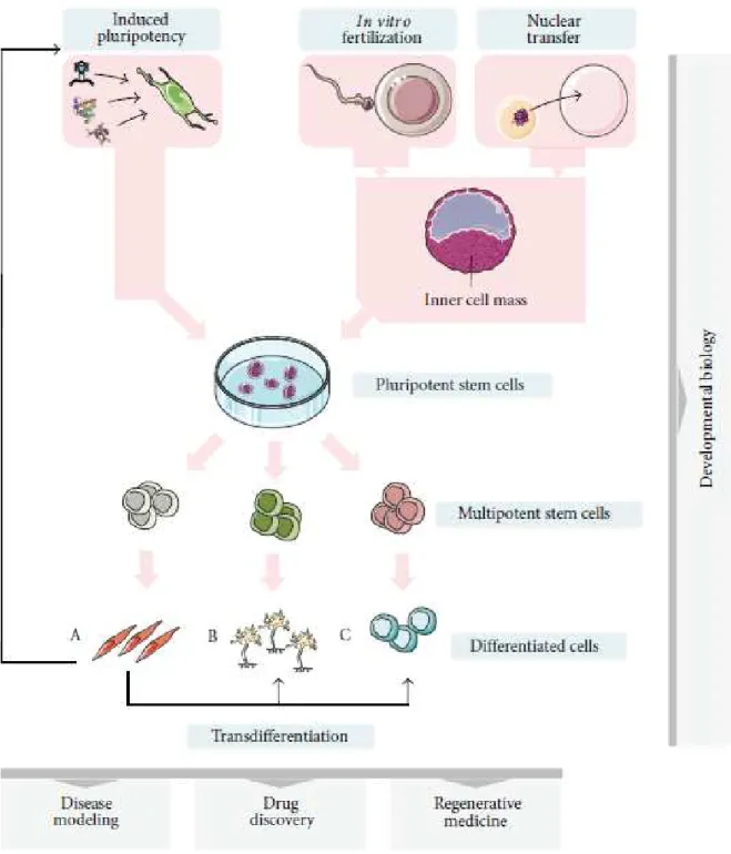
Murine embryonic stem cells (ESCs) were first isolated in 1981 [12] and their human
counterparts were derived by Thomson in 1998 [13]. ESCs were isolated from the inner cell
mass of blastocysts, pre-implanted embryos at day 5 of embryonic development (Figure 1.2).
Blastocysts comprise an outer layer of cells (trophoectoderm) and an inner cell mass. ESCs
are derived by removing the outer layer and isolating the inner cell mass [14].
ESCs can give rise to all cell types of the body, i.e. all three germ layers: endoderm,
ectoderm and mesoderm, and are thus called pluripotent. These cells present high
self-renewal capacity as they can proliferate continuously giving rise to undifferentiated cells
[15]. ESCs were the first stem cell source that could reliably give rise to cardiomyocytes in
vitro (reviewed in [4]). Despite their enormous potential for cell-based therapies, ESCs are
inevitably associated with ethical considerations due to the manipulation of embryos, are
technically difficult to derive and still present limitations for clinical applications, such as
immune rejection and possible teratoma formation (cell tumor containing tissue components
of the three germ layers) (reviewed in [3]).
Cell differentiation was recently shown to be a dynamic reversible process. In 2006,
Yamanaka and co-workers were able to reprogram mature cells to a pluripotent state [16].
The reverted cells were called induced pluripotent stem cells (iPSCs) and are generated by
reversing somatic cells to a state of pluripotency (embryonic-like state) by the induced
expression of specific reprogramming factors [17]. This insight has influenced all areas of
medicine and physiology, and was awarded the Nobel Prize in Physiology or Medicine in
2012 (to Dr. John B. Gurdon and Dr. Shinya Yamanaka) for the discovery that mature,
differentiated cells can be reprogrammed to a pluripotent stem cell state.
As mentioned above, the first study on derivation of iPSCs from murine fibroblast was
reported by Takahashi and Yamanaka in 2006, [16]. In this study, 4 transcription factors (
Oct-4, Sox2, Klf4 and c-Myc) were stated as being the minimal required for maintenance of
pluripotency and capable of yielding cells with characteristics similar to ESCs. After iPSC
3
factors can give rise to tumor formation after transplantation [18]. Therefore, in a later study
reported by Thomson and co-workers, c-Myc and Klf4, well known proto-oncogenes, were
replaced by Nanog and Lin28 [19]. It has also been shown that combinations of all these 6
factors are also able to generate iPSCs [7, 19].
4
The first reprogramming protocols relied on the use of integrating vectors to deliver
desired transgenes into the genome of somatic cells. These integrating vectors were mainly
retroviral or lentiviral [21, 22]. Even though reprogramming was possible, random transgene
insertion could interrupt existing genes causing tumor formation (after transplantation) and
disturb the maintenance of the cells undifferentiated state. This approach presents other
drawbacks such as low stability, difficult cellular targeting and possible insertion of
mutagenesis [23]. To overcome these limitations and establish safer iPSC lines, - methods
that allow the expression of pluripotency genes without integrating host genome are
currently being explored. For example, non-integrated virus (e.g. adenoviruses,
baculoviruses) have been used, despite suffering from low efficiencies [21]. Virus-free
reprogramming can also be achieved by using episomal vectors or excisable transposon
systems (e.g. PiggyBac or Cre/loxP systems), which integrate the cells but can be removed by
transposase, without leaving genetic material behind (reviewed in [21, 24]). Other techniques
for the generation of safer iPSC lines include the use of exogenous plasmids, protein factors,
small molecules and microRNAs. Nuclear reprogramming has been shown to be an inefficient
process. However, a novel method for reprogramming is able to successfully revert cells to a
pluripotent state, by deleting the Mbd3 gene, which is an important epigenetic regulator that
restricts expression of pluripotency genes. This method shows reprogramming efficiencies
near 100% for mouse and human cells [25].
In the last few years, iPSCs are boosting the field of regenerative medicine, offering
numerous advantages over adult stem cells and ESCs [3]. iPSCs can circumvent the need for
embryo use, being ethically less controversial, and thereby could potentially replace ESCs.
Also, differentiated cells derived from iPSCs have recently been shown to present limited
immunogenicity and could therefore be used in autologous [26] (patient specific) or
allogeneic therapies (universal donor-derived cells). iPSC-derived cardiomyocytes have
tremendous potential to be used in cardiac repair strategies.
Although recent approaches have proved promising, there are still challenges to
overcome in order to transfer PSCs to clinical applications. Selection of the adequate cell
source is crucial, namely whether to use autologous or allogenic therapies. Quantity and
purity of cells are essential for successful therapies, minimizing tumorigenic potential of
transplantation. Also, quality is a requirement, assuring the desired phenotype and potency
of the chosen cell source (reviewed in [15]).
Table 1.1 summarizes the major challenges and solutions facing the use of iPSCs in
5
Table 1.1: Major challenges and potential solutions facing the use of iPSCs for cardiac repair (adapted from [4])
Hurdles Possible solutions
Heterogeneous population of differentiated cells - Directed cardiac differentiation - Specific selection of cardiomyocytes Low cardiomyocyte yield - Directed cardiac differentiation
- Upscaling differentiation process
Survival, integration and maturation of cell-grafs in vivo
- Cell delivery approaches - Tissue engineering strategies - Genetic modifications
Tumorigenic potential
- Integration-free iPSCs generation - c-Myc-free reprogramming strategies - Appropriate selection process
Risk of genetic modifications - Genetic screening of undifferentiated iPSCs
1.2
Cardiomyocyte differentiation
1.2.1
Understanding cardiac differentiation
Cardiomyocyte differentiation of stem cells recapitulates aspects of cardiogenesis in
vivo, which involves complex processes highly controlled by positive and negative molecular
signals that guide stem cells towards cardiac fate [27]. As molecular mechanisms in human
cardiogenesis are still not thoroughly known, cardiac development in animal models (e.g.
mouse) has provided valuable information to improve in vitro cardiomyocyte differentiation
protocols (reviewed in [28]). Understanding how the cardiac lineage is established in these
models is crucial for developing efficient differentiation protocols. Cardiomyocyte
differentiation of murine iPSCs (miPSCs) was first described by Mauritz et al. [29] who
utilized the embryoid body (EB) system to obtain cardiomyocytes.
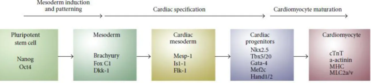
The mammalian heart is the first organ developed in embryogenesis and consists of
three mesodermal cell types: endothelial cells, vascular smooth muscle cells and
cardiomyocytes [30]. Cardiomyogenesis consists of four consecutive steps: i) mesoderm
formation, ii) mesoderm patterning toward anterior mesoderm or cardiogenic mesoderm, iii)
cardiac mesoderm formation and iv) cardiomyocyte maturation [31]. Cardiac development is
a dynamic process controlled by sequential expression of multiple signal transduction
proteins and transcription factors. Major signaling pathways have been implicated with
cardiac subtypes development, such as wingless/INT (WNTs) [32], nodal ([33]), bone
6
gastrulation (formation of gastrula in early embryonic development) signals mediated
through WNT/β-catenin and transcription growth factor β (TGF-β) family members promote differentiation of ESCs into mesoderm (reviewed in [36]). However, following mesodermal
induction, WNT/β-catenin signaling inhibits cardiac differentiation and may redirect cells to alternate mesodermal fates. This signaling pathway has a biphasic role during cardiac
differentiation in mouse ECS, being procardiac prior to primitive streak formation and
presenting an antagonizing role thereafter (reviewed in [36]). Due to this biphasic effect
during cardiac differentiation, the timing of the addition of specific cardiac factors is crucial
and must be carefully optimized in order to efficiently drive PSCs to a cardiac cell fate. Figure
1.3 presents a schematic overview of the steps required for obtaining PSC-derived
cardiomyocytes and typical markers expressed in each phase.
Figure 1.3: Schematic representation depicting sequential steps required for obtaining PSC-derived cardiomyocytes. Early mesoderm differentiates via cardiac mesoderm and committed cardiac progenitors to functional beating cardiomyocytes. Typical markers for each step are also indicated [37].
Currently, the main approaches for cardiomyocyte differentiation of PSCs are
spontaneous differentiation through embryoid body (EB) formation, co-culture with cells
presenting cardiac inductive activity and guided differentiation strategies (induced by
environmental factors) (reviewed in [3]). Figure 1.4 presents a schematic overview of current
differentiation methods used for generating PSC-derived cardiomyocytes.
Monolayers-based methods are the simplest approach for cardiomyocyte
differentiation. PSCs are plated on an extracellular matrix (ECM) or on a top of a monolayer of
feeder cells. These methods are focused primarily on the timing of application and
concentration of specific cardiac inductive growth factors (e.g. activin A and BMP4)
(reviewed in [3]). Other monolayer-based methods have focused on overlaying PSCs with
hydrogels or specific matrices [38], also called the matrix sandwich method. When combined
with sequential application of growth factors, this approach has been reported to generate
cardiomyocytes with high purity (up to 98%) [38]. Despite the increased efficiency of these
protocols, cell-cell interactions in EBs can stimulate the expression of early cardiac markers
(reviewed in [39]), which favors the use of 3D structures for cardiac differentiation, as
7
Figure 1.4: Current methods for cardiac differentiation of human PSCs. The 3 major approaches for differentiation of PSCs to cardiomyocytes are summarized: Embryoid bodies, monolayer cultures and inductive co-culture. StemPro34, APEL, DMEM/F12, KO-DMEM and RPMI/B27 are abbreviations of common culture medium formulations used for human PSC cultivation [40].
1.2.2
Embryoid body formation
When cultivated in non-adherent conditions (e.g. hanging drops, ultra low adherence
culture plates) cells assembled into spheroids, capable of differentiating into derivatives of all
three germ layers [41]. These three-dimensional (3D) aggregates are called embryoid bodies
(EBs), because of their ability to recapitulate the early events of embryogenesis [42, 43]. EBs
can spontaneously differentiate into contracting cardiomyocytes [44]. Early studies identified
critical culture parameters to optimize in vitro cardiogenesis such as cell inoculum
concentration, culture medium formulation (addition of serum or growth factors), culture
strategy and time [45]. Spontaneous differentiation of PSC-derived cardiomyocytes in EBs is
also highly dependent on cell line and is rather inefficient, usually yielding 10% of
cardiomyocytes exhibiting beating areas [41, 46]. Regardless of these issues, this method is
widely used for being simple and cost-effective [21].
Recent studies have shown that aggregate heterogeneity leads to adverse effects on
8
aggregation methods have been one of the most used strategies to control EB formation and
size. In this method, defined numbers of PSCs are centrifugated into U- or V-bottomed wells
(Figure 1.4), leading to the formation of aggregates (also called spin EBs) with the same size
in each well. Varying the number of cells per well allows the control of aggregate size with
precision [48, 49]. This method is practical, reproducible and efficient. Engineered microwells
(Figure 1.4) have also been used to form size-uniform EBs. It involves the microfabrication of
microwells with defined sizes that are coated with an extracellular matrix, such as Matrigel,
where cells attach and grow to fill the microwells. Removal of cell colonies from microwells
results in cell aggregates (also called Microwell EBs) with defined sizes [50, 51]. However,
microwells present reduced potential for scaling-up, requires appropriate and specialized
technology and are currently not commercially available (reviewed in [40]). An alternative
engineering approach is the use of micropatterned surfaces (Figure 1.4). In this approach,
cells are cultivated on a micropatterned ECM (e.g. Matrigel) islands. Size of the colonies can
be controlled by varying the size of these islands (e.g. 200μM, 400μM and 800μM) [47, 52]. Size-controlled EBs (Micropattern EBs) can be achieved after mechanical dissociation of
colonies. Protocols for spontaneous differentiation of PSC via EB formation have been
optimized aiming at improving cardiomyocyte differentiation yields, by adding specific
growth factors in combination with defined medium formulations [40].
1.2.3
Co-culture of PSCs with END2 cells
Another method for cardiomyocyte differentiation of stem cells is the co-culture of
PSCs with a visceral endoderm-like cell line (END-2) derived from mouse P19 embryonal
carcinoma cells [53]. This method was developed based on the discovery of cardiac inductive
activity of endoderm in early embryonic studies (reviewed in [54]). The inductive effect of
END-2 cells is not dependent on cell-cell contact, as studies have shown that conditioned
media derived from END-2 culture is sufficient to induce cardiac differentiation of human
ESCs (>10% cardiomyocytes) [55]. The presence of END-2 cells in culture causes rapid
depletion of insulin, a common media supplement that inhibits cardiogenesis [56]. END-2
cells also produce significantly higher levels of prostaglandin I2 (PGI2), a lipid molecule that
enhances cardiac induction [56]. Co-culture of PSCs with END-2 cells is a simple and rapid
protocol to differentiate hPSC into cardiomyocytes, as it requires few cells and generates
cardiomyocytes in sufficient quantity and quality to detect visible beating areas and identify
sarcomere structures [40]. Despite being a protocol with very low differentiation efficiency
(5-20% cardiomyocytes), optimization with serum- and insulin- free protocols results in
9
1.2.4
Guided differentiation with growth factors and small molecules
Many growth factors are used to enhance mesoderm formation and cardiomyogenesis
in cultures of PSC, as described above (Section 1.2.1). BMPs, members of the TGF-β family, play a key role in promoting mesoderm formation and specifying myocardial lineage
commitment during differentiation [57, 58]. Activin A, also a member of TGF-β, has been demonstrated to promote cardiomyogenesis in PSCs (reviewed in [57]). It has been shown
that sequential addition of these two growth factors (at specific concentrations) can generate
spontaneous contracting areas within 10 days and 30% of differentiated cardiomyocytes
within three weeks, under feeder-free and serum-free culture conditions [59]. The WNT
family, specially the WNT/β-catenin pathway, has stage dependent effects on cardiac differentiation. Activation of this pathway at the beginning of differentiation (prior to
gastrulation) enhances cardiomyogenesis, but in later stages, after mesoderm formation,
inhibitory effects of cardiomyogenesis are observed [60]. FGFs have also been described to
influence survival and proliferation of cardiac precursors [27]. More specifically, FGFs
cooperate with BMPs to further induce cardiomyogenesis; this cooperation was
demonstrated in chick embryos, as FGF2 and FGF4 induce cardiomyogenesis in
non-precardiac mesoderm, but differentiation is more efficient in the presence of BMP2 or BMP4
[61]. Efficient cardiomyocyte differentiation has also been verified when combining different
types of growth factors added into culture medium at specific time points [54]. As an
example, it was shown that the sequential addition of activin A, BMP4, basic fibroblast growth
factor (bFGF), Dickkopf homolog 1 (DKK1) and Vascular Endothelial Growth Factor (VEGF)
yields 40%-50% of cardiomyocytes [62, 63]. Indeed, guided differentiation protocols,
through addition of growth factors, usually assure higher yields of cardiomyocyte
differentiation. However, the high costs associated to the use of growth factors and their low
stability compromise scalability and reproducibility of differentiation protocols [40].
With the advent of high-throughput screening technologies, small molecule libraries
have been screened to identify molecular interactions leading to a particular stem cell fate
[40, 54]. The use of small molecules in guided differentiation protocols offer two major
advantages over growth factors and recombinant protein based methods. First, small
molecules can diffuse more efficiently throughout multiple cell layers within EBs to modulate
signaling, thus yielding more consistent results, whereas much larger proteins may not be
able to access the target cells. Second, a significant advantage of small molecules is that they
are less expensive than growth factors, offering greater flexibility and scale-up prospects to
guided differentiation protocols [64]. Several small molecules with potential to enhance
cardiomyogenesis have been identified (reviewed in [54]). Ascorbic acid, one form of vitamin
10
cardiomyocytes [65]. More specifically, the addition of ascorbic acid at early stages of culture
(between days 2 and 6) improves by 7.3-fold and 30.2-fold the yields of cardiomyocytes
derived from mouse and human iPSC, respectively. Ascorbic acid promotes cardiomyocyte
differentiation by increasing collagen synthesis, enhancing proliferation of cardiac progenitor
cells and upregulating late stage markers of cardiomyogenesis (e.g. cardiac troponin T,
sarcomeric myosin heavy chain, α-actinin) [65, 66]. Moreover, ascorbic acid induced cardiomyocytes showed better sarcomeric organization and enhanced responses of action
potentials and calcium transients during electrophysiological assays when compared to
cardiomyocytes differentiated in the absence of ascorbic acid. Like ascorbic acid, other small
molecules like 5-azacytidine (demethylating agent) or cyclosporin-A have been reported to
possess pro-cardiogenesis effects (reviewed in [54]).
1.2.5
Guided differentiation promoted by cyclic strains & oxygen
Stem cell fate is highly dependent on stimuli that lie in the extracellular environment
that drive specific cellular fates. Substantial effort has been made to identify relevant cues
governing stem cell fate. As mentioned above (Section 1.2.2-4) cell-cell interactions, the
extracellular matrix and soluble factors have impact on differentiation of stem cells into
cardiomyocytes. Stem cell fate can also be influenced by other extrinsic factors such as
physiochemical environment and physical forces. Since cardiomyocytes are exposed to
dynamic environments in vivo, these stimuli have potential to further drive differentiation in
vitro.
Physicochemical parameters such as dissolved oxygen concentration have
tremendous influence on cell cultures. Recent studies have shown that low oxygen tensions
(2-5%) enhance PSC proliferation [67, 68]. Also, during cardiomyogenesis, many cues and
processes are influenced by hypoxia conditions [69, 70].
Mechanical stimuli are translated into biological signals that mediate cell structure,
survival, proliferation and differentiation [71]. Advances in mechanical stimulation have
shown that fluid shear stress, cyclic strains and magnetically mediated strains present
potential to drive differentiation of cells that reside in mechanically dynamic environments,
such as cardiomyocytes [71], vascular smooth muscle cells [72] and endothelial cells [73].
Cardiomyocytes in the body are subjected to cyclic mechanical strain induced by the
rhythmic heart beating [74]. Thus, with the hypothesis that mechanical loading promotes
cardiomyogenesis of ESCs, a recent study has reported ESCs subjected to mechanical stimuli
11
1.2.6
Scalable production of cardiomyocytes derived from PSCs
As mentioned before, PSC-derived cardiomyocytes are powerful cells for cell
replacement therapies, tissue engineering, drug discovery and in vitro toxicology
applications. Cell-based therapies may require 108 to 109 hPSCs derived cardiomyocytes per
patient, reflecting the amount of working myocardial lost in myocardial infarction. Large
numbers of cardiomyocytes are also needed for drug screening pipelines and in vitro
toxicology tests (reviewed in [40]). To facilitate the implementation of these cells in clinic and
industry, there is a need to translate culture protocols developed at research laboratories into
validated bioprocesses that can guarantee reproducibility, scalability, standardization,
robustness and safety. The most attractive strategy for manufacturing cardiomyocytes
derived from PSCs consists in engineering stem cell niches by identifying key factors inducing
cardiomyocyte differentiation of PSC (as described above – Section 1.2.5) and creating culture approaches that allow 3D cell organization in a bioreactor-based system where key
environmental conditions are finely controlled.
3D cell culture strategies
In the last years, several two-dimensional (2D) monolayer protocols were described
for the differentiation of PSCs into functional cardiomyocytes (as described above – Section 1.2.1). However, 2D culture systems inappropriately resemble the in vivo microenvironment,
mislead in regards to cell-cell or cell-matrix interaction, tissue architecture and biochemical
signals [15, 76]. These systems also inherit uncontrollability, present low scalability and low
differentiation yields, making them unattractive and unsuitable for clinical and industrial
applications [15].
Transition from 2D cell monolayers to a 3D cell culturing approach is imperative to
fully enhance cell performance and potential. 3D culture systems can improve cell viability,
functionality and also offer higher degree of efficiency, robustness and predictability to
12
Figure 1.5: 2D and 3D strategies for cultivation of PSCs (adapted from [15]).
Formation of cell aggregates is one of the most common 3D cell culture strategies
used in stem cell bioprocessing (Figure 1.5). When cultured as aggregates, cells re-establish
mutual contacts allowing them to express a tissue-like structure, enhancing cell
differentiation and functionality [79, 80]. Cell aggregates offer easy handling, scalable and
reproducible opportunities to process development. The main limitation of this approach is
the need to control aggregate size, avoiding diffusion gradients inside the aggregate that lead
to necrotic centers and/or spontaneous differentiation [15]. Cell harvesting is also an issue as
dissociation of aggregates can compromise cell viability [81]. This approach has been widely
used for cardiac differentiation [69, 82]
One method for controlling cellular aggregation in suspension conditions is the use
microcarriers (Figure 1.5). A microcarrier is a support matrix that allows the growth of
anchorage-dependent cells in suspension systems. A wide range of microcarrier types have
been proposed for the cultivation of PSCs (porous, non-porous, composed by gelatin, glass,
collagen, cellulose) presenting dimensions within the range of 10–200 μm. A major advantage of microcarrier technology in PSC bioprocessing is the flexibility to easily adjust
the area available for cell growth, further facilitating the process scale-up. From
clinical/industrial perspectives, this attribute has a tremendous impact on reducing the costs
of cell manufacturing (reviewed in [15]). However, this approach presents disadvantages
such as microcarrier clumping [83] and harmful shear stress effects (cell damage due to
physical forces). Microcarrier technology has also been used for differentiation towards
13
Cell microencapsulation in hydrogels (Figure 1.5) ensures an environment free of
shear stress while avoiding cell clumping [83]. The main benefit of cell microencapsulation
technology is the possibility of designing the scaffold environment with specific biomaterials
that exhibit a wide range of mechanical/chemical properties, correlating to the properties of
native tissues. Nonetheless, microencapsulation also presents drawbacks such as additional
costs associated to the use of hydrogels and other materials, limited gas and mass diffusion
inside the capsule pores and difficult culture monitoring (reviewed in [15]). Studies have
shown that PSC-derived cardiomyocytes can be obtained using cell microencapsulation [86]
Bioreactors for PSCs cultivation
One of the most used strategies for scaling-up the production of PSC derivatives
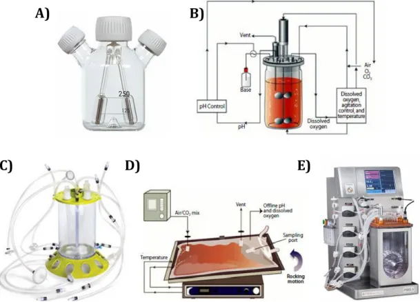
consists in cultivating cells using dynamic culture systems [40]. Spinner flasks (Figure 1.6A)
have been widely used for PSCs expansion [83] and differentiation into cardiomyocytes [87].
However this culture system lacks culture control over parameters like temperature, pH or
gas exchanges (e.g. oxygen).
Figure 1.6: Examples of different bioreactor designs used for stem cell bioprocessing.A) Spinner flask. B) Stirred tank bioreactor [88]. Single-use disposable bioreactors: C) Single-use stirred tank bioreactor (Mobius CellReady, Millipore) (www.millipore.com). D) Wave induced bioreactor (WAVE Bioreactor, GE Healthcare) [88]. E) Pneumatically mixed bioreactor (PBS 3, PBS Biotech) (www.pbsbiotech.com).
A)
B)
14
Bioreactors for PSC bioprocessing should accurately control and regulate cellular
microenvironment, aiming at supporting cell viability while providing spatial and temporal
control of signaling. Different bioreactor types have been used for PSC cultivation such as
microfluidic culture systems, rotatory culture systems and stirred tank bioreactors (reviewed
in [89]). Stirred tank bioreactors (Figure 1.6B-C) offer efficient gas/nutrient transfer, precise
control and monitoring of culture environment (e.g. pH, temperature, pO2, gas composition,
nutrients) and non-destructive sampling (Figure 1.6B-C). These bioreactors offer enormous
engineering prospects, as they are scalable, reproducible, versatile and fully automated. One
of the main limitations of stirred tank bioreactors is the hydrodynamic shear stress promoted
by stirring. Up to now, the minimal volume required to set up the experiments was very high
(approximately 50 mL), which compromised the use of stirred bioreactors for
high-throughput applications by demanding higher starting cell numbers and increasing the costs
associated to optimization studies. Large efforts have been made towards the development of
smaller scale systems (working volume 10–15 mL) and two options are available today including the ambr® systems (from TAP Biosystems) and the low volume spinner flasks
(from HexaScreen).
During the last decade, single-use disposable bioreactors have been developed and
applied in preclinical, clinical, and production-scale biotechnological facilities [90]. In
contrast to reusable bioreactors made from glass or stainless steel, single-use bioreactors are
made of FDA-approved (Food and Drug Administration) plastics [91]. Single-use bioreactors
offer several advantages such as reduced costs in construction of production facilities,
reduced risk of cross-contamination, less cleaning validation needed and rapid changeover of
processes [92]. However, these bioreactors also present limitations including lack of
instrumentation for single-use sensors, possible secretion of leachables and extractables from
the plastic cover and limited number of vendors available [91, 92].
Today, there are different disposable bioreactor types commercially available ([90,
91]). These include spinner vessels, such as the SuperSpinner D 1000 (Sartorius), and stirred
tank disposables: Mobius CellReady (Millipore; Figure 1.6C), Univessel SU (Sartorius) and
CelliGen BLU (Eppendorf); micro bioreactors are also available, the Ambr bioreactor.
Different bioreactor designs have also been developed as single-use disposables. One
example is the wave induced bioreactor (WAVE Bioreactor from GE Healthcare and BIOSTAT
CultiBag from Sartorious-Stedim). This bioreactor (Figure 1.6D) has a rocking platform
capable of inducing a wave motion in the culture media without an impeller or other invasive
mixer. This rocking system promotes rapid medium homogeneity and provides the optimal
15
lower shear stress when compared to the stirred tank bioreactor [94]. In addition, this
bioreactor has a simple design, appealing to either biological engineers or medical
professionals. Pneumatically mixed bioreactors (e.g. Air-Wheel® Bioreactor Systems from
PBS Biotech (Figure 1.6E)) are another example of disposable devices with unique
characteristics. This bioreactor provides mixing and optimal oxygen transfer via gas
buoyancy (air bubbles), eliminating the need for an external mechanical agitator [92, 95].
This pneumatic mixing type assures low shear stress [95], always an advantage for stem cell
bioprocessing.
In order to enhance stem cell metabolism and further improve cell viability,
proliferation and differentiation, different operation modes can be adopted, including
fed-batch and perfusion [15]. The fed-fed-batch strategy is considered the most adequate for
optimizing cell metabolism, enabling a more efficient uptake and consumption of nutrients,
resulting in reduced accumulation of metabolites in culture supernatant [96]. Perfusion
strategies have been developed in stem cell bioprocesses to ensure continuous renewal of
nutrients and growth factors and constant removal of toxic byproducts [70, 96]. Growth
factors play an important role in stem cell processes, providing survival, proliferation and
differentiation signals. This feature of continuous medium addition and removal has potential
to be integrated in expansion, differentiation and/or cell lineage selection steps. Addition of
different medium formulations and removal of cell debris are also possible using perfusion
strategies.
Optimization of PSC bioprocessing should result in large cell yields through process
intensification, specialization and integration, rather than just scale-up technologies [15]. The
establishment of platforms capable of integrating isolation and reprogramming, inoculation,
expansion, differentiation, purification and harvesting would result in the scale-up of well
differentiated cells to clinical relevant numbers [15]. Several bioprocesses combining PSC
(human and mouse) expansion and cardiomyocyte differentiation have been reported in
recent years. Table 1.2 presents results of recent studies integrating PSC-derived expansion
and differentiation in computer controlled bioreactors, operating in batch and perfusion
16
Table 1.2: Studies involving cultivation of mESCs in scalable 3D approaches involving expansion and differentiation into cardiomyocytes.
Stirred Tank Bioreactor Operating mode Culture volume (ml) Initial cell number Final CM number CM/ESC ratio Ref
Standard Spinner vessel Batch 250 5x10⁷ 5.86x10⁷ 1.2 [97]
DASGIP Cellferm-Pro
(DASGIP Tech.) Perfusion 250 0.9x10
6 3.5x10⁶ 3.8 [70]
Biostat MD (Sartorius) Batch 2000 2x10⁸ 1.28x10⁹ 6.4 [98]
Biostat MD (Sartorius) Perfusion 2000 2x10⁸ 4.6x10⁹ 23 [99]
1.3
Purification of iPSC-derived cardiomyocytes
The use of cardiomyocytes derived from PSCs in cell-based therapy has been
hampered by the inability of differentiation protocols to obtain homogenous, pure and
functional cardiomyocytes. To prevent unwanted side effects, like tumor formation or
inefficient therapies, it is necessary to eliminate the high degree of heterogeneity of the final
cell population (reviewed in [3]). Therefore developing effective purification protocols is
imperative to obtain populations highly enriched in cardiomyocytes. There are several
purification/enrichment strategies, including genetic and non-genetic approaches for
selection based on distinct cellular and molecular characteristics of cardiomyocytes.
1.3.1
Genetic selection of cardiomyocytes
Genetic-based purification protocols allow efficient enrichment of cardiomyocytes in
mESC cultures (purity >99.6% [100]). In this approach, undifferentiated cells are genetically
modified to integrate and stably express a transgene, such as a fluorescent marker (e.g. eGFP,
dsRed) or an antibiotic-resistant gene (e.g. resistance against geneticin, puromycin) under
control of a specific cardiac promoter. A number of cardiac specific promoters have been
used, such as human myosin light chain ventricular isoform (Myl2) [101] and mouse [102]
and human [103] cardiac α-myosin heavy chain (α-MHC). Since differentiated cardiomyocytes express these selection markers, these cells can be selected from a
heterogeneous population by fluorescence-activated cell sorting (FACS) or by addition of
antibiotics, if the transgene encodes antibiotic resistance genes (reviewed in [54]). Although
this approach results in a highly enriched population, it presents some disadvantages, as
genetic modification of cells can promote/induce tumor formation, cell lines have to stably
express the transgene, which is a time consuming process, and the enrichment process is
17
1.3.2
Non-genetic purification of cardiomyocytes
Cell lineage purification from heterogeneous populations can also be achieved using
non-genetic methods. These strategies range from mechanical to biochemical methods, based
on cells physical and structural properties (reviewed in [3]).
Micro dissection and Percoll gradient centrifugation are the most widely used
methods for cardiomyocyte enrichment [21]. Micro dissection is based on the isolation of
contracting areas, enriched with cardiomyocytes, through the manual dissection of these
areas from a cell culture plate [104]. In Percoll separation methods, dissociated cells are
loaded onto two layers of Percoll and centrifuged, enabling separation of cardiomyocytes
from non cardiac cells, who will reside in a upper layer (lower density than cardiomyocytes)
[105]. Both these techniques are simple and inexpensive, however they lack scalability and
are time-consuming (reviewed in [40, 54]).
Enrichment of cardiomyocytes can also be carried out by targeting specific surface
markers of cardiomyocytes. Using specific antibodies, enrichment is possible by selecting
differentiated cardiomyocytes (positive selection) or non-cardiac cells (negative selection),
since their surface marker expression is distinct (reviewed in [54]). Three surface markers
are associated with stem-cell derived cardiomyocytes: signal-regulatory protein alpha
(SIRPA) [106], vascular adhesion molecule 1 (VCAM1) [107] and activated leukocyte cell
adhesion molecule (ALCAM) CD166, which is a specific marker for early murine
cardiomyocytes [108]. Cell sorting can be achieved with fluorescence-activated cell sorting
(FACS) or magnetic-assisted cell separation (MACS) techniques. FACS can simultaneously
target multiple markers, while MACS can only target a single marker although presenting
higher throughput. These methods can assure enriched populations, containing over 90% of
cardiomyocytes [54]. However studies have shown that these approaches result in the loss of
some cardiomyocytes, as not all express these markers [106, 107]. Cardiomyocytes have a
unique feature of presenting high content of mitochondria. Thus, labeling the mitochondria of
cardiomyocytes with fluorescent dyes provides another means of purification.
Tetramethylrhodamine methyl ester perchlorate (TMRM) has been used for this purpose, as
it is non-toxic to cells and the labeling is reversible [109]; using this method cardiomyocytes
can be efficiently selected (99% of purity) using FACS, as they will be part of the high
fluorescence intensity fraction [3, 109].
Recently, novel purification methods have been developed based on distinct
metabolic flows of cardiomyocytes. Exploiting distinct metabolic flow methods, studies have
stated that 99% purity can be achieved using glucose depleted culture medium containing
18
cardiomyocytes enrichment protocol is achieved without the use of complex devices or
labor-intensive steps, being adequate for clinical applications [111].
1.4
Characterization of cardiomyocytes
Structural and functional characterization of differentiated cardiomyocytes is
essential to evaluate the outcome of differentiated protocols and quality of cell-based
products. For cell-based therapies, characterization of cell-based product or cells to be used
in transplantation is important to minimize the risk of host rejection, tumor formation or
arrhythmias (reviewed in [3]). In the last few years, extensive characterization of
PSC-derived cardiomyocytes have been carried out to evaluate and confirm their cardiac
phenotype [40, 112].
The cardiomyocyte differentiation process of PSCs can be monitored by following the
temporal gene expression pattern. Pluripotency markers are down-regulated soon after
differentiation followed by up-regulation of cardiac mesoderm markers and then
cardiomyocyte-associated genes (reviewed in [54]). To confirm the phenotype of the final
differentiated population, it is essential to detect the expression of cardiomyocyte-associated
genes [113]. Some of these cardiac proteins are described in Table 1.3.
Table 1.3: Cardiac markers used for the characterization of cardiomyocytes (adapted from
[54]).
Structural proteins α-Actinin; cTnT; cTnI; α/β-MHC; Myl2; Myl7; Desmin; Titin
Transcription factors NKX2-5; GATA4; MEF2C; TBX5; TBX20
Gap junction proteins N-cadherin; Connexin 43
Surface proteins and ion channels α1/β1-adrenergic receptors; L-type calcium channel, HCN4 * cTnT – cardiac troponin T; cTnI – cardiac troponin I; MHC – myosin heavy chain; Myl2/Myl7 – myosin light chain (ventricular/atrial); NKX2-5 – NK2 transcription factor related locus 5; Gata4 – Zinc finger transcription factor 4; MEF2C – myocyte enhancer factor; TBX5/TBX20 – T box protein 5/20; HCN4 – Hyperpolarization Activated Cyclic Nucleotide-Gated Potassium Channel 4.
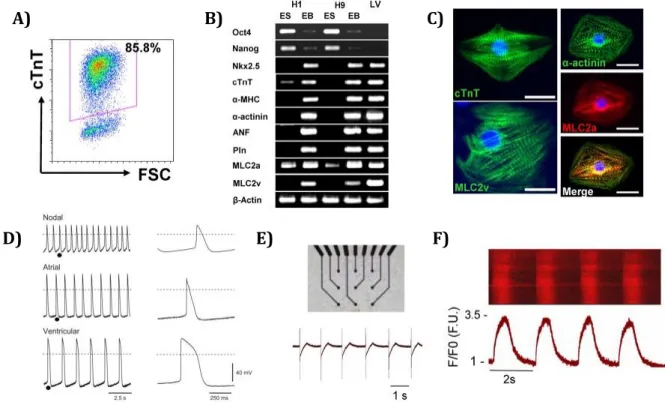
There are some techniques available to ensure a robust and efficient characterization
of PSC-derived cardiomyocytes. These techniques enable characterization based on the
morphologic, structural and electrophysiological properties of cardiomyocytes. Flow
cytometry (Figure 1.7A) provides a quantitative method to evaluate the purity of the
differentiated and enriched population, by measuring the number of cells expressing
cardiac-specific proteins [3]. Reverse transcription-polymerase chain reaction (RT-PCR, Figure 1.7B)
![Figure 1.1: Causes of death in Europe, 2011 (adapted from [2]).](https://thumb-eu.123doks.com/thumbv2/123dok_br/16672892.742789/23.893.149.753.436.714/figure-causes-death-europe-adapted.webp)
![Figure 1.5: 2D and 3D strategies for cultivation of PSCs (adapted from [15]).](https://thumb-eu.123doks.com/thumbv2/123dok_br/16672892.742789/34.893.112.787.96.453/figure-d-d-strategies-cultivation-pscs-adapted.webp)