Promotion of Expansion and Differentiation
of Hematopoietic Stem Cells by
Interleukin-27 into Myeloid Progenitors to Control
Infection in Emergency Myelopoiesis
Jun-ichi Furusawa1, Izuru Mizoguchi1, Yukino Chiba1, Masayuki Hisada2, Fumie Kobayashi3, Hiroki Yoshida4, Susumu Nakae5, Akihiko Tsuchida2, Tetsuya Matsumoto6, Hideo Ema7, Junichiro Mizuguchi8, Takayuki Yoshimoto1*
1Department of Immunoregulation, Institute of Medical Science, Tokyo Medical University, Tokyo, Japan,
2Department of Gastrointestinal and Pediatric Surgery, Tokyo Medical University, Tokyo, Japan,
3Department of Infectious Diseases, Kyorin University of Medicine, Tokyo, Japan,4Department of Biomolecular Sciences, Faculty of Medicine, Saga University, Saga, Japan,5Laboratory of Systems Biology, Center for Experimental Medicine and Systems Biology, Institute of Medical Science, University of Tokyo, Tokyo, Japan,6Department of Microbiology, Tokyo Medical University, Tokyo, Japan,7Department of Cell Differentiation, The Sakaguchi Laboratory of Developmental Biology, Keio University School of Medicine, Tokyo, Japan,8Department of Immunology, Tokyo Medical University, Tokyo, Japan
*yoshimot@tokyo-med.ac.jp
Abstract
Emergency myelopoiesis is inflammation-induced hematopoiesis to replenish myeloid cells in the periphery, which is critical to control the infection with pathogens. Previously, pro-inflammatory cytokines such as interferon (IFN)-αand IFN-γwere demonstrated to play a critical role in the expansion of hematopoietic stem cells (HSCs) and myeloid progenitors, leading to production of mature myeloid cells, although their inhibitory effects on hematopoi-esis were also reported. Therefore, the molecular mechanism of emergency myelopoihematopoi-esis during infection remains incompletely understood. Here, we clarify that one of the interleukin (IL)-6/IL-12 family cytokines, IL-27, plays an important role in the emergency myelopoiesis. Among various types of hematopoietic cells in bone marrow, IL-27 predominantly and con-tinuously promoted the expansion of only Lineage−Sca-1+c-Kit+(LSK) cells, especially
long-term repopulating HSCs and myeloid-restricted progenitor cells with long-term repopu-lating activity, and the differentiation into myeloid progenitors in synergy with stem cell fac-tor. These progenitors expressed myeloid transcription factors such asSpi1,Gfi1, and Cebpa/bthrough activation of signal transducer and activator of transcription 1 and 3, and had enhanced potential to differentiate into migratory dendritic cells (DCs), neutrophils, and mast cells, and less so into macrophages, and basophils, but not into plasmacytoid DCs, conventional DCs, T cells, and B cells. Among various cytokines, IL-27 in synergy with the stem cell factor had the strongest ability to augment the expansion of LSK cells and their dif-ferentiation into myeloid progenitors retaining the LSK phenotype over a long period of time. The experiments using mice deficient for one of IL-27 receptor subunits, WSX-1, and IFN-γ revealed that the blood stage of malaria infection enhanced IL-27 expression through IFN-γ OPEN ACCESS
Citation:Furusawa J-i, Mizoguchi I, Chiba Y, Hisada M, Kobayashi F, Yoshida H, et al. (2016) Promotion of Expansion and Differentiation of Hematopoietic Stem Cells by Interleukin-27 into Myeloid Progenitors to Control Infection in Emergency Myelopoiesis. PLoS Pathog 12(3): e1005507. doi:10.1371/journal. ppat.1005507
Editor:Mary M. Stevenson, McGill University, CANADA
Received:August 17, 2015
Accepted:February 24, 2016
Published:March 18, 2016
Copyright:© 2016 Furusawa et al. This is an open access article distributed under the terms of the
Creative Commons Attribution License, which permits unrestricted use, distribution, and reproduction in any medium, provided the original author and source are credited.
Data Availability Statement:All relevant data are within the paper and its Supporting Information files.
production, and the IL-27 then promoted the expansion of LSK cells, differentiating and mobilizing them into spleen, resulting in enhanced production of neutrophils to control the infection. Thus, IL-27 is one of the limited unique cytokines directly acting on HSCs to pro-mote differentiation into myeloid progenitors during emergency myelopoiesis.
Author Summary
Emergency myelopoiesis is inflammation-induced hematopoiesis that is critical for con-trolling infection with pathogens, but the molecular mechanism remains incompletely understood. Here, we clarify that one of the interleukin (IL)-6/12 family cytokines, IL-27, plays an important role in emergency myelopoiesis. Among various types of hemato-poietic cells in bone marrow, IL-27 predominantly and continuously promoted expansion
of only Lineage−Sca-1+c-Kit+(LSK) cells, especially long-term repopulating hematopoietic
stem cells, and differentiation into myeloid progenitors in synergy with stem cell factor.
These progenitors expressed myeloid transcription factors such asSpi1,Gfi1, andCebpa/b
through activation of signal transducer and activator of transcription 1 and 3, and had enhanced potential to differentiate into neutrophils, but not into plasmacytoid dendritic cells. Among various cytokines, IL-27 in synergy with stem cell factor had the strongest ability to augment the expansion of LSK cells and their differentiation into myeloid pro-genitors. The blood stage of malaria infection was revealed to enhance IL-27 expression
through interferon-γproduction, and IL-27 then promoted the expansion of LSK cells,
dif-ferentiating and mobilizing them into the spleen, resulting in enhanced production of neu-trophils to control the infection. Thus, IL-27 is one of the limited unique cytokines directly acting on hematopoietic stem cells to promote differentiation into myeloid progenitors during emergency myelopoiesis.
Introduction
Emergency myelopoiesis is inflammation-induced hematopoiesis, which is critical for
control-ling systemic infection with pathogens such as a virus, bacteria, or parasite [1,2]. In contrast to
adaptive immune cells such as T cells and B cells, which can vigorously proliferate in response to their specific antigens, innate immune cells need to be replenished from hematopoietic stem cells (HSCs) and progenitors in bone marrow (BM) because of their low proliferative activity. However, the molecular mechanism of emergency myelopoiesis during infection remains incompletely understood. HSCs and hematopoietic progenitors can directly sense the presence of pathogens via pattern recognition receptors (Rs) such as Toll-like receptors (TLRs), and they can also respond to pro-inflammatory cytokines such as interferon (IFN)-α, IFN-γ,
inter-leukin (IL)-1, tumor necrosis factor (TNF)-α, and granulocyte colony-stimulating factor
(G-CSF) [1]. IFN-αand IFN-γhave pleiotropic effects on many cell types, including HSCs and
hematopoietic progenitors [1]. Recently, these cytokines were demonstrated to induce an
expansion of HSCs and myeloid progenitors, leading to the production of mature myeloid cells
[3–6], although their inhibitory effects on hematopoiesis were previously reported [7–9].
Cur-rently, thus, there are several conflicting positive and negative effects of IFN-αand IFN-γin
hematopoiesis [10,11]. However, these discrepancies may be explained by compensatory
mech-anisms, including IFN-γ-mediated secretion of other cytokines such as IL-6 [12] and
fms-related tyrosine kinase 3 ligand (Flt3L) [13].
Role of IL-27 in Emergency Myelopoiesis
Competing Interests:The authors have declared
IL-27 is one of the IL-6/IL-12 family cytokines; it plays important roles in immune
regula-tion with both pro-inflammatory and anti-inflammatory properties [14–16]. IL-27 consists of
p28 and Epstein-Barr virus-induced gene 3 (EBI3), and its receptor is composed of WSX-1 and glycoprotein (gp)130, which is a common receptor subunit in many of the IL-6 family cyto-kines. We previously demonstrated that IL-27 plays a role in HSC regulation, and that IL-27
expands HSCs and promotes their differentiationin vitro[17]. Moreover, transgenic (Tg) mice
expressing IL-27 showed enhanced myelopoiesis in BM and extramedullary hematopoiesis in
the spleen [17].
In the present study, we further examined the effects of IL-27 on hematopoiesis, the molecu-lar mechanisms, and the physiological role of IL-27 in the control of mamolecu-laria infection. IL-27
acted on and expanded Lineage (Lin)−Sca-1+c-Kit+(LSK) cells, which are highly enriched in
HSCs together with very primitive hematopoietic progenitors [18,19], in BM cells in synergy
with stem cell factor (SCF, c-Kit ligand) and differentiated HSCs into myeloid progenitors through activation of signal transducer and activator of transcription 1 (STAT1) and STAT3.
Moreover, malaria infection induced IFN-γproduction, which augmented IL-27 expression,
and the IL-27 then promoted the expansion and mobilization of LSK cells into the spleen, resulting in enhanced myelopoiesis to resolve the infection. Our results revealed that IL-27 is one of the limited unique cytokines directly acting on long-term HSCs (LT-HSC), which repre-sent the true stem cells capable of self-renewing, and promotes the expansion and differentia-tion of them into myeloid progenitors.
Results
IL-27 and SCF expand only LSK cells among various kinds of BM
progenitors
Previously, we demonstrated that stimulation of LSK cells with IL-27 and SCF induces an expansion of HSCs and hematopoietic progenitors, including short-term repopulating cells
[17]. Moreover, we found that only the combination of IL-27 and SCF, but not either alone,
vigorously and continuously expands BM cells to produce LSK cells and CD11b+c-Kit−cells
[17]. To examine which cell populations in BM cells respond to IL-27 and SCF in more detail,
BM cells were divided into two populations positive or negative for Lin markers except CD11b,
and the Lin−population was further divided into four populations positive for either c-Kit or
CD11b, or both positive, or both negative. Each population purified by sorting was then
stimu-lated with IL-27 and SCF. Among these five populations, only the Lin−c-Kit+population
greatly expanded (Fig 1A). Next, the BM cells were divided into respective hematopoietic
pro-genitors according to the expression of cell surface markers, as reported previously [20–22],
and stimulated with IL-27 and SCF. Only the LSK cell population vigorously and continuously expanded over more than 6 weeks, although transient and slight expansion was seen in the cell populations of granulocyte/macrophage progenitor (GMP), common myeloid progenitor
(CMP), and megakaryocyte/erythrocyte progenitor (MEP) (Fig 1B–1E). The expanding cells in
the LSK cell population were further analyzed for the expression of cell surface markers. In line with the preliminary results, there seemed to be two populations, the phenotypical LSK
popula-tion and the Lin+c-Kit−population (Fig 1E).
We previously demonstrated thatIL-27Tg mice, which express high amounts of IL-27 in
blood, show an increased number of LSK cells in the BM and spleen [17]. To further examine
thein vivoeffects of IL-27 on the expansion of LSK cells, LSK cells were purified by sorting
from BM cells ofGFPTg mice and transferred into wild-type (WT) andIL-27Tg mice. The
transferred GFP+LSK cells vigorously expanded in the BM and spleen ofIL-27Tg mice, but
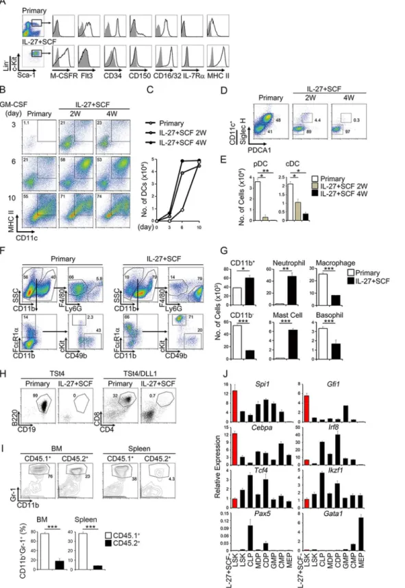
Fig 1. IL-27 and SCF most strongly expand only LSK cells among various kinds of BM progenitors and differentiate them into myeloid progenitors retaining the LSK phenotype.(A) Expansion of only the c-Kit+CD11b−Lin−population by IL-27 and SCF in BM cells. Total BM cells were divided into two populations positive or negative for Lin markers, and the Lin−population was further divided into four populations positive for either c-Kit or CD11b, both positive, or both negative. Each population (5 × 10³) purified by sorting was stimulated by either IL-27 or SCF alone, or both, in a round 96-well plate and photographed 2 weeks later. (B-E) Expansion of only the LSK cell population by IL-27 and SCF among various BM progenitors. BM progenitors (4–5 × 10³)
purified by sorting were stimulated with IL-27 and SCF. Cell expansion was photographed at indicated periods (B) and cell number of the expanded LSK cell population retaining the LSK phenotype was counted at 1 week (C). Time kinetic analysis of cell numbers in the LSK population retaining the LSK phenotype
markers for LSK phenotype (Fig 1F). These results suggest that IL-27 vigorously and
continu-ously augments the expansion of LSK cells bothin vitroandin vivo.
IL-27 has the strongest ability to augment the expansion of LSK cells
and their differentiation into myeloid progenitors retaining the LSK
phenotype
Because it was previously reported that IFN-αand IFN-γinduce proliferation of HSCsin vivo
[3–5], we next explored the effects of various cytokines in collaboration with SCF on the
expan-sion of LSK cellsin vitro. However, IFN-αand IFN-γaugmented the expansion of LSK cells
very little, and only IL-27 enhanced it vigorously over 4 weeks (Fig 1G and 1H). Moreover,
although there are several cytokines, such as IL-3, IL-11, G-CSF, and TPO, that are known to
transiently expand and differentiate HSCs [1], none showed an ability superior to that of IL-27
in expanding LSK cells retaining the LSK phenotype over a long period of time (S1 Fig). Thus,
IL-27 has the strongest ability to augment the expansion of LSK cells.
LSK cells expanded by IL-27 and SCF are multipotent myeloid
progenitors
The LSK cells expanded by IL-27 and SCF were further analyzed for the cell surface expression of various markers, and the expression levels were compared with those of primary LSK cells freshly prepared from BM of WT mice. The expression levels of macrophage colony-stimulat-ing factor receptor (M-CSFR), CD16/32, and MHC class II in the LSK cells expanded by IL-27
and SCF were much higher than those in primary LSK cells (Fig 2A). The expression levels of
CD34 and CD150 in the expanded LSK cells were slightly less than those in primary LSK cells (Fig 2A). In contrast, the expanded LSK cells were almost completely negative for Flt3
expres-sion, whereas primary LSK cells were positive for Flt3 (Fig 2A). Thus, IL-27 and SCF expand
and differentiate primary LSK cells into M-CSFR+Flt3−CD16/32+LSK cells (myeloid
progeni-tor cells).
Next, multipotency of the LSK cells expanded by IL-27 and SCF were examined under vari-ous differentiation conditions for migratory dendritic cells (mDCs) by granulocyte/macro-phage (GM)-CSF, plasmacytoid DCs (pDCs) and conventional DCs (cDCs; lymphoid-resident
DCs) were examined using Flt3L and thrombopoietin (TPO) [23], myeloid cells were examined
using IL-3 and SCF, and T cells and B cells were examined by using thymic stromal cells (TSt4)
with and without expressing Notch ligand Delta-like 1 (DLL1), respectively [24]. The expanded
LSK cells much more rapidly proliferated and differentiated into MHC class II+CD11c+mDCs
than primary LSK cells in response to GM-CSF, although the total number of mDCs achieved
seemed to be similar for both (Fig 2B and 2C). However, LSK cells stimulated with IL-27 and
SCF rapidly lost the ability to differentiate into pDC and cDC (Fig 2D and 2E). These
phenom-ena are highly consistent with the almost complete abolishment of Flt3 expression on the
expanded LSK cells (Fig 2A). Under myeloid differentiation conditions, the expanded LSK cells
differentiated much more greatly into neutrophils and slightly into mast cells, but less so into
(D). Representative flow cytometry dot plot analysis of c-Kit and Lin (upper) and of c-Kit and Sca-1 in the Lin−c-Kit+population (lower) of expanded LSK cells and progenitor cells at 1 week (E). (F) Expansion of LSK populationsin vivoby IL-27 inIL-27Tg mice. LSK cells were purified by sorting from BM cells ofGFP
Tg mice and transferred into non-lethally irradiated WT andIL-27Tg mice. Twenty days later, BM and spleen in these recipient mice were analyzed for GFP+ LSK populations. Data are shown as mean±SEM (n = 2–3) and are representative of at least two independent experiments.*P<0.05. (G-H) Augmented
and prolonged expansion of the LSK cell population by only IL-27 and SCFin vitro. LSK cells (1 × 10³) from WT mice were stimulated by various cytokines together with SCF for 1 to 4 weeks, the stimulated cells were analyzed by flow cytometry (G), and cell number was counted with time course (H). Data are representative of at least two independent experiments.
Fig 2. LSK cells expanded by IL-27 and SCF are multipotent myeloid Progenitors.(A) Flow cytometry histogram analysis of cell surface markers in the LSK cells expanded by IL-27 and SCF for 4 weeks and primary BM cells using antibodies as indicated (solid line) and their control antibodies (plane line with shading). (B-C) Augmented potential of the LSK cells expanded by IL-27 and SCF to differentiate into mDCs. LSK populations (3 × 10³) purified from the LSK cells expanded by IL-27 and SCF for 2 and 4 weeks and primary BM cells were stimulated with GM-CSF. After the indicated time, these stimulated cells were analyzed for the expression of MHC class II and CD11c (B), and the cell numbers of mDC (MHC class II+CD11c+) were counted (C). (D-E) Decreased
macrophages and basophils (Fig 2F and 2G). Similar enhanced differentiation into myeloid
cells was observed using the LSK cells obtained from WT andIL-27Tg mice (S2 Fig). In
con-trast, the ability to differentiate into B cells and T cells was almost completely abrogated in the
expanded LSK cells (Fig 2H).
Moreover, the ability of the LSK cells expanded by IL-27 and SCF to differentiate into
mye-loid cellsin vivowas explored by using mixed BM chimeras. The equal cell numbers of the LSK
cell population expanded from CD45.1 congenic mice and BM cells from CD45.2 congenic mice were mixed and transferred into sublethally irradiated CD45.2 congenic mice. After 9 days, cell populations of neutrophils in the BM and spleen were analyzed. In agreement with thein vitroresults described, the percentages of neutrophils derived from the expanded LSK
cells were markedly higher than those from primary LSK cells in both BM and spleen (Fig 2I).
To explore the molecular mechanisms whereby IL-27 and SCF expand LSK cells, total RNA was prepared from the expanded LSK cells and respective hematopoietic progenitors and ana-lyzed with real-time reverse transcriptase-polymerase chain reaction (RT-PCR). The expanded cells highly expressed the transcription factors critical for differentiation into myeloid cells
such asSpi1,Gfi1, andCebpa, but expression was much less for those important for the other
types of cells such asIrf8,Tcf4, andIkzf1[25–27] (Fig 2J). No expression of transcription
fac-tors important for B cells and erythrocytes,Pax5[28] andGata1[29], respectively, was
observed. In addition, the expression ofCbepb, which is a transcription factor recently
demon-strated to be regulated by cytokines and control emergency granulopoiesis [30–32], was also
increased (S3 Fig).
These results suggest that LSK cells expanded by IL-27 and SCF are multipotent myeloid progenitors that have unique potential to differentiate into mDCs, neutrophils, and mast cells, and less so into macrophages, and basophils, but not into pDCs, cDCs, T cells, and B cells.
STAT1 and STAT3 are important for expansion and differentiation of
LSK cells by IL-27 and SCF
We and others previously demonstrated that IL-27 activates both STAT1 and STAT3 through
WSX-1 and gp130, respectively [14,15]. Consistent with these reports, real-time RT-PCR
anal-ysis revealed that LSK populations purified from LSK cells expanded by IL-27 and SCF and
pri-mary BM cells were positive for mRNA expression ofSTAT1andSTAT3(Fig 3A).
Phosphorylation of STAT1 and STAT3 was also detected in primary WT LSK cells, but not
WSX-1-deficient LSK cells (Fig 3B), in response to IL-27 and SCF, which were analyzed by flow cytometry. Furthermore, IL-27 alone induced phosphorylation of both STAT1 and STAT3, whereas SCF alone failed to induce phosphorylation of either one, as discussed
previ-ously [33] (S4 Fig).
To further investigate the roles of STAT1 and STAT3 in the expansion of LSK cells and the
ability to differentiate into myeloid progenitors by IL-27 and SCF, we usedSTAT1-deficient
LSK cells and conditionalSTAT3-knockout (STAT3cKO) LSK cells. LSK cells from WT (129)
potential of the LSK cells expanded by IL-27 and SCF to differentiate into pDCs (Siglec H+PDCA1+CD11c+) and cDCs (Siglec H−PDCA1−CD11c+). (F-G) Enhanced potential of the LSK cells expanded by IL-27 and SCF for 2 weeks to differentiate into neutrophils. Neutrophil; Ly6G+CD11b+, macrophage; F4/ 80+CD11b+, mast cell; c-Kit+Fc
εR1α+CD11b−, basophil; CD49b+FcεR1α+CD11b−. (H) Abrogated potential of the LSK cells expanded by IL-27 and SCF for 2 weeks to differentiate into T (day 18) and B (day 15) cells. (I) Enhanced potential of the LSK cells expanded by IL-27 and SCF to differentiate into neutrophils
in vivo. LSK populations purified from the LSK cells from CD45.1 congenic mice expanded by IL-27 and SCF for 2 weeks were transferred into sublethally irradiated CD45.2 recipient mice with the same congenic BM cells. Development of Gr-1+CD11b+neutrophils in the BM and spleen were analyzed by flow cytometry after 9 days, and the percentage of neutrophils in each CD45.1+and CD45.2+cells was compared. (J) Increased expression of transcription factors critical for the differentiation into myeloid cells in the LSK cells expanded by IL-27 and SCF. RNA was prepared from the LSK population purified from LSK cells expanded for 2 weeks together with other progenitors and subjected to real-time RT-PCR. Data are shown as mean±SEM (n = 2–4) and are
representative of two to three independent experiments.*P<0.05,**P<0.01,***P<0.005.
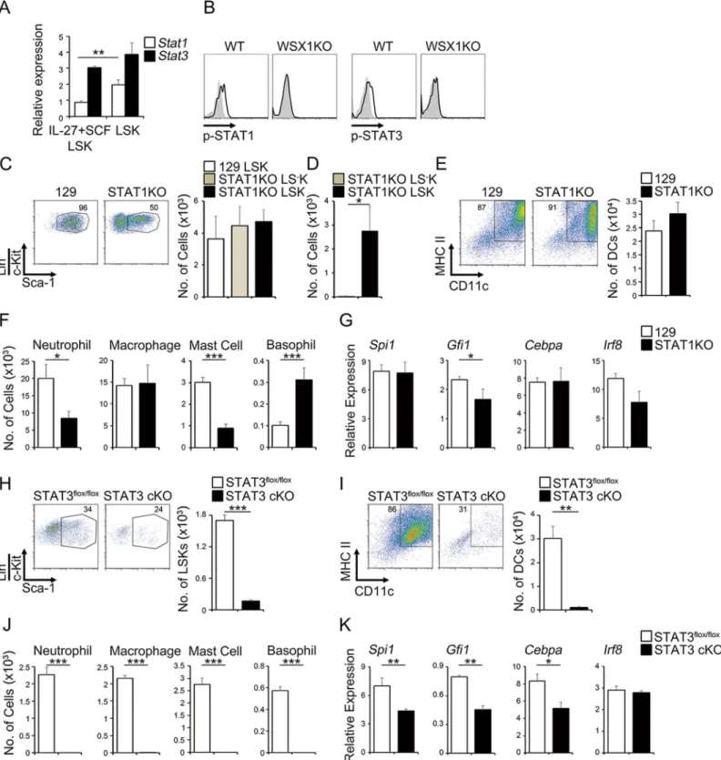
Fig 3. STAT1 and STAT3 are important for expansion and differentiation of LSK cells by IL-27 and SCF.(A) mRNA expression ofSTAT1andSTAT3in the LSK cells expanded by IL-27 and SCF for 2 weeks and in primary LSK cells. (B) Flow cytometry histogram analysis of primary LSK cells after stimulation with IL-27 and SCF for 60 min using anti-pY-STAT1 or anti-pY-STAT3 (solid line) and control antibody (plain line with shading). (C-D) Dispensable role for STAT1 in the expansion of LSK cells in response to IL-27 and SCF. LSK cells (1 × 10³) purified from BM cells of WT (129) mice andSTAT1-deficient mice were expanded by IL-27 and SCF for 2 weeks and analyzed for expression of LSK phenotype (C). The cell numbers of LSK and LS−K cell populations were counted. Purified cells of theSTAT1-deficient LSK and LS−K cells (1 × 10³) were further stimulated by IL-27 and SCF for more 1 week and the cell number of expanded cells was counted (D). (E-G) Contribution of STAT1 to the differentiation into mDC (E) and myeloid cells (F) of LSK cells expanded by IL-27 and
andSTAT1-deficient mice were stimulated with IL-27 and SCF. Although WT LSK cells
com-prised more than 90% of cells with the LSK phenotype after 7 days,STAT1-deficient LSK cells
comprised half LSK phenotype cells and half Sca-1−LK (LS−K) (Fig 3C). Nevertheless, the
number of expanded cells was comparable among them, probably due to the anti-proliferative
effects by STAT1 signaling [34]. However, the purifiedSTAT1-deficient LS−K cells failed to
survive thereafter, even in the presence of IL-27 and SCF (Fig 3D), although the purified
STAT1-deficient LSK cells expanded well (Fig 3D), as did WT 129 LSK cells (Fig 3C).
More-over, WT andSTAT1-deficient LSK cells had similar abilities to differentiate into MHC class
II+CD11c+mDC cells (Fig 3E) and macrophages (Fig 3F).STAT1-deficient LSK cells showed
reduced ability to differentiate into neutrophils and mast cells, but increased ability to
differen-tiate into basophils (Fig 3F). In line with the reduced ability to differentiate into neutrophils,
mRNA expression of the critical transcription factorGfi1was significantly reduced inSTAT1
-deficient LSK cells compared with that in WT LSK cells (Fig 3G). In contrast,STAT3cKO LSK
cells expanded very little in response to IL-27 and SCF (Fig 3H). Moreover, the residual
surviv-ing LSK cells almost completely lost the ability to differentiate into mDCs (Fig 3I) and myeloid
cells (Fig 3J). Consistent with the abrogated abilities, these cells showed reduced expression of
the critical transcription factors such asSpi1,Gfi1, andCebpa, but notIrf8(Fig 3K). These
results suggest that both STAT1 and STAT3 are necessary for LSK cells to fully expand and dif-ferentiate into myeloid progenitor cells in response to IL-27 and SCF.
IL-27 and SCF expand CD34
−CD150
+LSK cells into myeloid progenitor
cells
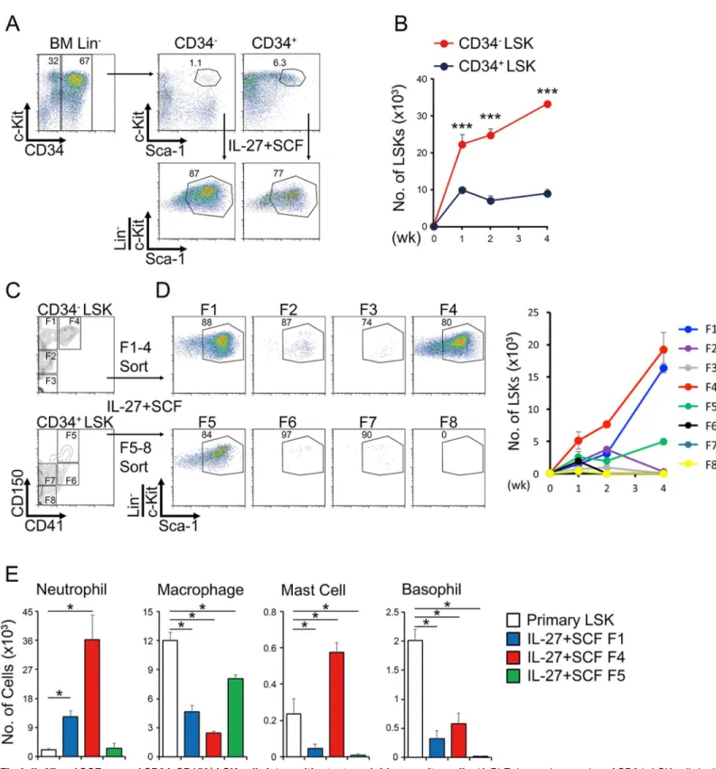
To more precisely define which cell population responds to stimulation with IL-27 and SCF, LSK cells were further divided into two populations according to CD34 expression. Although the
per-centage of the more primitive population of CD34−LSK cells was much less than that of CD34+
LSK cells, the CD34−LSK cells responded much better to stimulation with IL-27 and SCF and
expanded more vigorously than CD34+LSK cells (Fig 4A and 4B). Then, the LSK cells were
fur-ther divided into eight populations, F1 to F8, including LT-HSCs (CD34−CD150+CD41−LSK,
F1) and myeloid-restricted progenitor cells with long-term repopulating activity (MyRPs,
CD34−CD150+CD41+LSK, F4) according to the recently revised criteria [19]. Respective
popula-tions purified by sorting (Fig 4C) were stimulated with IL-27 and SCF. Only two populations, F1
and F4, vigorously expanded. F5, which corresponds to populations more differentiated toward
myeloid cells such as macrophages, slightly expanded (Fig 4D and 4E). The F1 and F4
popula-tions expanded by IL-27 and SCF had great abilities to differentiate into myeloid cells,
particu-larly neutrophils (Fig 4E). Thus, IL-27 and SCF expand CD34−CD150+LSK cells, including
LT-HSCs and MyRPs, and differentiate them into myeloid progenitor cells, which have great potential to differentiate mainly into neutrophils.
IL-27 plays an important role in expansion, differentiation, and
mobilization of LSK cells to control malaria infection
We previously demonstrated that in the blood stage of malaria infection with the attenuated
variantPlasmodium (P) bergheiXAT derived from the lethal strainP.bergheiNK65 IFN-γ
SCF for 2 weeks together with their mRNA expression of transcription factors (G). (H) Indispensable role for STAT3 in the expansion of LSK cells in response to IL-27 and SCF. Purified GFP−STAT3flox/floxLSK cells and GFP+STAT3cKO LSK cells (1 × 10²) were expanded by IL-27 and SCF for 10 days and analyzed for expression of LSK phenotype and their cell numbers. (I-K) Critical role for STAT3 in the differentiation into mDC (I) and myeloid cells (J) of LSK cells expanded by IL-27 and SCF for 10 days together with their mRNA expression of transcription factors (K). Data are shown as mean±SEM (n = 3–4) and
representative of two to three independent experiments.*P<0.05,**P<0.01,***P<0.005.
Fig 4. IL-27 and SCF expand CD34−CD150+LSK cells into multipotent myeloid progenitor cells.(A-B) Enhanced expansion of CD34−LSK cells by IL-27 and SCF. LSK cells from WT mice were divided into two populations according to the expression of CD34, CD34−LSK, and CD34+LSK cells, and each population (1 × 10²) was stimulated with IL-27 and SCF. One to 4 weeks later, these stimulated cells were analyzed by flow cytometry; representative dot plots of c-Kit and Sca-1 in the Lin−population at 2 weeks are shown (A). Cell numbers of these stimulated cells were counted with time course (B). (C-E) Augmented expansion of CD34−CD150+LSK cells by IL-27 and SCF. LSK cells were further divided into eight populations (F1-F8) according to the
expression of CD34, CD150, and CD41 (C), and each population (50 cells) purified by sorting was stimulated with IL-27 and SCF. One to 4 weeks later, these stimulated cells were analyzed by flow cytometry. Representative dot plots of c-Kit and Sca-1 in the Lin−population at 4 weeks are shown, and the cell number of the LSK cell population in these stimulated cells was counted with time course (D). LSK populations (1 × 10³) purified from primary or
production induced by IL-12 and phagocytic cells in the spleen are critical for controlling
para-sitemia [35,36]. Recently, it was reported that the blood stage ofP.chabaudiinfection induces
mobilization of early myeloid progenitor cells out of BM, thereby transiently establishing
mye-lopoiesis in the spleen through IFN-γto resolve the infection [6,37]. In line with these results,
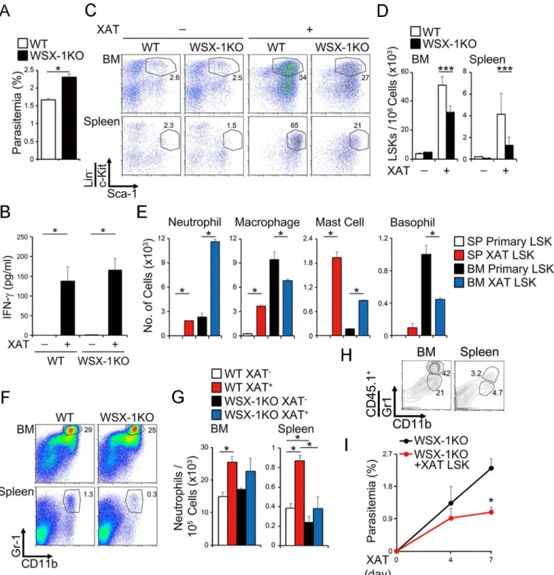
WSX-1-deficient mice showed more increased parasitemia than WT mice at 7 days (Fig 5A),
just prior to when the parasitemia reaches its peak after infection withP.bergheiXAT (S5A
Fig). In contrast, no significant difference was observed in the serum IFN-γlevels in WT and
WSX-1-deficient mice (Fig 5B). The infection markedly induced the enhanced percentage and
number of LSK cells in the BM and spleen of WT mice (Fig 5C and 5D). The LSK cells in the
BM showed greatly augmented abilities to differentiatein vitrointo neutrophils and mast cells,
but had slightly reduced abilities to differentiate into macrophages and basophils after infection (Fig 5E). Moreover, the LSK cells in the spleen exhibited a much more enhanced ability to
dif-ferentiate into neutrophils, macrophages, and mast cells (Fig 5E). In contrast, of note,WSX-1
-deficient mice showed significantly reduced percentage and number of LSK cells in the BM
and spleen compared with WT mice after infection (Fig 5C and 5D). In particular, the cell
number of the neutrophils in the spleen was increased very little inWSX-1-deficient mice (Fig
5F and 5G). Consistent with this, LSK cells from BM ofWSX-1-deficient mice showed reduced
abilities to differentiatein vitrointo neutrophils after infection compared with those of WT
mice (S6 Fig). In addition, more greatly reduced abilities to differentiatein vitrointo various
myeloid cells were observed when LSK cells from spleen were used (S6 Fig). Moreover, mixed
bone marrow chimera experiments using bone marrow cells from WT andWSX-1-deficient
mice revealed that the effect of IL-27 on the expansion of LSK cells and neutrophils is actually a
cell-autonomous direct effect (S7 Fig).
To further elucidate the protective role of LSK cells in malaria infection, LSK cells purified
from BM cells of infected WT CD45.1 mice were injected into the infectedWSX-1-deficient
CD45.2 mice. Consistent with the increased percentage of neutrophils differentiated from the
WT LSK cells in the BM and spleen of transferredWSX-1-deficient mice (Fig 5H), parasitemia
was significantly decreased in theWSX-1-deficient mice by the transfer of WT LSK cells
com-pared with that in non-transferredWSX-1-deficient mice (Fig 5I). Thus, the blood stage of
malaria infection induces expansion, differentiation, and mobilization of LSK cells into the spleen to produce myeloid cells such as neutrophils in an IL-27-dependent manner.
Malaria infection enhances IL-27 expression through IFN-γ
production to
promote the expansion, differentiation, and mobilization of LSK cells
Although it was previously reported that IFN-αand IFN-γinduce proliferation of HSCsin
vivo[3–5], IFN-αand IFN-γaugmented the expansion of LSK cells very littlein vitro, and only
IL-27 enhanced it vigorously over 4 weeks (Fig 1G and 1H). However, similar to the work
pre-viously reported [37,38],IFN-γ-deficient mice showed increased parasitemia with almost no
increase in the number of LSK cells in BM and spleen (Fig 6A and 6B). To clarify the molecular
mechanism whereby IFN-γinduces the expansion of LSK cells, the expression of IL-27
sub-units EBI3 and p28 was examined. Although the infection did not increaseEBI3mRNA
expres-sion in the BM and spleen of both WT andIFN-γ-deficient mice (S8 Fig), intriguingly, the
infection greatly enhancedp28mRNA expression in WT mice but failed to enhance it in
IFN-γ-deficient mice (Fig 6C). In agreement with this, p28 protein levels in the serum were greatly
expanded F1, F4, and F5 LSK cells were differentiated into myeloid cells by IL-3 and SCF, and cell number was counted (E). Data are shown as mean±SEM
(n = 3–4) and are representative of two to three independent experiments.*P<0.05,***P<0.005.
Fig 5. IL-27 plays an important role in expansion, differentiation, and mobilization of LSK cells to control malaria infection.(A-D) Reduced induction of LSK cell population inWSX-1-deficient mice after malaria infection, accompanied by increased parasitemia and comparable production of IFN-γ. WT or
WSX-1-deficient mice were infected with the blood stage ofP.bergheiXAT. Seven days later, parasitemia (A) and serum IFN-γlevel (B) were determined,
and LSK populations in the BM and spleen were analyzed by flow cytometry, and representative dot plots of c-Kit and Sca-1 in the Lin−population are shown (C). Cell number of the LSK cell population was counted (D). (E) Augmented potential of LSK cells to differentiate into myeloid cells by malaria infection. LSK cells (1 × 10³) in the BM and spleen of the malaria-infected or non-infected WT mice were purified and differentiated into myeloid cellsin vitroby IL-3 and SCF, and cell number of differentiated cells was counted. (F-G) Reduced cell number of neutrophils inWSX-1-deficient mice after malaria infection. The BM and spleen cells were analyzed for expression of Gr-1 and CD11b at 7 days after the infection (F), and cell number of neutrophils (Gr-1+CD11b+) was
increased by the infection in WT mice but not inIFN-γ-deficient mice (Fig 6D). Consistent
with thein vivorole of IFN-γ, we also observed the augmentation of mRNA expression ofp28,
but notEBI3, and p28 protein production in the culture supernatants of WT BM cells
stimu-lated with IFN-γin vitro(S9 Fig).
To further clarify the role of IL-27 downstream of IFN-γ, we next performed the experiment
to see the effects of forced expression of IL-27 on the susceptibility to malaria infection in
IFN-γ-deficient mice. The hydrodynamic injection of IL-27 expression vector into the infected
IFN-γ-deficient mice showed significantly decreased parasitemia compared with that of control
vec-tor (Fig 6E). This phenomenon was accompanied by the enhanced percentage of LSK cells and
augmented numbers of LSK cells and neutrophils in both BM and spleen (Fig 6F and 6G).
Thus, the blood stage of malaria infection augments the expression of IL-27 through IFN-γ, and IL-27 then promotes the expansion, differentiation, and mobilization of LSK cells into the spleen to control parasitemia.
Discussion
Previously, we found that IL-27, which is in the IL-6/IL-12 family of cytokines, plays a role in
the regulation of HSCsin vitroandin vivo[17]. Here, we have further elucidated that IL-27 is a
unique cytokine that directly acts on LSK cells to promote their differentiation into myeloid
progenitor cells called M-CSFR+Flt3−CD16/32+LSK cells, which still retain the LSK phenotype
(Fig 2A–2I). These progenitors have great potential to give rise to neutrophils, mDCs, and mast cells, but not to pDCs, cDCs, T cells, and B cells. Interestingly, among various BM pro-genitor cells, IL-27 and SCF vigorously and continuously expand only HSCs and primitive myeloid progenitor cells with long-term repopulating activity, such as LT-HSCs and MyRPs,
respectively [19], for more than 4 weeks (Fig 4). Consistent with the ability to differentiate into
myeloid progenitor cells, the LSK cells expanded by IL-27 and SCF expressed transcription
fac-tors such asSpi1,Gfi1,Cebpa, andCebpb, which are critical for myeloid differentiation [25–
27,30–32] (Fig 2JandS3 Fig). AlthoughCebpbwas reported to be an important transcription
factor for emergency granulopoiesis [30–32], STAT3 signaling was revealed to be important
for its upregulation, whereas STAT1 signaling unexpectedly suppressed its expression (S10
Fig). This phenomenon seems to correlate to the expression level of the anti-apoptotic gene
Bcl-2[39,40], but not the transcription factorE2-2, which is critical for pDC differentiation
[41]. Further studies are necessary to elucidate the precise roles of each STAT in the regulation
ofCebpbexpression. Thus, IL-27 is one of the limited unique cytokines that directly acts on the most primitive LT-HSCs; it promotes their expansion and differentiation into myeloid
progen-itor cells, presumably through MyRPs [19], to replenish myeloid cells such as neutrophils in
the periphery during emergency myelopoiesis.
Sca-1 is an IFN-responsive molecule that is highly upregulated in many hematopoietic cells
following exposure to IFNs [10,11,37]. Consequently, myeloid-restricted progenitor cells
nor-mally identified as Lin−Sca-1−c-Kit+(LS−K) become positive for Sca-1 and can no longer be
distinguished from the real multipotent LSK cells, resulting in overestimation of the latter pop-ulation, and this is a problem. IL-27 was previously reported to enhance the expression of
Sca-1 on T cells [42]. However, to alleviate the problem, we initially identified the cell population
counted (G). (H-I) Decreased parasitemia in theWSX-1-deficient mice transferred with LSK cells purified from BM cells of the malaria-infected WT mice. LSK cells purified from BM cells of the infected WT CD45.1 mice were transferred into non-lethally irradiatedWSX-1-deficient CD45.2 mice 7 days before infection. Neutrophil population in the BM and spleen was analyzed by flow cytometry, and representative dot plots of CD11b and Gr-1 in the CD45.1+ population are shown (H). Parasitemia was measured 4 and 7 days after the infection (I). Data are shown as mean±SEM (n = 3–9) and are representative of
at least two independent experiments.*P<0.05,***P<0.005.
Fig 6. Malaria infection enhances IL-27 expression through IFN-γproduction to promote the expansion, differentiation, and mobilization of LSK cells.(A-B) Indispensable role for IFN-γin the expansion of LSK cells after malaria infection. WT andIFN-γ-deficient mice were infected with the blood stage
ofP.bergheiXAT. Seven days later, parasitemia was determined (A) and LSK populations were analyzed by flow cytometry (B). (C-D) IFN-γ-dependent
induction of IL-27 p28 subunit expression by malaria infection. RNA was prepared 7 days after the infection and analyzed for expression ofp28by real-time RT-PCR (C), and serum p28 levels were determined by ELISA (D). (E-G) Decreased parasitemia and augmented expansion of LSK cell population inIFN-γ
-deficient mice by IL-27.IFN-γ-deficient mice were hydrodynamically injected with IL-27-expression vector or control vector at days 0 and 4 after infection; at
day 7, parasitemia was measured (E), LSK population was analyzed by flow cytometry (F), and cell numbers of LSK cells and neutrophils were counted (G). Data are shown as mean±SEM (n = 3–5) and are representative of at least two independent experiments.*P<0.05,**P<0.01,***P<0.005.
doi:10.1371/journal.ppat.1005507.g006
responsive to IL-27 and SCF among BM cells by using LSK cells and various hematopoietic progenitor cells purified by sorting. Intriguingly, it turned out that the SCF and LSK cell
popu-lations expanded vigorously and continuously in response to IL-27, and that the LS−K cell
pop-ulations including GMP, CMP, and MEP only transiently and slightly responded during the
first week and then disappeared thereafter (Fig 1A–1E). Moreover, in almost allin vitro
experi-ments, we used primary LSK cells that were purified by sorting. The high responsiveness of the
LSK cells to IL-27 seems to be partially due to the higher mRNA expression ofWSX-1in the
LSK cells compared to that of other hematopoietic progenitor cells (S3 Fig) [17].
We previously demonstrated that during the blood stage of malaria infection with attenu-atedP.bergheiXAT, IL-12-mediated IFN-γproduction and phagocytic cells (including
neutro-phils) in the spleen are critical for controlling parasitemia [35,36,43]. Previous studies
demonstrated that neutrophils play an important role in killing malaria parasites in mice, rats,
and humans [43–45]. In marked contrast, regarding infection with lethalP.bergheiNK65,
IL-12-mediated IFN-γproduction was shown to contribute to T-cell-dependent
immunopathol-ogy [46]. However, a major role of IL-27 in infection is its suppression of excess immune
responses against infection by controlling the production of pro-inflammatory cytokines [14–
16]. Consistent with this, WSX-1/IL-27 was recently demonstrated to have a critical role in
lim-iting the effector CD4+T-cell-mediated immunopathology caused by IL-12-dependent IFN-γ
production during infection with lethalP.bergheiNK65 [47–49]. The present study clearly
revealed that WSX-1/IL-27 contributes to clearance of parasites due to enhanced myelopoiesis
during the early phase of infection with attenuatedP.bergheiXAT (Fig 5andS5A Fig).
How-ever, during the late phase of infection, WSX-1/IL-27 seems to play a role in limiting the
pro-duction of pro-inflammatory cytokines such as IFN-γ(S5B Fig), leading to augmented
reduction of parasitemia (S5A Fig), as in the case of infection with lethalP.bergheiNK65 [47–
49]. Moreover, our preliminary data revealed that there were no apparent differences observed
in parasitemia or expansion of LSK cells and neutrophils in the BM and spleen of WT and
WSX-1-deficient mice 7 days after infection with lethalP.bergheiNK65 (S11 Fig). It is conceiv-able that pro-inflammatory cytokines other than IL-27 were abundantly produced in the
absence of IL-27 during infection with lethalP.bergheiNK65 and that late-phase infection
with attenuatedP.bergheiXAT may have redundantly compensated for the loss of IL-27 to
promote myelopoiesis. However, other studies have shown that IL-27 limits migration of neu-trophils from the BM to the site of inflammation by reducing production of cytokines and
che-mokines during influenza infection [50] and septic peritonitis [51]. IL-27 was also reported to
be a negative regulator of neutrophil function [52]. Although IL-27 directly promotes
myelo-poiesis to produce myeloid progenitors in BM, as shown in the present study, IL-27 may indi-rectly regulate migration of these progenitors and neutrophils to the site of inflammation and limit neutrophil function. Thus, IL-27 has both positive and negative effects on neutrophils; therefore, the overall outcome of the effects of IL-27 is likely to be governed by the balance between these effects, depending on the disease model.
It was recently demonstrated thatP.chabaudiinfection induces mobilization of early
mye-loid progenitor cells out of BM, thereby transiently establishing myelopoiesis in the spleen
through IFN-γ[37]. However, the expression of IFN-γR in the hematopoietic compartment
was dispensable, whereas its expression in the irradiation-insensitive cellular compartment,
including endothelial cells and stromal cells, was important [37]. Secretion of IFN-γ-induced
chemokines such as CCL2 and CCL7 by non-hematopoietic cells plays a critical role in the
mobilization of CCR2-expressing HSCs [37]. In this study, however, there is no experimental
evidence regarding how IFN-γregulates the activation of HSCs. In the present study,WSX-1
-deficient mice showed significantly reduced numbers of LSK cells and neutrophils compared
andS5A Fig). These results suggest that endogenous IL-27 greatly contributes to the clearance
of parasitemia through augmentation of myelopoiesis. Moreover, similar toP.chabaudi
infec-tion,P.bergheiXAT infection could not increase the number of LSK cells inIFN-γ-deficient
mice with increased parasitemia (Fig 6A and 6B). Of note, afterP.bergheiXAT infection,p28
mRNA expression and its serum protein level were markedly upregulated in an
IFN-γ-depen-dent manner (Fig 6C and 6DandS9 Fig). This is consistent with previous reports indicating
thatp28gene transcription in macrophages is induced by IFN-γand TLR ligands [53], and
that IFN-γlimits Th17-mediated and Th9-mediated autoimmune inflammation through IL-27
production [54,55]. In addition, the hydrodynamic injection of the IL-27 expression vector
into infectedIFN-γ-deficient mice greatly recovered the number of LSK cells and neutrophils
in the BM and spleen and eventually reduced parasitemia (Fig 6E–6G). Thus, during malaria
infection, it is highly conceivable that the proliferative effects on LSK cells by IFN-γare
indi-rectly mediated by IL-27. In our study, we could not observe any direct proliferative effect of
IFN-αand IFN-γon LSK cellsin vitro, as recently pointed out by others [10,11], and only
IL-27 augmented the proliferative effect for more than 4 weeks (Fig 1G and 1H).
Recently, IL-27 was reported to have a polyglutamic acid domain in the p28 subunit, which is unique among cytokines, and to confer hydroxyapatite-binding and bone-binding properties
and bone tropism to bone sialoprotein and the endosteal bone surface [56]. This location in the
BM has been identified as a niche for HSCs [57], and these properties support the idea that
IL-27 plays a critical role in the regulation of HSCs in that niche. We detected much higher
expressions of IL-27 subunits (bothp28andEBI3) at mRNA levels in BM than in the spleen
during the steady state, andP.bergheiXAT infection greatly augmented the expression ofp28
mRNA in both BM and spleen, and also its serum protein level (Fig 6C and 6D). Further
stud-ies are necessary to clarify which BM cells produce IL-27 during malaria infection; mesenchy-mal stromesenchy-mal cells might be a candidate because of their reported IL-6 production during viral
infection [12], as described in the next section.
It was recently demonstrated that specific cytotoxic CD8+T cells during an acute viral
infec-tion with lymphocytic choriomeningitis virus secrete IFN-γ, thus enhancing the production of
IL-6 in BM mesenchymal stromal cells and resulting in an increased number of early multipo-tent progenitors and committed myeloid precursors in the BM and accumulation of myeloid
cells in the periphery [12]. The IL-6Rαchain is only expressed at the stage of early multipotent
progenitors and downstream myeloid precursors, and it is lacking HSCs [12]. In contrast,
IL-27 most predominantly acts on only HSCs, as shown in the present study. Both IL-6 and IL-IL-27 share gp130, which is ubiquitously expressed as a common receptor subunit. Therefore, down-stream of IFN-γ, both IL-27 and IL-6 may be necessary to induce the maximum myelopoiesis to control infection. However, the mode of IL-6 action is complex and there are two major
mechanisms: IL-6 classic signaling through membrane IL-6Rαand IL-6trans-signaling
through soluble IL-6Rα[58,59]. It was recently reported thatIL-6Rα-deficient mice show
increased resistance toP.chabaudiinfection and that IL-6trans-signaling, but not IL-6 classic
signaling, contributes to a lethal outcome of infection [60]. In contrast to the viral infection, we
could not detect any increased mRNA expression ofIL-6in the BM or spleen of WT and
IFN-γ-deficient mice withP.bergheiXAT infection (S8 Fig). A similar inability of IFN-γto enhance
IL-6mRNA expression in WT BM cellsin vitrowas also observed (S9A Fig). In addition,IL-6
-deficient mice showed little increased susceptibility to theP.bergheiXAT infection, reduced
cell numbers in the LSK cell population, and reduced neutrophils in the BM and spleen (S12
Fig). Thus, individual pathogens may utilize different mechanisms to induce emergency
myelo-poiesis through IL-27, IL-6, and others.
In conclusion, the present results provide a novel role and mechanism for the action of
IL-27 downstream of IFN-γin the efficient expansion of myeloid progenitor cells from LT-HSCs
and MyRP cells and their mobilization into the spleen during acute malaria infection.
Materials and Methods
Ethics statement
The animal study was approved by the Animal Care and Use Committee of Tokyo Medical University (S-230043, S-24012, S-25059, S-26003, and S-27009) and was performed in accor-dance with our institutional guidelines and the Fundamental Guidelines for Proper Conduct of Animal Experiment and Related Activities in Academic Research Institutions under the juris-diction of the Ministry of Education, Culture, Sports, Science and Technology, 2006.
Mice
C57BL/6 (CD45.2) mice and C57BL/6 (CD45.1) mice were purchased from Sankyo Lab
(Tokyo, Japan). The 129/Sv mice andSTAT1-deficient mice (129/Sv background) were
pur-chased from Taconic Farms (Germantown, NY, USA).IL-6-deficient mice (C57BL/6
back-ground) were purchased from Jackson Laboratory (Bar Harbor, ME, USA).IFN-γ-deficient
mice (C57BL/6 background),STAT3flox/floxmice (a mixed background of 129/Sv and C57BL/
6), andGFPTg mice (C57BL/6 background) [61] were provided by Dr. Iwakura (Tokyo
Uni-versity of Science), Dr. Takeda (Osaka UniUni-versity), Dr. Okabe (Osaka UniUni-versity) and Dr. Ito
(Tokyo Medical University), respectively. In addition to these mice,IL-27Tg mice (C57BL/6
background) [17], andWSX-1-deficient mice (C57BL/6 background) [62] were maintained in
specific pathogen-free conditions under the care of the Laboratory Animal Center of Tokyo
Medical University.STAT3cKO cells were obtained by infectingSTAT3flox/floxcells with
Cre-expressing retrovirusin vitro.
Antibodies and reagents
Monoclonal antibodies (mAbs) for mouse c-Kit (2B8), Sca-1 (D7), CD3ε(145-2C11), CD4
(GK1.5), CD8α(53–6.7), CD19 (6D5), CD49b (DX5), Gr-1 (RB6-8C5), TER119/erythroid cell
(TER-119), CD11c (N418), CD11b (M1/70), F4/80 (BM8), NK1.1 (PK136), B220 (RA3-6B2),
FcεRIα(MAR-1), M-CSFR (AFS98), Flt3 (A2F10), CD16/32 (2.4G2), CD150 (TC15-12F12.2),
CD41 (MWReg30), IL-7Rα(A7R34), MHC class II I-A/I-E (M5/114.15.2), Siglec H (551),
Ly6G (1A8), CD45.1 (A20), and CD45.2 (104) were purchased from BioLegend (San Diego, CA). mAbs against mouse pY701-STAT1 (4a) and pY705-STAT3 (4/P-STAT3) were chased from BD Pharmingen (San Diego, CA). mAbs against mouse CD34 (RAM34) was pur-chased from eBioscience (La Jolla, CA). mAbs against mouse PDCA1 (JF05-1C2.4.1) was purchased from Miltenyi Biotec (Bergisch Gladbach, Germany). APC-Cy7-conjugated strepta-vidin, PerCP/Cy5.5-conjugated streptastrepta-vidin, and Brilliant Violet 510-conjugated streptavidin were purchased from BioLegend and used to reveal staining with biotinylated Abs. Mouse recombinant IL-27 and hyper-IL-6 were prepared as a tagged single-chain fusion protein by
flexibly linking EBI3 to p28 and soluble IL-6Rαto IL-6, respectively, using HEK293-F cells
(Life Technologies, Carlsbad, CA) as described previously [63,64]. Mouse recombinant SCF,
IL-1β, IL-3, IL-6, IL-7, IL-11, IL-12, thymic stromal lymphopoietin (TSLP), G-CSF, M-CSF,
GM-CSF, TNF-α, and human recombinant TPO were purchased from PeproTech (Rocky Hill,
NJ). Human recombinant Flt3L was purchased from Miltenyi Biotec. Mouse recombinant
was purchased from PBL Biomedical Laboratories (Piscataway, NJ). Mouse recombinant
IFN-γwas provided from Shionogi Pharmaceutical Co., Ltd. (Osaka, Japan),
Preparation of cells
Spleen and BM Lin−cells were enriched by negative selection using an autoMACS Pro
(Milte-nyi Biotec) with a combination of magnetic beads conjugated with mAbs against CD3ε, CD4,
CD8α, Gr-1, TER119, CD11b, CD11c, NK1.1, B220, and FcεRIα. Subsequently, cells were stained with mAbs against c-Kit, and Sca-1 was used for LSK. For multiple fractions of HSC,
cells were stained with CD34, c-Kit, Sca-1, CD150, and CD41 mAbs [19]. In the case of
com-mon lymphoid progenitor (CLP), macrophage-DC progenitor (MDP), and comcom-mon DC
pro-genitor (CDP), cells were stained with c-Kit, Sca-1, IL-7Rα, M-CSFR, and Flt3 mAbs [20–22].
For GMP, CMP, and MEP, cells were stained with CD34, c-Kit, Sca-1, and CD16/32 mAbs
[20–22]. Sorting was performed on FACS Aria or FACS Aria III (BD Bioscience).
Culture of cells
Cells were cultured at 37°C under 5% CO2/95% air in RPMI-1640 (SIGMA, St. Louis, MO)
containing 10% fetal calf serum, 50μM 2-mercaptoethanol (GIBCO, Grand Island, NY), and
100μg/ml kanamycin (Meiji Seika, Tokyo, Japan). To proliferate progenitors, sorted cells were
cultured with 10 ng/ml IL-27 and/or 10 ng/ml SCF. To examine the effect of various cytokines
on proliferation of LSK, IL-1β, IL-3, IL-6, hyper-IL-6, IL-11, IL-12, IL-23, IL-25, IL-33, TSLP,
G-CSF, TPO, TNF-α, and IFN-αwere used as a final concentration of 10 ng/ml. IFN-γwas
used as a final concentration of 100 U/ml. Half of the medium was changed every 3 days, with cytokines added.
In vitro
differentiation assay
For evaluation of mDC potential, sorted cells (1–5 × 10³) in 96-well plates were cultured with
20 ng/ml GM-CSF for 3 to 10 days. For pDC and cDC potential, sorted cells (5 × 10³) were
cul-tured with 100 ng/ml Flt3L and 20 ng/ml TPO for 10 days. For analysis of multipotent myeloid
potential, sorted cells (1 × 10²–5 × 10³) in 96-well plates were cultured with 10 ng/ml SCF and
10 ng/ml IL-3 for 6 days. Half of the medium was replaced every 3 days, with cytokines added.
For detection of B-cell potential, sorted cells (2 × 10²) were cultured with a monolayer of
thy-mic stromal cells (TSt4) containing 2 ng/ml IL-7 for 15 days [24]. For detection of T-cell
poten-tial, sorted cells (2 × 10²) were cultured with TSt4 cells expressing DLL1, TSt4/DLL1 cells,
containing 2 ng/ml IL-7 and 2 ng/ml Flt3L for 18 days [24]. Half of the medium was replaced
every 7 days, with cytokines added. The following cell surface markers were used to identify
respective cells: mDC; MHC class II+CD11c+, pDC; Siglec H+PDCA1+CD11c+, cDC; Siglec
H−PDCA1−CD11c+, neutrophil; Ly6G+CD11b+or Gr-1+CD11b+, macrophage; F4/
80+CD11b+, mast cell; c-Kit+FcεR1α+CD11b−, basophil; CD49b+FcεR1α+CD11b−, B cell;
B220+CD19+, double positive T cell; and CD4+CD8+.
Flow cytometry
Flow cytometry was performed on a FACS Canto II (BD Bioscience, San Jose, CA) and data were analyzed using FlowJo Software (Tree Star, Ashland, OR). Cell number was counted using flow cytometry unless otherwise indicated. For intracellular cytokine staining, cells were fixed with Fixation Buffer (BD Bioscience) for 30 min and permeabilized with Perm Buffer II (BD Bioscience) for 30 min. Then, samples were stained with antibodies for pY701-STAT1 and pY705-STAT3.
Quantitative real-time RT-PCR
Total RNA was prepared using RNeasy Mini Kit (QIAGEN, Hilden, Germany), and cDNA was prepared using oligo(dT) primer and SuperScript III RT (Invitrogen, Carlsbad, CA, USA). Real-time quantitative PCR was performed using SYBR Premix Ex Taq II and a Thermal cycler Dice real-time system according to the manufacturer’s instructions (TAKARA, Otsu, Shiga,
Japan).Glyceraldehyde-3-phosphate(GAPDH) was used as housekeeping gene to normalize
mRNA. Relative expression of real-time PCR products was determined by using theΔΔCt
method to compare target gene andGAPDHmRNA expression. Primers used in this study are
listed inS1 Table.
Adoptive cell transfer
Forin vivoproliferation analysis, BM LSK cells (8 × 10³) purified fromGFPTg mice were
intravenously transferred into irradiated (4 Gy) WT andIL-27Tg mice. To evaluatein vivo
development, IL-27/SCF-cultured BM LSK cells (2 × 10⁴) from CD45.1 congenic mice were
transplanted into sublethally irradiated (6 Gy) CD45.2 recipient mice with the same congenic
BM cells (2 × 10⁶). As in the case of malaria infection, BM LSK cells (5 × 10⁴) were sorted from
malaria-infected CD45.1 mice and intravenously transferred into irradiated (4 Gy)WSX1
-defi-cient mice 7 days before infection.
Retroviral transduction
The retroviral vector pMX-Cre-GFP (from Dr. M. Kubo) was transfected into Platinum-E
packaging cells [65] using FuGENE 6 (Promega, Madison, WI), and supernatants of these
cul-tures were used as the source of viral particles. LSK cells sorted from BM cells ofSTAT3flox/flox
mice were stimulated with IL-27 and SCF (10 ng/ml each) and transduced with viral particles
by spin infection (2,000 rpm, 90 min, 25°C) using 8μg/ml Polybrene at 24 hr and 48 hr later.
The next day, GFP+LSK cells were sorted and used asSTAT3cKO LSK cells.
Malaria infection
Mice were injected intravenously with a red blood cell (RBC) suspension containing parasitized
RBC (1 × 10⁴) with the nonlethal strainP.bergheiXAT [35], which is an irradiation-induced
attenuated variant of the lethal strainP.bergheiNK65, or theP.bergheiNK65 [46]. Parasitemia
was assessed by the microscopic examination of Giemsa-stained smears of tail blood after infection. The percentage of parasitemia was calculated as follows: parasitemia (%) = [(number of infected RBC) / (total number of RBC counted)] × 100.
Hydrodynamic tail-vein injection
IFN-γ-deficient mice were intravenously injected with 25μg of p3xFLAG-CMV (Sigma
Chem-ical Co., St. Louis, MO), or p3xFLAG-IL-27 plasmids at days 0 and 4 after malaria infection.
ELISA
Amounts of IL-27 p28 in culture supernatants or serum were determined by using Quantikine kits (R&D) according to the manufacturer’s instruction.
Statistical analysis
Data are represented as mean ± SEM. Statistical analyses were performed by two-tailed
tests for multiple groups.P<0.05 was considered to indicate a statistically significant
difference.
Supporting Information
S1 Table. Primers used in this study. (TIF)S1 Fig. IL-27 possesses the strongest ability to augment the expansion of LSK cells retaining the LSK phenotype among various cytokines.LSK cells (5 × 10²) from WT mice were stimu-lated by various cytokines together with SCF. The stimustimu-lated and expanded cells were analyzed
for expression of c-Kit and Sca-1 in the Lin−population by flow cytometry 1 week (A) and 3
weeks (B) later, and cell number of the LSK cell population was counted. Data are shown as mean ± SEM (n = 3–4) and are representative of two independent experiments.
(TIF)
S2 Fig. LSK cells in the BM cells ofIL-27Tg mice have enhanced ability to differentiate into neutrophils.(A) An increased number of LSK cells inIL-27Tg mice. BM and spleen cells
of WT andIL-27Tg mice were analyzed for the expression of c-Kit and Sca-1 in the Lin−
popu-lation by flow cytometry, and cell number of the LSK cell popupopu-lation was counted. (B-D)
Aug-mented potential of LSK cells inIL-27Tg mice to differentiate into neutrophils, but markedly
reduced differentiation into pDCs and cDCs. LSK populations (3 × 10³) purified from BM cells
of WT andIL-27Tg mice were stimulated with GM-CSF. After 10 days, stimulated cells were
analyzed for the expression of MHC class II and CD11c, and cell number of mDC (MHC class
II+CD11c+) was counted (B). LSK populations (5 × 10³) were also stimulated with Flt3L and
TPO. Ten days later, stimulated cells were analyzed for the expression of Siglec H and PDCA1
in the CD11c+population, and the cell numbers of pDC and cDC were counted (C). The LSK
populations were also stimulated with IL-3 and SCF. Six days later, these stimulated cells were
analyzed regarding their multipotency in differentiating to Ly6G+CD11b+neutrophils, F4/
80+CD11b+macrophages, c-Kit+FcεR1α+CD11b−mast cells, and CD49b+FcεR1α+CD11b−
basophils, and the cell numbers of respective cells were counted (D). Data are shown as
mean ± SEM (n = 3) and are representative of two to four independent experiments.P<0.05,
P<0.01,P<0.005.
(TIF)
S3 Fig. Increased expression ofCebpbin the LSK cells expanded by IL-27 and SCF, and the highest expression ofWSX-1in primary LSK cells among hematopoietic progenitors.RNA was prepared from the LSK cells expanded by IL-27 and SCF for 2 weeks together with primary LSK cells and other progenitors, and subjected to real-time RT-PCR. Data are shown as mean ± SEM (n = 2–4) and are representative of two independent experiments. (TIF)
S4 Fig. IL-27 alone, but not SCF alone, induces phosphorylation of STAT1 and STAT3. Flow cytometry histogram analysis of primary LSK cells after stimulation with the combination
with IL-27 (10 ng/ml) and SCF (10 ng/ml), IFN-γ(100 U/ml) and SCF (10 ng/ml), or each
alone for 60 min using anti-pY-STAT1 or anti-pY-STAT3 (solid line) and control antibody (plain line with shading). Data are shown as mean ± SEM (n = 3).
(TIF)
S5 Fig. Time course of parasitemia and serum IFN-γlevel in WT andWSX-1-deficient mice after infection with the blood stage ofP.bergheiXAT.WT mice andWSX-1-deficient
mice were infected with the blood stage ofP.bergheiXAT and parasitemia was measured with
time course after infection (A). Serum IFN-γlevel was determined 14 days later (B). Data are shown as mean ± SEM (n = 3–5) and are representative of at least two independent
experi-ments.P<0.05,P<0.01.
(TIF)
S6 Fig. Reduced potential of LSK cells fromWSX-1-deficient mice to differentiate into myeloid cells compared with those from WT mice after malaria infection.LSK cells in the
BM and spleen of WT andWSX-1-deficient mice infected with malaria for 7 days were purified
and differentiated into myeloid cellsin vitroby IL-3 and SCF, and cell number of differentiated
cells was measured. Data are shown as mean ± SEM (n = 3) and are representative of at least
two independent experiments.P<0.05,P<0.01,P<0.005.
(TIF)
S7 Fig. Promotion of expansion of LSK cells by IL-27 in a cell-autonomous direct manner.
BM cells (1 × 10⁶) from CD45.1 congenic mice and BM cells (1 × 10⁶) fromWSX-1-deficient
mice (CD45.2) were equally mixed and transferred into lethally (9 Gy) irradiated CD45.2
recip-ient mice. After 7 days, these mice were infected with the blood stage ofP.bergheiXAT; an
additional 7 days later, and populations of LSK cells (A) and neutrophils (B) in the BM and
spleen were analyzed by flow cytometry. Representative dot plots of CD45.1+and CD45.2+
cells in these populations are shown and percentages of these CD45.1+and CD45.2+cells in
each population were compared. Data are shown as mean ± SEM (n = 3–4).P<0.05,
P<0.005.
(TIF)
S8 Fig. Expression of cytokine and chemokine mRNA in the BM and spleen after malaria infection.WT andIFN-γ-deficient mice were infected with the blood stage ofP.bergheiXAT,
and RNA was prepared from BM and spleen of WT andIFN-γ-deficient mice 7 days after the
infection, and the mRNA expression levels of cytokines were analyzed as indicated by real-time RT-PCR. Data are shown as mean ± SEM (n = 3) and are representative of two independent
experiments.P<0.05,P<0.005.
(TIF)
S9 Fig. Augmentation of mRNA expression ofIL-27 p28, but notIL-6, by IFN-γin WT BM cellsin vitro.Total BM cells of WT mice were stimulated with IFN-γ(100 U/ml) for 48 hr and
the mRNA expression ofIL-27 p28,EBI3, andIL-6was analyzed by real-time RT-PCR (A).
IL-27 p28 levels in culture supernatants were also determined by ELISA (B). Data are shown as
mean ± SEM (n = 3) and are representative of two independent experiments.P<0.05,
P<0.005.
(TIF)
S10 Fig. mRNA expression of transcription factors and molecule critical for cell differentia-tion and proliferadifferentia-tion inSTAT1KO andSTAT3cKO LSK cells.(A) LSK cells purified from
BM cells of WT (129) mice andSTAT1-deficient mice were expanded by IL-27 and SCF for 2
weeks, and the LSK population was then purified by sorting and subjected to real-time
RT-PCR. (B) Purified GFP−STAT3flox/floxLSK cells and GFP+STAT3cKO LSK cells were
expanded by IL-27 and SCF for 10 days, and the LSK population was then purified by sorting and subjected to real-time RT-PCR. Data are shown as mean ± SEM (n = 3–4) and
representa-tive of two to three independent experiments.P<0.05,P<0.005.
(TIF)
NK65 infection.WT orWSX-1-deficient mice were infected with the blood stage ofP.berghei
NK65. Seven days later, parasitemia was determined (A), and populations of LSK cells (B) and neutrophils (C) in the BM and spleen were analyzed by flow cytometry, and representative dot
plots of c-Kit and Sca-1 in the Lin−population and CD11b and Gr-1 are shown. Cell number
of these populations in the BM and spleen was also counted. Data are shown as mean ± SEM
(n = 2–4).P<0.05.
(TIF)
S12 Fig. Dispensable role of IL-6 in infection with the blood stage ofP.bergheiXAT.(A)
Comparable susceptibility of WT andIL-6-deficient mice to malaria. WT andIL-6-deficient
mice were infected with the blood stage ofP.bergheiXAT and parasitemia was counted at 7
days after infection. Data are shown as mean ± SEM (n = 5) and are representative of at least
two independent experiments.P<0.05,P<0.01,P<0.005. (B-C) Similar cell numbers
of the LSK cell population and neutrophils in the BM and spleen of WT andIL-6-deficient
mice compared with mice infected with malaria. BM and spleen cells were analyzed by flow
cytometry 7 days after malaria infection; representative dot plots of c-Kit and Sca-1 in the Lin−
population (B) and Gr-1 and CD11b (C) are shown. The cell numbers of the LSK cell popula-tion and neutrophils were counted (B-C). Data are shown as mean ± SEM (n = 3) and are
rep-resentative of two independent experiments.P<0.05,P<0.01,P<0.005.
(TIF)
Acknowledgments
The authors thank Dr. K. Takeda (Osaka University), Dr. Y. Iwakura (Tokyo University of Sci-ence), Dr. M. Kubo (Tokyo University of SciSci-ence), Dr. K. Ikawa (RIKEN) and Dr. H. Kawa-moto (University of Kyoto), Dr. M. Okabe (Osaka University) and Dr. M. Ito (Tokyo Medical
University), and the Animal Research Center of Tokyo Medical University forSTAT3flox/flox
mice,IFN-γ-deficient mice, pMX-Cre-GFP, TSt-4 and TSt-4/DLL1,GFPTg mice, and animal
care.
Author Contributions
Conceived and designed the experiments: TY JF MH AT. Performed the experiments: JF IM YC TM HE JM. Analyzed the data: JF IM YC. Contributed reagents/materials/analysis tools: FK HY SN. Wrote the paper: TY JF. Directed the study: TY. Provided technical support and discussed results: MH HE TM AT JM.
References
1. Takizawa H, Boettcher S, Manz MG (2012) Demand-adapted regulation of early hematopoiesis in infection and inflammation. Blood 119: 2991–3002. doi:10.1182/blood-2011-12-380113PMID:
22246037
2. King KY, Goodell MA (2011) Inflammatory modulation of HSCs: viewing the HSC as a foundation for the immune response. Nat Rev Immunol 11: 685–692. doi:10.1038/nri3062PMID:21904387
3. Sato T, Onai N, Yoshihara H, Arai F, Suda T, et al. (2009) Interferon regulatory factor-2 protects quies-cent hematopoietic stem cells from type I interferon-dependent exhaustion. Nat Med 15: 696–700. doi:
10.1038/nm.1973PMID:19483695
4. Essers MA, Offner S, Blanco-Bose WE, Waibler Z, Kalinke U, et al. (2009) IFNalpha activates dormant haematopoietic stem cells in vivo. Nature 458: 904–908. doi:10.1038/nature07815PMID:19212321
5. Baldridge MT, King KY, Boles NC, Weksberg DC, Goodell MA (2010) Quiescent haematopoietic stem cells are activated by IFN-gamma in response to chronic infection. Nature 465: 793–797. doi:10.1038/
nature09135PMID:20535209