Computational modeling
of prefrontal cortex
circuits
from neurons to networks
Jacinto José Fonseca Pereira
Dissertation presented to obtain the Ph.D degree in Biology
Instituto de Tecnologia Química e Biológica | Universidade Nova de Lisboa
Computational modeling
of prefrontal cortex
circuits
from neurons to networks
Jacinto José Fonseca Pereira
Dissertation presented to obtain the Ph.D degree in Biology
Instituto de Tecnologia Química e Biológica | Universidade Nova de Lisboa
Research work coordinated by:
Computational modeling
of prefrontal cortex circuits:
from neurons to networks
A Dissertation Presented to
Instituto de Tecnologia Qu´ımica e Biol´ogica
Universidade Nova de Lisboa
to Obtain the Ph.D Degree in Biology
In Partial Fulfillment of the
Ph.D Program in Computational Biology
Instituto Gulbenkian de Ciˆencia
Prof. Jos´e Pereira Leal, co-Advisor
With Scientific Work Developed at
Department of Neurobiology
Yale University School of Medicine
Prof. Xiao-Jing Wang, Advisor
by
The Preparation of this Dissertation was
Supported by:
Acknowledgements
I leave a note of appreciation to:
My advisor Xiao-Jing Wang for being an inspiration and a model of scientific integrity;
My collaborators Amy Arnsten, Min Wang and David McCormick, for challenging me with new ideas and for the overall great science;
All members of the Wang lab, past and present, for creating a
good work environment, especially Alireza Soltani, Moran Furman, Alberto Bernacchia, Salva Ardid and my
semi-office-mate John Murray;
All my US and expat friends, especially
Catarina Nogueira, Sofia Fertuzinhos and Jon Arellano; The “New Haven bike crew”.
E agradecimentos igualmente para: A organiza¸c˜ao do PDBC, em particular
Jorge Carneiro, Marie-France Sagot
e o meu co-orientador Jos´e Leal;
Toda a minha fam´ılia (alargada) que me recebe de bra¸cos abertos
quando estou com eles e me faz sempre sentir “da casa”; As minhas manas, que me incentivaram a pesquisar desde
pequeno o significado de conceitos abstractos (jibi).
Um muito obrigado aos meus pais M´ario e Teresa, por me terem dado tudo o que sou e serem os melhores exemplos de vida a dois.
E finalmente um agradecimento especial para a Mafalda, a minha
Abstract
The most outstanding feature of the human brain is its abil-ity to perform highly complex cognitive tasks and one key region of
the brain involved in these elaborated tasks is the prefrontal cortex. However, little is known about the basic neuronal processes that sus-tain these capacities. This dissertation describes the computational
study of the biophysical properties of neurons in the prefrontal cor-tex that underlie complex cognitive processes with special emphasis in working memory, the ability to keep information online in the
brain for a short period of time while processing incoming external stimuli. The goal of this study is to link basic mechanisms occur-ring at the cellular level with the activity of the neuronal network
that generates the memory trace, and ultimately to understand the mechanisms underlying working memory function.
The current models built to simulate working memory are
sus-ceptible to drifts in the memory representation that contribute to deviate it from the original stimulus properties. To improve the sta-bility of the working memory trace, we investigated three slow
suppression of inhibition, calcium-dependent nonspecific cationic
current and short-term facilitation. We found that these processes on one hand enhance the memory accuracy by counteracting the impact created by noise on the mnemonic representation. On the
other hand, they make it harder to erase a memory trace with short transient inputs. We characterize this trade-off between accuracy and flexibility and suggest that it can be adjusted according to
be-havioral demands.
The second part of this dissertation describes how the nonselec-tive hyperpolarized-activated H-current (IH) modulates prefrontal
cortex functions. The pharmacological blockage of IH in the
pre-frontal cortex has been reported to augment neural firing in pyra-midal cells, induce stronger persistent activity of the network and
improve working memory task performance. Additional studies in vitro have shown a similar increase in excitability when an IH
an-tagonist was applied to cortical slice preparations. We modeled
these results in single cell compartment models and found that IH
alone could not account for these changes. We proved that IH is a
mostly depolarizing current and its blockage leads to a decrease in
the generation of action potentials.
Lastly, we provide two alternative hypotheses that could account for the experimental results of the blockage of IH. First, IH may
interact with an outward, hyperpolarizing current that is mediated by channels that are sensitive to the IH antagonist. Second, we
propose that the presence of IH in local interneurons promotes the
decrease of activity in pyramidal cells. Conversely, the blockage of
IH results in a reduced excitability of interneurons and an increased
Sum´
ario
A caracter´ıstica mais extraordin´aria do c´erebro humano ´e a sua capacidade para realizar tarefas cognitivas de elevada complexidade,
e uma regi˜ao do c´erebro essencial nestas tarefas ´e o c´ortex pr´e-frontal. No entanto, pouco se sabe acerca dos processos neuronais b´asicos que sustentam estas faculdades mentais. Esta disserta¸c˜ao
descreve o estudo computacional das propriedades biof´ısicas de neu-r´onios do c´ortex pr´e-frontal que definem os processos cognitivos complexos, com especial ˆenfase na mem´oria de trabalho, ou seja, a
capacidade de guardar informa¸c˜ao on-line no c´erebro por um curto per´ıodo de tempo enquanto se faz o processamento de est´ımulos externos. O objetivo deste estudo ´e fazer a liga¸c˜ao entre
mecanis-mos b´asicos que ocorrem ao n´ıvel celular com a atividade da rede neuronal que produz o sinal da mem´oria e, em ´ulima an´alise, com-preender os mecanismos que sustentam a mem´oria de trabalho.
Os modelos atuais constru´ıdos para simular a mem´oria de tra-balho s˜ao suscet´ıveis de sofrer desvios na representa¸c˜ao da mem´oria o que contribui para distanci´a-la das propriedades iniciais do
de actividade e que est˜ao presentes em neur´onios do c´ortex
pr´e-frontal: supress˜ao de inibi¸c˜ao dependente de despolariza¸c˜ao, cor-rente cati´onica n˜ao espec´ıfica dependente de c´alcio e facilita¸c˜ao de curta dura¸c˜ao. Descobrimos que estes processos, por um lado
au-mentam a exactid˜ao da mem´oria, neutralizando o impacto criado por ru´ıdo na representa¸c˜ao mnem´onica. Por outro lado, tornam mais dif´ıcil apagar o sinal da mem´oria com est´ımulos transit´orios
breves. Caracteriz´amos este compromisso entre exactid˜ao e flexi-bilidade e sugerimos que ele pode ser ajustado de acordo com as exigˆencias comportamentais.
A segunda parte desta disserta¸c˜ao descreve como a corrente H (IH), que ´e ativada por hiperpolariza¸c˜ao e ´e n˜ao-seletiva, modula
as fun¸c˜oes do c´ortex pr´e-frontal. Tem sido descrito que o bloqueio
farmacol´ogico da IH no c´ortex pr´e-frontal aumenta o disparo
neu-ronal em c´elulas piramidais, induz uma forte atividade persistente da rede e melhora o desempenho em tarefas que testam a mem´oria
de trabalho. Estudos adicionais in vitro demonstraram um semel-hante aumento na excitabilidade celular quando um antagonista da IH foi administrado a prepara¸c˜oes de tecido cortical. Simul´amos
estes resultados em modelos compartimentais de c´elulas ´unicas e descobrimos que a IH sozinha n˜ao pode ser respons´avel por essas
mudan¸cas. Prov´amos que a IH´e uma corrente maioritariamente
de-spolarizante e que o seu bloqueio leva a uma diminui¸c˜ao da gera¸c˜ao de potenciais de ac¸c˜ao.
Por ´ultimo, sugerimos duas hip´oteses alternativas que poder˜ao
lugar, a IH pode interagir com uma corrente hiperpolarizante, que
promove o fluxo de cati˜oes para o exterior da c´elula e que ´e medi-ada por canais sens´ıveis ao antagonista da IH. Em segundo lugar,
propomos que a presen¸ca da IHem interneur´onios vizinhos promove
a gera¸c˜ao de potenciais de ac¸c˜ao nestas c´elulas inibit´orias, levando a uma diminui¸c˜ao da atividade nas c´elulas piramidais. Por outro lado, o bloqueio da IH resulta numa excitabilidade reduzida dos
Contents
Acknowledgements iv
Abstract vi
Sum´ario ix
List of Figures xvi
1 Introduction 1
2 A trade-off between accuracy and flexibility in a
work-ing memory circuit endowed with slow feedback
mech-anisms 12
2.1 Abstract . . . 13
2.2 Introduction . . . 15
2.3 Materials and Methods . . . 18
2.3.1 Single neuron model . . . 18
2.3.2 Synaptic interactions . . . 19
2.3.3 Network connectivity . . . 21
2.3.5 Slow calcium-dependent nonspecific cationic
current . . . 22
2.3.6 Depolarization-induced suppression of inhibi-tion . . . 23
2.3.7 Short-term facilitation . . . 25
2.3.8 Parameter change . . . 25
2.3.9 Analysis of simulation data . . . 26
2.3.10 Bistability analysis and bifurcation diagrams . 27 2.3.11 Simulation method . . . 27
2.4 Results . . . 28
2.4.1 Dominant time constant determines memory accuracy . . . 29
2.4.2 ICANincreases memory stability but decreases system flexibility . . . 32
2.4.3 DSI shows trade-off between accuracy and flex-ibility . . . 36
2.4.4 ICANand DSI enhance the robustness of work-ing memory . . . 39
2.4.5 ICAN and DSI counteract heterogeneity . . . . 42
2.4.6 Short-term facilitation increases memory ac-curacy . . . 44
2.4.7 Slow mechanisms protect memory against dis-tractors . . . 51
2.5 Discussion . . . 55
2.5.1 Random drifts . . . 56
2.5.3 Memory flexibility . . . 57
2.5.4 Slow mechanisms modulate dynamics of a work-ing memory system . . . 58 2.5.5 Accuracy-flexibility trade-off . . . 60
3 Physiological function of IH in pyramidal cells 63
3.1 Abstract . . . 64 3.2 Introduction . . . 66 3.3 Materials and Methods . . . 73
3.3.1 Single and multi compartment models of pyra-midal cells . . . 73 3.3.2 Synaptic Input . . . 75
3.3.3 Ionic currents . . . 76 3.3.4 Network model of spatial working memory . . 80 3.3.5 Simulation method and Analysis of data . . . 80
3.4 Results . . . 81 3.4.1 IH has a depolarizing net effect on membrane
potential . . . 81
3.4.2 Resistance of spine neck influences electrical filtering . . . 87 3.4.3 Presence of IH in dendrites increases the
so-matic EPSP peaks . . . 89 3.4.4 IMmodulates cellular excitability in single
neu-rons and working memory circuit . . . 92
3.5 Discussion . . . 98 3.5.1 IH depolarizes membrane potential and
3.5.2 IM has a relevant role in working memory but
does not interact with IH . . . 100
3.5.3 Neck resistance significantly increases electri-cal compartmentalization of spine . . . 101
3.5.4 Complete mechanism of ZD7288-related in-crease in excitability is still unknown . . . 102
4 A novel understanding of IH function 104
4.1 Abstract . . . 105
4.2 Introduction . . . 106 4.3 Materials and Methods . . . 109 4.3.1 Network model with slow oscillatory activity . 109
4.3.2 Network model of spatial working memory . . 113 4.3.3 IHL, a ZD7288-sensitive leak current . . . 114
4.3.4 Simulation method . . . 114
4.4 Results . . . 115 4.4.1 Network model simulates slow oscillations . . 115 4.4.2 Slow oscillatory model is influenced by
chan-nel repertoire . . . 115 4.4.3 IHin interneurons determines the working
memory-related persistent activity . . . 119
4.5 Discussion . . . 123
5 Concluding remarks 127
List of Figures
1.1 Sequence of events and neural responses in the ocu-lomotor delayed-response task . . . 4
2.1 Persistent activity and random drifts of a memory trace in a spiking network model for spatial working memory . . . 30
2.2 Trade-off between memory accuracy and flexibility with ICAN . . . 33
2.3 Trade-off between memory accuracy and flexibility
with DSI . . . 37 2.4 Multistability analysis of the working memory model
as a dynamical system reveals that ICAN and DSI
increase the robustness of memory function . . . 41 2.5 DSI and ICAN stabilize the memory trace in the
pres-ence of heterogeneity across neurons in the network . 43
2.6 Short-term facilitation of recurrent excitatory synapses reduces random drifts . . . 46 2.7 A simplified model with fixed F profile shows that
2.8 Short-term facilitation stabilizes the remembered cue
locations in the presence of heterogeneity across neu-rons in the network . . . 50 2.9 Slow mechanisms preserve cue representation and
de-crease the influence of long distractor stimuli . . . 53 2.10 Summary phase-plane diagram of our working
mem-ory model, during three stages of a shutdown process 59
3.1 Blockage of HCN channels strengthens working memory-related firing of PFC neurons . . . 67 3.2 A Model ofα2A-cAMP-HCN Regulation of PFC
Mi-crocircuits . . . 69 3.3 Schematic representation of the single cell multi-compartment
model of a neocortical pyramidal cell . . . 74
3.4 Representation of IH and IM kinetics . . . 78
3.5 IH in a single compartment model increases the peak
height . . . 81
3.6 In the action potential threshold Vm range, IH is
de-polarizing . . . 83 3.7 Presence of IM inhibits generation of action potentials 86
3.8 Spine neck resistance affects EPSP amplitude mea-sured at different cell locations . . . 88 3.9 IH on the spine reduces the EPSP amplitude (left
panel) but increases the peak height . . . 90 3.10 Presence of IH and IM increases the somatic EPSP
3.11 Increase of IM and spine neck resistance reduces the
EPSP peak at the soma . . . 93 3.12 A small reduction in the excitatory drive can have
a high impact on the persistent activity required to
maintain information in a circuit . . . 94 3.13 Persistent activity during a spatial working memory
task depends on the magnitude of IM in pyramidal
cells of the network model . . . 96
4.1 Mechanism of the slow oscillation . . . 112 4.2 The network model reproduces the slow Up and Down
states recorded in experiments . . . 116 4.3 Incorporation of IH raises resting Vm and the
ex-citability of all neurons in the model, disrupting
os-cillations . . . 117 4.4 Simultaneous blockage of IH and IHL increases
ex-citability . . . 118
4.5 The performance of working memory in a PFC circuit depends on the magnitude of IH in pyramidal cells
Chapter 1
Introduction
The scientific study of the brain is one of the most challenging tasks currently faced by humanity. The nervous system is not only our gateway to the external world and social interactions but also
underlies our internal state of mind, thoughts, decisions and mem-ories. Such a multifaceted system requires a variety of investigative approaches in order to be fully comprehended. While that goal is
still a distant prospect, several significant contributions have been made to the field of Neuroscience.
Like all other systems in living beings, the nervous system is
framed by the expression of genes coded in the DNA. However neu-ral function is only partially explained by genetics and the same could be said about physical and chemical interactions, cellular
behav-ioral responses. Each of these dimensions can, and should, be
stud-ied separately to attain their detailed components and mechanisms. Nevertheless, a more complete understanding of neural processes may only be achieved through a cross-examination and integration
of the different levels of abstraction.
Neural circuits dynamics have a more immediate relationship with cognition, in a sense that it is the output of networks of
neu-rons that drive such functions as motor response, memory storage, learning and decision-making. Notwithstanding, low-level synap-tic and cellular mechanisms determine the properties and behavior
of each of those neurons and provide invaluable substrates for the manipulation of the brain activity and treatment of psychiatric dis-orders, as proven by many compounds currently used in medicine,
such as fluoxetine, valproic acid and guanfacine.
The neural functions are performed by the interplay between several brain regions. The prefrontal cortex (PFC), in the anterior
part of the frontal lobes, plays a crucial role in supervising higher cognitive functions (Fuster 1997; Miller and Cohen 2001). It is vastly connected to other anatomical regions, which allows it to
in-tegrate information from several brain sources and exert top-down control over most processes in the central nervous system. A dis-tinct property of the neural circuits of the PFC is the capacity to
maintain persistent neural activity for several seconds without di-rect stimulation (Fuster and Alexander 1971; Funahashi et al. 1989; Miller et al. 1996; Romo et al. 1999). This duration is much longer
therefore, it has been proposed that persistent activity is sustained
by slow reverberatory dynamics within a neural circuit (Hebb 1949; Amit 1995; Goldman-Rakic 1995; Wang 2001). Studies in monkeys found evidence of this type of circuit in the superficial layers of the
dorsolateral PFC (Levitt et al. 1993; Kritzer and Goldman-Rakic 1995). The theoretical modeling of persistent activity suggested that the driving force behind it lies in recurrent synaptic
excita-tion that depends on the N-methyl-d-aspartate (NMDA) receptors (Wang 1999b). This prediction has recently garnered direct support from experimental results obtained in primates (Wang et al. 2013).
Persistent activity in the PFC has been hypothesized to be the basis of working memory (Hebb 1949; Fuster and Alexander 1971; Miyashita and Chang 1988; Amit 1995; Goldman-Rakic 1995; Wang
2001), which is the active maintenance of information during peri-ods of a few seconds in the absence of direct external inputs. Work-ing memory allows a variety of information, such as sensory stimulus
or internal thoughts, to be stored and retrieved during planning and execution of behavioral tasks (Miller 1960).
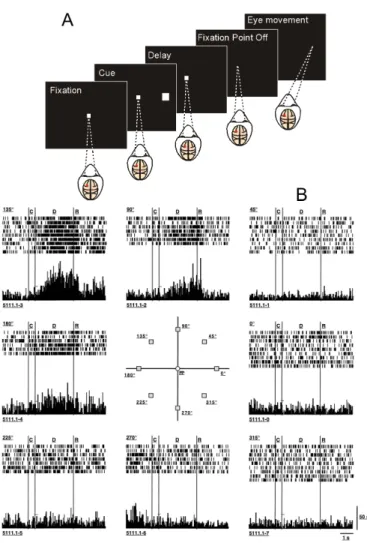
A well-known paradigm to study working memory is the
oculo-motor delayed-response (ODR) task. In this experiment, a subject is required to retain information of a visual cue location (direc-tional angle) throughout a delay period between the stimulus and
memory-guided behavioral response (Fig. 1.1A). In a version of this experiment, previously-implanted electrodes in the dorsolateral pre-frontal cortex record the activity of single neurons while the subject
Figure 1.1. (Cont.) This neuron exhibited the strongest directional delay period activity following presentation of the cue in the upper left quadrant (135 degrees). Adapted from Constantinidis and Wang (2004) and from Funahashi et al. (1989).
Rakic 1998; Constantinidis and Goldman-Rakic 2002; Constantini-dis and Wang 2004). These recordings show that certain neurons produce high levels of persistent firing during the delay period only
after the presentation of cues at a given angle (Fig. 1.1B). Assuming that different neurons are tuned to different locations, similarly to what happens in the primary visual cortex (Hubel and Wiesel 1968),
it is believed that a circuit in the PFC is capable of representing the continuous 360 degree space.
Based on these observations, theoretical models of spatial
work-ing memory have been developed. A basic concept in some of these models is the representation of persistent activity as dynamical at-tractors (Amari 1977; Amit 1995; Wang 2001). The term attractor
refers to a self-sustained and stable state of a dynamic system, such as a neural network (Wang 2013). A spatial working memory sys-tem can be either in the spontaneous state or in a continuum of
location-selective memory states. Each of these states is considered an attractor and transitions between them can be achieved through transient inputs.
The theoretical simulation of working memory can take into ac-count different levels of biophysical detail. The most basic prop-erties incorporated in some models are the overall firing rates of
and Wang 1998; Durstewitz et al. 1999; Itskov et al. 2011). While
these models can simulate the main input-output function of a cir-cuit, they do not provide a clear link to physiological data. In order to unravel interdependencies between cellular properties and neural
circuit dynamics, it is preferable to include more detailed biophysi-cal properties and neural spiking dynamics (Amit and Brunel 1997; Durstewitz et al. 2000a; Compte et al. 2000; Tegn´er et al. 2002;
Renart et al. 2003; Machens et al. 2005; Carter and Wang 2007; Hansel and Mato 2013). These type of models, which are more rel-evant to the work described in this dissertation, allow the modeler
to propose predictions that could be tested in electrophysiological experiments.
The model proposed by Compte et al. (2000) to simulate the
ODR task has a population of excitatory pyramidal cells figuratively arranged in a ring-like fashion. Each of these neurons is selectively-tuned to a subsection of the 360-degrees space (like the neuron in
Fig. 1.1B). A crucial feature is that neurons not only receive exter-nal inputs, but are also connected to each other with weights that are inversely dependent on their distance in the ring. When a cue
at a certain angle is presented, the group of neurons tuned to that angle elevates their firing rates and stimulate each other recipro-cally. This positive feedback through recurrent synaptic excitation
is primarily mediated by NMDA receptors (Wang 1999b) and gives rise to a localized persistent activity, creating an attractor memory state. The overall activity of the network is kept from escalating
of GABAergic, inhibitory interneurons.
The hypothesis that explains working memory maintenance through states with stable activity is not consensual. A study pro-posed that persistent activity is a costly consumption of
metaboli-cal energy and is not required for working memory (Mongillo et al. 2008). According to their modeling analysis, neocortical networks encode and maintain information through slow calcium-mediated
synaptic facilitation (Tsodyks and Markram 1997; Hempel et al. 2000). This mechanism consists in a state of increased neurotrans-mitter release that will allow the memory to be reactivated.
How-ever, this reactivation requires either an unrealistic readout stimulus or an artificial increase in the background input that effectively re-sults in persistent activity. Another study proposed that memory
maintenance relies on positive feedforward instead of feedback be-tween neurons, even in anatomically recurrent networks (Goldman 2009). This idea can explain some experimental data observations
but, on the other hand, the proof of its biological realism is still tenuous. Barak et al. (2013) recently compared three paramet-ric working memory models of a delayed vibrotactile
discrimina-tion task. The models were comprised of neurons with different degrees of tuning and dynamics: ranging from a system with prede-termined connectivity and stable neural representations (Machens
et al. 2005) to a random network that evolves according to the readout. They found that an intermediate model was the one that best simulated the data at their disposal.
activity (in line with Compte et al. (2000)) because it relies on
biophysically-realistic parameters, it can explain relevant electro-physiological data of working memory tasks, and allows to formu-late testable predictions at both cellular and circuit level. We also
analyze simpler models of pyramidal cells to determine more basic properties of these neurons. As a result, a recurring theme in this dissertation is the attempt to connect several levels of abstraction
within the PFC.
Chapter 2 deals with the robustness of the above mentioned spatial working memory model. A characteristic of this type of
networks, in its current state of description, is the accumulation of small deviations to the memory trace over time, which can result in a final memory representation that is distant from the original cue
stimulus. The cellular and synaptic properties of these networks are mostly homogeneous. The presence of heterogeneous properties can disrupt the continuous family of attractors, i.e. the network is
no longer able to encode all 360 degrees and quickly drifts to one of just a few privileged locations (Ben-Yishai et al. 1995; Tsodyks and Sejnowski 1995; Zhang 1996; Renart et al. 2003; Itskov et al.
2011). Finally, if a distractor signal is presented while the nework is encoding a previous stimulus, the remembered cue location may move towards the distractor. All these phenomena can have
nega-tive implications on the accuracy of the memory trace.
As stated before, the stability of the persistent activity dur-ing de delay period of workdur-ing memory is sustained by slow
this question, we tested how three cellular and synaptic biophysical
mechanisms affect the memory representation: short-term facilita-tion, depolarization-induced suppression of inhibition and calcium-activated inward currents. Briefly, short-term facilitation (STF) is
the increase in neurotransmitter release and postsynaptic response after repetitive stimulation of a synapse (Tsodyks and Markram 1997; Hempel et al. 2000). Depolarization-induced suppression of
inhibition (DSI) is a mechanisms that decreases the inhibition re-ceived by pyramidal cells when they are very active (Ohno-Shosaku et al. 2001; Wilson et al. 2001; Wilson and Nicoll 2001). A
calcium-activated inward current (ICAN) is independent of the membrane
potential and depolarize cells after calcium influx. All three mecha-nisms were found in the PFC, are activity-dependent, provide
pos-itive feedback and operate under a slow time course of activation. The incorporation of these mechanisms in the network increased the memory robustness and decreased the variability observed across
trials. On the other hand, their slow nature resulted in costlier transitions between memory and resting state, which decreased the flexibility of the overall system. We were able to explain these
ob-servations through a detailed analysis of the network model. The project described in chapter 3 was done in collaboration with experimentalists. One group carried out electrophysiological
recordings in monkeys, who were performing ODR tasks. At the same time, they applied pharmacological compounds that block or activate receptors and cellular machinery in specific regions of the
them to associate aspects of the monkey behavior with firing
ac-tivity of neurons and molecular pathways (Wang et al. 2007). A second group studied the modulation of slow oscillations by ionic currents (Shu et al. unpublished observations). Both studies
ob-served that the blockage of the channels that mediate H-current (IH) increases the excitability of pyramidal cells. This current is
ac-tivated by hyperpolarized membrane potentials and is inward
(de-polarizing) at sub-threshold potentials. In order to understand the basic mechanism of IH, we incorporated it in compartment neural
models. We demonstrate that the inactivation of this current alone
was not enough to produce the results observed with the pharma-cological blocking of IH channels in experiments. We also tested the
properties of a second potassium current (IM) in the single cell and
working memory models.
Finally, in chapter 3 we propose two alternative hypotheses that can explain the effects of blocking the IH channels. The first
con-sists of the interplay of IHwith an outward (hyperpolarizing) current
(Migliore and Migliore 2012). In the second, IH is present not only
in excitatory pyramidal cells as currently assumed, but also in
in-hibitory cells as reported in some studies (Maccaferri and McBain 1996; Kawaguchi and Kubota 1997; Lupica et al. 2001; Notomi and Shigemoto 2004; Aponte et al. 2006; Hughes et al. 2013).
We demonstrate how these two hypotheses are compatible with the experimental procedures and observations.
A main goal of this work is to contribute for the understanding
find-ing links between the cellular properties and the circuit dynamics
Chapter 2
A trade-off between
accuracy and flexibility in a
working memory circuit
endowed with slow
2.1
Abstract
Recent studies have led to the recognition that reverberation underlying mnemonic persistent activity must be slow, in order to ensure the stability of a working memory system and to give rise
to long neural transients capable of accumulation of information over time. Is the slower the underlying process, the better? To ad-dress this question, we investigated three biophysical mechanisms
operating on slow timescales, all are activity-dependent and promi-nently present in neurons of the prefrontal cortex: depolarization-induced suppression of inhibition (DSI), calcium-dependent
non-specific cationic current (ICAN) and short-term facilitation (STF).
Using a spiking network model for spatial working memory, we found that these slow biophysical processes enhance the accuracy
of memory representation by counteracting noise-induced drifts of a memory trace, heterogeneity-induced systematic loss of stored in-formation and distractors. Furthermore, the incorporation of DSI
and ICAN enlarges the range of network’s properties required for
memory states. However, when a progressively slower process dom-inates the network, it becomes increasingly more difficult to erase a
memory trace and reset the network by brief external inputs, which is required for proper function of a working memory circuit. We demonstrate this basic trade-off between accuracy and flexibility
quantitatively and provide an explanation of it using a state-space analysis. Our results support the scenario in which the NMDA-receptor dependent recurrent excitation is the workhorse for the
cellular processes contribute to the robustness of mnemonic
2.2
Introduction
Working memory is thought to be represented by persistent ac-tivity (Fuster and Alexander 1971; Gnadt and Andersen 1988; Fu-nahashi et al. 1989; Amit 1995; Goldman-Rakic 1995; Miller et al.
1996; Romo et al. 1999; Wang 2001; Major and Tank 2004). Such activity patterns are likely sustained by positive feedback processes in a neural circuit, but the precise mechanisms remain unresolved.
Computational models stressed the role of recurrent synaptic ex-citation (Amit 1995; Camperi and Wang 1998; Amit and Brunel 1997; Brunel and Wang 2001; Durstewitz et al. 2000b) that
de-pends on the NMDA receptors (Wang 1999b; Compte et al. 2000; Lim and Goldman 2013), a prediction supported by findings from a recent experiment (Wang et al. 2013).
Other synaptic and cellular process, present in the prefrontal cortex (PFC), are likely involved in mnemonic persistent activity, including short-term facilitation (STF) (Hempel et al. 2000; Wang
et al. 2006; Mongillo et al. 2008; Szatmary and Izhikevich 2010; Hansel and Mato 2013), depolarization-induced suppression of inhi-bition (DSI) (Carter and Wang 2007) and calcium-activated inward
currents (ICAN) (Tegn´er et al. 2002; Egorov et al. 2002; Frans´en
et al. 2006; Yoshida and Hasselmo 2009; Kulkarni et al. 2011; Kalmbach et al. 2013). STF and ICAN provide feedback excitation,
whereas DSI is a disinhibition process. All are activity-dependent, thus become selective for neurons that show elevated persistent ac-tivity. Furthermore, these mechanisms operate with biophysical
synaptic excitation. Therefore, the long-standing question
(Ma-jor and Tank 2004) has gained urgency: what may be the relative contributions to working memory function of these slow synaptic and cellular processes versus the recurrent network mechanism?
We analyzed the role of slow biophysical processes in mnemonic persistent activity, using a biologically-based continuous spiking cir-cuit model for spatial working memory. This model system is
en-dowed with a resting state and a continuum of spatially tuned per-sistent activity patterns (“bump attractors”) for memory storage of an analog quantity such as spatial location (Camperi and Wang
1998; Compte et al. 2000; Gutkin et al. 2001; Laing and Chow 2001; Renart et al. 2003; Carter and Wang 2007; Wei et al. 2012; Murray et al. 2012). During a mnemonic delay period, a bump
attractor drifts over time (Compte et al. 2000; Carter and Wang 2007; Murray et al. 2012), resulting in random deviations of the memory away from the to-be-remembered sensory cue.
Addition-ally, heterogeneity in single neurons disrupts the continuous family of attractors (Ben-Yishai et al. 1995; Tsodyks and Sejnowski 1995; Zhang 1996), leading to systematic drifts of memory trace (Renart
et al. 2003; Itskov et al. 2011). Furthermore, the system may be perturbed by external distractor stimuli. Interestingly, we found that while STF, DSI and ICAN enhance the accuracy of a memory
trace, they hinder rapid memory erasure and network reset. The latter is not functionally desirable, since behavior demands that brief transient inputs should be sufficient to switch a working
(Compte et al. 2000; Gutkin et al. 2001). Therefore, our study
2.3
Materials and Methods
In an oculomotor delayed-response (ODR) task, the sensory stimulus is a visual cue and the motor response is a saccade to the cued location. A subject is briefly shown a visual cue that must
be remembered during a delay period of a few seconds. This mem-ory is subsequently used to perform a memmem-ory-guided behavioral response (the saccade). During the delay period, many neurons in
the dorsolateral PFC show high persistent activity that is spatially selective (Funahashi et al. 1989). The present work uses a spiking network model for the ODR task that has been tested thoroughly
(Compte et al. 2000; Carter and Wang 2007; Wei et al. 2012; Murray et al. 2012). The parameters were modified starting with the original “control parameter set” in Compte et al. (2000). The
model consists of a population of excitatory pyramidal cells and a population of inhibitory interneurons. Pyramidal cells are arranged in a ring-like fashion and labeled by their preferred cue direction,
from 0 to 360 degrees. A schematic of the network structure is shown in Fig. 2.1A.
2.3.1
Single neuron model
Both pyramidal cells and interneurons are modeled as leaky in-tegrate and fire units (Tuckwell 1988). Each type of cell is char-acterized by total capacitance Cm, total leak conductance gL, leak
reversal potential VL, threshold potential Vth, reset potential Vres
are Cm = 0.5 nF, gL = 25 nS, VL = −70 mV, Vth =−50 mV, Vres
= −60 mV, and τref = 2 ms for pyramidal cells; and Cm = 0.2 nF,
gL = 20 nS, VL = −70 mV, Vth = −50 mV, Vres =−60 mV, and
τref = 1 ms for interneurons. The subthreshold membrane potential,
V(t), follows:
Cm
dV(t)
dt =−gL(V(t)−VL)−Isyn(t)
where Isyn(t) is the total synaptic current to the cell.
2.3.2
Synaptic interactions
The network consists of NE = 2048 pyramidal cells and NI =
512 inhibitory interneurons. Neurons receive recurrent, background, and external inputs. Excitatory synaptic currents are mediated by
2-amino-3-(3-hydroxy-5-methyl-isoxazol-4-yl) propanoic acid recep-tors (AMPARs) and NMDARs, and inhibitory synaptic currents are mediated by γ-aminobutyric acid type A receptors (GABAARs).
The total synaptic current to each neuron is
Isyn =IN M DA+IAM P A+IGABA+Iext
where Iext delivers stimulus input to pyramidal cells. The dynamics
of synaptic currents for neuron i follow:
Ii,AM P A = (Vi−VE) X
j
Ii,N M DA = (Vi−VE)
P
j gji,N M DA sj,N M DA
1 + [M g2+]exp(−0.062V
i/mV)/3.57
Ii,GABA = (Vi−VI) X
j
gji,GABA sj,GABA
where VE= 0 mV and VI=−70 mV and gji,syndenotes the synaptic
conductance strength on neuron i from neuron j. NMDAR-mediated
currents exhibit voltage dependence controlled by the extracellular magnesium concentration [Mg2+] = 1 mM (Jahr and Stevens 1990). Given a spike train {tk} in the presynaptic neuron j, the gating
variables sj,AMPA and sj,GABA for AMPAR- and GABAR-mediated
currents, respectively, are modeled as:
ds dt =
X
k
δ(t−tk)−
s τs
The gating variable sj,NMDA for NMDAR-mediated current is
modeled as:
dx dt =αx
X
k
δ(t−tk)−
x τx
ds
dt =αsx(1−s)− s τS
decay time constant τs is 2 ms for AMPA, 10 ms for GABA, and
100 ms for NMDA. For simplicity, background inputs are mediated entirely by AMPARs, and recurrent excitatory inputs are mediated entirely by NMDARs, as they are critical for the stability of
persis-tent activity (Wang 1999b; Compte et al. 2000; Wang et al. 2013). All cells receive background excitatory inputs from other cortical areas. This overall external input is modeled as uncorrelated
Pois-son spike trains to each neuron at a rate ofνext = 1800 Hz per cell,
with AMPAR maximal conductances of 3.1 nS on pyramidal cells and 2.38 nS on interneurons.
2.3.3
Network connectivity
As stated above, pyramidal cells are organized in a ring architec-ture and are tuned to the angular location on a circle (0–360◦
, Fig.
2.1A), with uniform distribution of their preferred angles. The net-work structure follows a columnar architecture, such that pyramidal cells with similar stimulus selectivity are preferentially connected to
each other. The synaptic conductance on neuron i from neuron j, gji,syn= W(θj−θi)Gsyn, where θi is the preferred angle of neuron i,
and W(θj−θi) is the connectivity profile normalized such that:
1 360◦
Z 360◦
0◦
W(θ)dθ = 1
For pyramidal-to-pyramidal connections, W(θj−θi) = J
−
+J+exp[−
(θj − θi)2/2σ2]. We use J+ = 1.62 and σ = 14.4
◦
conduc-tance strengths are given by GEE = 0.381 nS, GEI = 0.292 nS, GIE
= 1.336 nS, GII = 1.024 nS.
2.3.4
Stimulus
Inputs are modeled as injected current with a Gaussian profile,
I(θ) = I0 exp[−(θ−θc)2/2σI2], where the maximum current I0 = 200
pA, except otherwise noted. θc is the stimulus location, and the
width parameter σI= 18
◦
.
2.3.5
Slow calcium-dependent nonspecific cationic
current
ICANcan trigger a sustained depolarization outlasting the
stimu-lus for several seconds (Haj-Dahmane and Andrade 1998; Str¨ubing et al. 2001; Egorov et al. 2002; Tegn´er et al. 2002). The
activa-tion of this current requires a rise in intracellular calcium. In some simulations (results in Fig. 2.2, 2.4, 2.5), ICAN was added to the
network model (described above) according to the following:
ICAN =−gCANm2CAN(V −ECAN)
dmCAN
dt =φCAN ×
m∞([Ca
2+])−m
CAN
τCAN([Ca2+]) !
m∞([Ca
2+
]) = α[Ca
2+]2
τCAN([Ca2+]) =
1
α[Ca2+]2+β
with gCAN = 1.5 nS, ECAN =−20 mV,β = 0.002 ms
−1
,α = 0.0056
[(ms(µM)2]−1
. φCAN is used to adjust the effective time constant of
ICAN, without changing the steady state levels of activity.
Calcium influx to pyramidal cells is triggered by spikes and obeys
first-order kinetics as follows (Liu and Wang 2001):
d[Ca2+]
dt =αCa
X
i
δ(t−ti)−
[Ca2+]
τCa
When an action potential fires (at time ti), [Ca2+] is incremented
by αCa (0.2 µM). The calcium concentration decays back to zero
exponentially, with a time constant τCa (240 ms).
2.3.6
Depolarization-induced suppression of
in-hibition
Depolarization-induced suppression of inhibition is detected in various regions of the brain (Llano et al. 1991; Pitler and Alger 1992; Trettel and Levine 2003). DSI is dependent on
endocannabi-noids that are released by active pyramidal cells, triggered by cal-cium influx (Ohno-Shosaku et al. 2001; Wilson et al. 2001; Wil-son and Nicoll 2001). These endogenous cannabinoids retrogradely
(CCK) (Marsicano and Lutz 1999; Katona et al. 1999). The
acti-vation of CB1R results in the suppression of transmitter release to postsynaptic pyramidal cells.
DSI was added to the network model (Fig. 2.3, 2.4, 2.5, 2.9) as
previously described in Carter and Wang (2007) and the same pa-rameters were used, unless noted otherwise. Briefly, the inhibitory synaptic conductance gGABA to a pyramidal cell is multiplied by a
factorD, which is proportional to the fraction of inhibitory synapses that are sensitive to cannabinoid and their presynaptic release prob-ability. D varies between 0 and 1. There is no DSI effect if D is
set to 1. DSI is the fractional reduction in inhibitory event size or frequency. The dynamics of D are described by the following equation:
dD
dt =φD×
1−D τD
−βD ×[Ca2+]×(D−Dmin) !
where [Ca2+] represents the intracellular calcium concentration in
the pyramidal cell and has the same kinetics as ICAN. When [Ca2+]
accumulates, D decreases with a rate controlled by βD (1.66×10
−5
(µM ms)−1
), leading to disinhibition. Dis bounded below at Dmin,
which determines the maximum disinhibition and biophysically
cor-responds to the maximum number of synapses that are cannabinoid sensitive multiplied by the maximal reduction in release probability at each synapse due to DSI. Unless stated otherwise, Dmin was set to
time constant τD (16.7 s). The factor φD accounts for temperature
sensitivity and was used to adjust the effective time constant of DSI without changing the steady state levels of activity.
2.3.7
Short-term facilitation
In simulations where we incorporated short-term facilitation (re-sults in Fig. 2.6, 2.7, 2.8), only the recurrent excitatory synapses are facilitatory. To implement short-term facilitation, the
parame-ter αx is multiplied by F, which is the facilitation factor and obeys
the following dynamical equation (Matveev and Wang 2000):
dF dt =αF
X
i
δ(t−ti)(1−F)−
F τF
The parameter αF controls the facilitation potency and was set
at 0.6. Starting a paired-pulse facilitation simulation with F0 = 0,
it is possible to demonstrate that, for the first two spikes, with an inter-spike interval 1/r:
F1 = 1−e
−αF
; F2 = 1−(1−F
−
)e−αF
; F2
F1
= 1 +erτF−1−αF
2.3.8
Parameter change
A key manipulation in our study is to gradually change the timescale of a biophysical process. For ICAN, we varied the
pa-rameter φCAN, which scales the speed of the channel kinetics
mCAN. Similarly, we varied the parameter φD to systematically
change the time constant of DSI while preserving the average level of the activity variableD. Unlike ICAN or DSI, for STF the activity
variable F undergoes discrete jumps in time and what matters is
its value immediately after each jump due to a presynaptic spike, rather than the temporal average. For this reason, we varied τF
directly (see Results for more details).
When a slow mechanism is added to a network model, the overall level of activity of the excitatory population changes significantly, to a degree correlated to the nature and strength of the mechanism.
This changes the shape of a population activity pattern and may even disrupt its stability. For this reason, when ICAN, DSI or STF,
were present in the model, GEE was adjusted from 0.381 to 0.378,
0.379 or 0.383 nS, respectively. This way, the network maintained consistently a fixed steady-state activity across all simulations, al-lowing a fair comparison between scenarios.
2.3.9
Analysis of simulation data
To determine the remembered cue location at any given time, we used the population vector, which is a simple readout of the
peak location of a spatially tuned persistent activity pattern (Geor-gopoulos et al. 1982).
The minimum time to shutdown (tSHUT,MIN), in Figs. 2E, 3E
and 6A was determined as follows. For each time constant (τ), a range of shutdown pulse durations (tSHUT) was considered. For
inhibitory input current lasting for tSHUT was applied when the
network was in a bump attractor state. At the end of each simulated trial (seconds after pulse offset), whether the bump state was still present or not was judged through the maximum of the firing rate
profile. If more than 95% of simulations of a set yielded successful shutdowns, the corresponding pulse duration was accepted. Finally, for each τ, tSHUT,MIN was chosen as the minimum of those accepted
pulse durations.
2.3.10
Bistability analysis and bifurcation
dia-grams
To plot the bifurcation diagrams in Fig. 2.4 and Fig. 2.7B, we ran simulations across a range of values for the varied parameter
(GEE and F profile, respectively) with and without cue input and
measured the firing rate during the delay. The maximum firing rate across the network indicated whether the system had evolved to the
memory state (typically >20 Hz) or remained at the baseline state (<5 Hz).
2.3.11
Simulation method
The model was implemented in python in the Brian simulator (Goodman and Brette 2009). The equations were integrated us-ing a second-order Runge-Kutta algorithm (timestep = 0.02 ms).
2.4
Results
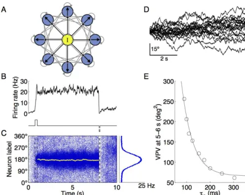
Our working memory model was designed for an ODR task, which proceeds from cue (angle) presentation, to a delay period and memory-guided behavioral response. The cue stimulus activates a
group of pyramidal neurons with preferred directions around the sensory cue (first step current, lower panel of Fig. 2.1B). If the firing rate of this subpopulation of neurons is sufficiently elevated
and mutual excitation among them is strong enough, reverberation can give rise to self-sustained persistent activity after the stimulus offset (plateau in Fig. 2.1B, upper panel) (Wang 2001). At the end
of the delay, a negative input is applied to all excitatory neurons in the network (Fig. 2.1B, lower panel, second step current). This shutdown pulse should be sufficient long to switch the network back
to the baseline resting state.
The spatiotemporal activity pattern of the network model is shown in Fig. 2.1C (left panel). The memory trace is encoded as
a population activity pattern that persists during the delay period. The spatial profile of the bump state, corresponding to the activity during the delay period, has a typical Gaussian shape (Fig. 2.1C,
right panel). The population vector (shown in yellow) quantifies the peak location of the bump attractor as the internal representation of the sensory cue at any instant. In this example, the
remem-bered cue location fluctuates slightly around the initial cue (180◦
) and remains reasonably close to it at the end of the delay period. Consequently, in this trial, the PFC circuit model successfully
readout.
2.4.1
Dominant time constant determines
mem-ory accuracy
The analysis of simulations across trials reveals that the remem-bered cue (the population vector) as encoded by the network activ-ity pattern displays random drifts over time (Fig. 2.1D). This is
because the system is endowed with a continuous family of bump attractors, each for a directional angle as an analog quantity. Dur-ing a delay period, irregular neural activity leads to random
shift-ing of the network state among those bump states. At the end of a trial, if the drifts have grown over time greatly, the remembered cue location could be located significantly away from the sensory cue
angle. This is shown in some trials of Fig. 2.1D, with deviations of more than 20 degrees. These simulations therefore show a relatively low accuracy of memory representation, which implies poor
perfor-mance. Note that, across trials, the average of random drifts is zero (i.e. there is no systematic drift), whereas the variance increases roughly linearly over time (Camperi and Wang 1998; Compte et al.
2000; Renart et al. 2003; Carter and Wang 2007). This variance of population vector (VPV) quantifies the magnitude of random drifts, which we used as a measure to assess the network’s function:
the smaller is the VPV, the more accurate is the representation of a memory trace and the better is the behavioral performance.
Figure 2.1. (Cont.) The yellow line is the population vector, which traces the peak of the bell-shaped persistent activity pattern (bump attractor) as the internal representation of the cue location. Right panel: population firing profile, averaged over the delay period. D, remembered cue as measured by the population vector from 20 sample trials with the same cue location. The memory traces drift away from the initial cue during the delay, the variance of population vector (VPV) across trials quantifies this deviation so that the smaller is the VPV, the more accurate is the memory readout. E, drift magnitude at 5–7 s of the delay period, as measured by VPV (N = 500 trials), is plotted as a function of the time constant of the NMDA receptor mediated synaptic excitation
τS. The VPV decreases steeply with increasing τS; the fitting line is an exponential function for ease of eye inspection.
activity is stabilized by slow reverberation mediated by the NMDA
receptors at the recurrent excitatory synapses (Wang 1999b). The NMDA receptor dependent synaptic current has a time constant
τS on the order of 50–100 milliseconds. We hypothesized that, the
longer is τS, the more robust will be the memory trace. To test
this possibility, we gradually varied the value of the NMDAR decay time constant, and measured the variance of the remembered cue
location during a delay interval across hundreds of trials. The VPV decreases inversely with increasingτS(Fig. 2.1E). The VPV is 206.2
deg2 with τ
S equal to 100 ms. A substantial reduction in the VPV
is observed whenτSis increased three-fold (300 ms,σ2 = 61.5 deg2).
This result serves as a proof-of-principle of the idea that extending the dominant time constants decreases random drifts of persistent
activity and improves the accuracy of memory representation. In the following, we will consider three slow, biophysically-plausible
memory function.
2.4.2
I
CANincreases memory stability but
de-creases system flexibility
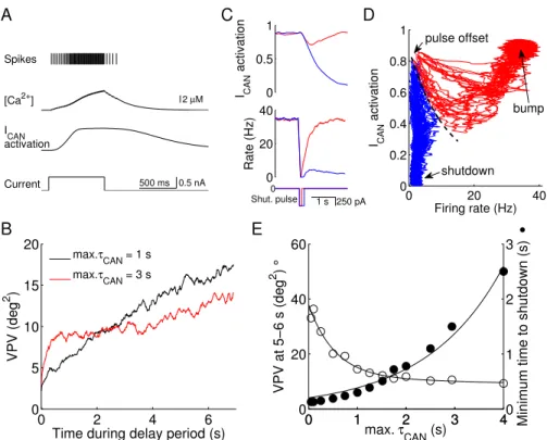
Fig. 2.2A shows the spiking activity of an integrate-and-fire sin-gle neuron model endowed with the slow inward current ICAN. An
external current results in action potentials that induce calcium
in-flux, which in turn activates ICAN. After the stimulus offset, the
activation of ICAN decays slowly, which allows it to provide positive
feedback that is enough to trigger a few additional spikes
(after-discharges). It is worth noting that we assumed that ICAN is not
sufficiently strong to produce stable persistent activity in an iso-lated neuron (Fig. 2.2A), and we were interested in examining the
contribution of the activity-dependent ICAN in single neurons to the
maintenance of a persistent firing pattern in a recurrent working memory circuit.
We ran simulations with ICAN present in excitatory cells and
measured the VPV of the delay-period memory trace across trials. We tested two different values of max τCAN that lie within the
ex-perimentally measured range (Partridge and Valenzuela 1999; Faber et al. 2006; Gross et al. 2009; Sidiropoulou et al. 2009). With a shorter maxτCAN (1 second), the VPV increases quasi-linearly with
time (Fig. 2.2B, black curve). By contrast, with maxτCAN = 3
Spikes
[Ca2+] 2 µM
ICAN activation
Current 500 ms 0.5 nA
0 2 4 6
0 5 10 15 20
Time during delay period (s)
VPV (deg
2 )
max.τCAN = 1 s max.τ
CAN = 3 s
0 0.5 1 ICAN activation 0 20 40 Rate (Hz) 0
Shut. pulse 1 s 250 pA 0 20 40
0 0.2 0.4 0.6 0.8 1
Firing rate (Hz)
ICAN
activation
pulse offset
shutdown bump
0 1 2 3 4
0 20 40 60
max. τ
CAN (s)
VPV at 5−6 s (deg
2 )
°
0 1 2 3 40
1 2 3
Minimum time to shutdown (s)
•
A
B
C D
E
Figure 2.2. (Cont.) the two conditions (the same color scheme, N = 10 trials). With 100 ms, ICAN activation decays by a small amount but immediately increases after the shutdown input is over, providing the necessary positive feedback for the return of the high-firing mem-ory state. After a longer shutdown pulse (200 ms) the activation decays to such an extent that ultimately leads to the resting state. D, state space analysis with the population rate and the ICAN activation shown in C plotted against each other in phase space. Each trajectory corre-sponds to a trial and starts immediately at the shutdown pulse offset. Red trajectories evolve to the bump attractor; blue proceed to shutdown (resting state). There is a clear diagonal boundary that separates the two attractors (dashed black curve), suggesting the presence of an unsta-ble manifold. E, trade-off between decrease in variance of remembered cue location (VPV) and minimum time to shutdown (tSHUT,MIN), with increasing max τCAN. Open circles were determined as in Fig. 2.1E, with maxτCAN between 50 ms and 4 s. Filled circles express tSHUT,MIN (see Methods) (N = 500 trials). The two data sets are fitted as a sum of two exponentials (VPV) or as a simple exponential (tSHUT,MIN). A compromise corresponds to an optimal value of max τCAN ≃1.5 sec.
2.2B, red curve). A possible explanation for the initial rise in drifts
(which is not visible for max τCAN = 1 s) is that with a slower time
constant, the ICAN takes longer to be activated and does not
pro-vide robustness against drifts as promptly. The crossover between
the two time courses shows that shorterτCAN is more advantageous
for shorter delay periods, whereas slower τCAN increases memory
accuracy in longer delays.
The increase in memory robustness provided by ICAN, however,
is just one of the effects this current has in the working memory model; the incorporation of a slow mechanism also makes it harder
which completely silences the network. If this pulse is not
suffi-ciently long, the network returns to the memory state, with high ICAN activation and elevated neural firing (Fig. 2.2C, red traces,
100 ms pulse). With a longer shutdown pulse, in contrast, ICAN
de-activates to a sufficiently low level that does not allow the return of the high spiking activity and the network is switched off from a bump attractor state (Fig. 2.2C, blue traces, 200 ms pulse).
To further demonstrate the role of ICAN in the memory erasure
process, we recorded simultaneously the activation variable of this current and the firing rate of the network and plotted them in a
state space, for several trials (Fig. 2.2D). We only recorded neu-rons around the cue location and in simulations that successfully maintained a memory during the delay. All trajectories initiate
immediately after the shutdown (“pulse offset”). There is a clear divergence between two kinds of traces: in a given trial the sys-tem’s trajectory either revert back to the memory state (red traces,
“bump”) or decays to the resting state (blue traces, “shutdown”). A boundary (dashed line) separates the regions of attraction of the two states. This result shows that even though a relative weak ICAN
(which by itself does not yield persistent activity in a single neuron (Fig. 2.2A)) does not determine whether a network generates per-sistent activity per se, it can have a remarkably significant impact
on the network’s behavior.
Therefore, ICANstabilizes the memory trace by reducing memory
drifts over time; at the same time it renders the network less flexible,
This accuracy-flexibility trade-off was demonstrated more explicitly
when we varied maxτCAN parametrically (Fig. 2.2E). The increase
in maxτCAN decreases the variance of the remembered cue location
(the VPV, open circles) but increases the minimum time required to
shutdown the network (filled circles). A “sweet spot” corresponds to the crossover point of the two curves (maxτCAN = 1–2 s), where the
VPV is close to the minimum while tSHUT,MIN is reasonably short
(a few hundreds of milliseconds). However, an optimal compromise for a working memory circuit could be different depending on the functional demand that may emphasize either accuracy or flexibility.
2.4.3
DSI shows trade-off between accuracy and
flexibility
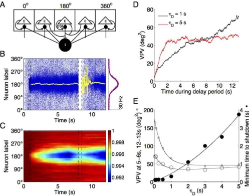
DSI is a cannabinoid-dependent process through which synaptic inhibition to excitatory neurons is reduced by the magnitude of DSI, which in turn is controlled by the activity of the same E cells (Fig.
2.3A). Thus, for each neuron, a higher level of excitation leads to a weaker inhibition, resulting in an effective positive feedback.
The cells that are most active during an ODR task are those
around the peak of the bump activity pattern (Fig. 2.3B, cue lo-cation at 180 deg). Therefore, due to its activity-dependence, DSI is the strongest in this group as well. This is depicted in the blue
Figure 2.3. Trade-off between memory accuracy and flexibility with DSI.A, schematic of network model of spatial working memory endowed with DSI. This mechanism is implemented as a cell-specific reduction in inhibitory input conductance. Adapted from Carter and Wang (2007).
B, left panel: spatiotemporal pattern of excitatory cells endowed with DSI (τD = 5 s). Cue was presented at 180 degrees during the 0.75–1 s interval. A shutdown pulse of 500 ms was applied at 8 s. The yellow lines represent the remembered cue location during delay and after shutdown pulse. Right panel: population firing profiles, averaged over the delay period (blue) or over the last second of the simulation (red), showing that the bump state survives the shutdown input and the memory trace is not erased. C, spatiotemporal representation of the activation variable
D of DSI (inverted scale, 1 means no DSI) of the same trial. Only the
Figure 2.3. (Cont.) D, the accuracy-flexibility trade-off with DSI. The variance of the remembered cue location (VPV) during the delay period with effective τD of 1 (black trace) and 5 (red trace) seconds (N = 500 trials). In the former scenario, the VPV keeps increasing almost linearly. By contrast, in the latter, it stabilizes after an initial period of 2 seconds.
E, trade-off between decrease in the VPV (open symbols) and tSHUT,MIN (closed circles), as τD is increased from 50 ms to 5 s (N = 500 trials). The VPV was determined during two intervals of the delay period: 5–6 s (open circles, same as Fig. 2.1E and Fig. 2.2E) or 12–13 s (open squares). The data sets were fitted by solid curves for eye inspection.
of the sensory cue, thereby reducing spontaneous drift and
stabiliz-ing the neural representation of the remembered cue (Carter and Wang 2007).
In order to quantify this DSI-induced effect, we determined the variance of the remembered cue location (the VPV). We proceeded in a similar way as described above, and the results are remarkably
similar. When DSI is controlled by a long time constant (5 s), there is an initial period of rise in drifts (Fig. 2.3D, red trace, first 2 sec-onds of delay), similar to a network without DSI. However, once the
mechanism is fully activated (with a longer delay), the VPV does not grow any longer, reaching a plateau instead. For the shorter time constant (1 s), the variance increases almost monotonically
(Fig. 2.3D, black trace).
Another notable feature in the particular sample trial of Fig, 3B-C is the persistence of the inhibition suppression. Given the slow
nature of its decay (τD = 5 s), DSI does not have sufficient time
restart the memory bump at approximately the same angle, without
a new cue presentation (Fig. 2.3B, right panel, red profile).
As shown in Fig. 2.3E (open symbols), the duration of a step current required to reset the network increases dramatically with
τD (0.5 s: tSHUT,MIN = 130 ms; 5 s: tSHUT,MIN = 3.75 s). On the
other hand, the variance of the remembered cue location, the VPV, is larger in simulations with shortτDand decreases for progressively
longerτD, reaching a low plateau for τD larger than 1 s. Compared
to the control (Fig. 2.1E with τS = 100 ms, VPV = 206 deg2) with
the same delay period duration of 5–6 seconds, a circuit endowed
with DSI displays a smaller variance of drifts overall (VPV = 70.5 deg2 with τ
D = 50 ms, 42.1 deg2 with τD = 5 s) (Fig. 2.3E, open
circles). With larger delays (12–13 s), the simulations show higher
variance due to the accumulation of drifts over a longer time (Fig. 2.3E, open squares). However, in agreement with the traces in Fig. 2.3D, this relative increase of VPV due to longer delays is mostly
observed for shorter τD and is minimal for longer ones. Therefore,
our analysis shows a trade-off between the ease of shutdown and memory accuracy which is the same with DSI as that observed
with ICAN.
2.4.4
I
CANand DSI enhance the robustness of
working memory
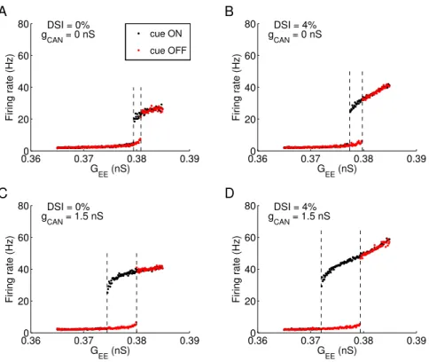
we quantified the network states in a bifurcation diagram (Fig.4).
The desirable behavior corresponds to multistability (the coexis-tence of a resting state with a low rate and memory states with a high rate), which is realized only in a range of the strength for the
recurrent connection between excitatory neurons (GEE). When DSI
and ICAN are not present, the multistability range (bounded by two
dashed lines) is restricted to a narrow range around GEE = 0.38 nS
(Fig. 2.4A). If either DSI or ICAN is incorporated (same as in
pre-vious simulations: 4% DSI or gCAN = 1.5 nS), the lower boundary
of the range is extended to smaller GEE values (Fig. 2.4B-C). The
maximum broadening effect occurs when both slow mechanisms are present (Fig. 2.4D). This is readily understood: with the help of DSI and ICAN, less recurrent excitation is required to generate
per-sistent activity.
A second noteworthy feature of Fig. 2.4 is that the slow bio-physical mechanisms increase the firing rate of memory states while
that of the resting state remains roughly the same. This is because DSI and ICAN are activity-dependent, therefore minimal in the
low-firing spontaneous activity but significant in the high-rate memory
states. This leads to a larger separation between the resting and memory states. Consequently, a random fluctuation in spontaneous spiking activity will be less prone to give rise to a “false” memory,
and the network function is more reliable.
To conclude, ICANand DSI are beneficiary to the system by
mak-ing it less sensitive to variations of the network properties (such as
0.360 0.37 0.38 0.39 20 40 60 80 G EE (nS)
Firing rate (Hz)
DSI = 0% g
CAN = 0 nS cue ON
cue OFF
0.360 0.37 0.38 0.39 20
40 60 80
G EE (nS)
Firing rate (Hz)
DSI = 4% g
CAN = 0 nS
0.360 0.37 0.38 0.39 20
40 60 80
G EE (nS)
Firing rate (Hz)
DSI = 0% gCAN = 1.5 nS
0.360 0.37 0.38 0.39 20
40 60 80
G EE (nS)
Firing rate (Hz)
DSI = 4% gCAN = 1.5 nS
A B
C D
Figure 2.4. Multistability analysis of the working memory model as a dynamical system reveals that ICAN and DSI increase the robustness of memory function. Simulations were ran with (black dots) or without (red dots) cue presentation, for a range of recurrent excitatory conductance (GEE) values. The maximum firing rate among all excitatory cells, at the end of the delay period, is either low (2–6 Hz) corresponding to the resting state or higher than 20 Hz corresponding to a memory sate. The resulting state diagram is shown for the control network without slow mechanisms (A), with only DSI (B) or ICAN(C) or both (D). The range of GEE values for multi stability is delimited by 2 vertical dashed lines. The presence of DSI (B) and ICAN(C) alone increased the multistability range and also the firing rate separation between memory and resting states. These effects are larger when both mechanisms are combined (D).
2.4.5
I
CANand DSI counteract heterogeneity
Network models endowed with a continuum of attractor states
require that its constituent neurons have identical properties (Ben-Yishai et al. 1995). Under this condition, if a localized pattern of activity is spatially displaced, it will lead to another identical
pattern centered at the new location. However, any neural net-work shows a certain degree of variability across cells (Marder and Goaillard 2006). Can DSI and ICAN remedy the system’s
vulnera-bility to heterogeneity, by virtue of creating a privileged location in the network in an activity-dependent manner? To investigate this question, we implemented a modest amount of heterogeneity, by
assuming that the leak potential VL varies from cell to cell
accord-ing to a Gaussian distribution (mean VL =−70 mV and standard
deviation SD(VL) = 1 mV).
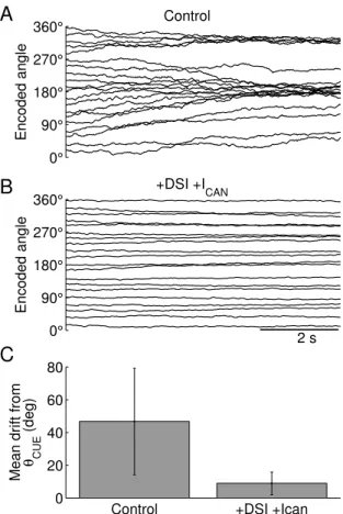
Across a large number of trials, the input cues are presented at 20 angle locations equally distributed along the 360 degrees of a circle. When both mechanisms are absent, the remembered cue
locations display systematic drifts and, as previously reported (Re-nart et al. 2003), tend to converge to a few privileged locations (Fig. 2.5A, θ = 180 and 320 deg). These locations are determined
by the heterogeneous distribution of the cellular excitability across the network, which disrupts the continuous family of bump attrac-tors. The mean drift from the cue location is minimal in networks
with DSI and ICAN (8.9 ± 6.9 deg) and significantly different
(2-sample t-test, p = 5×10−110
) from that of the control network (46.7
0° 90° 180° 270° 360° Time (s) Encoded angle Control 0° 90° 180° 270° 360° Time (s) Encoded angle +DSI +I CAN 2 s
Control +DSI +Ican
0 20 40 60 80
Mean drift from θ
CUE
(deg)
A
B
C
Intuitively, when DSI and ICAN are included, the remembered
cue locations show much smaller drifts (Fig. 2.5B). Both mecha-nisms are activity-dependent, so they create a privileged location in the network that “traps” a bump attractor encoding the sensory
cue. These slow mechanisms are powerful enough to overcome the disrupting effect of heterogeneity.
2.4.6
Short-term facilitation increases memory
accuracy
Finally, we considered the effect of short-term facilitation (STF)
in our working memory model. STF shares similar features with ICAN and DSI, namely activity-dependence, positive feedback and
slow time course of activation (Zucker 1989; Tsodyks and Markram
1997; Fisher et al. 1997; Abbott and Regehr 2004). It is espe-cially prevalent in excitatory synapses between pyramidal cells in the frontal cortex (Hempel et al. 2000; Wang et al. 2006).
The implementation of STF in the model reduced random drifts of the memory trace during the delay. Compared to the control network (Fig. 2.1E, τS = 100 ms, VPV = 206 deg2) the variance of
the remembered cue location was lower for any τF (Fig. 2.6A, open
circles, VPV ranges 71–126 deg2). However, contrary to DSI and
ICAN, the VPV increased rather than decreases with longerτF. This