R E S E A R C H
Open Access
Clinical, hematological and biochemical
alterations in hamster (
Mesocricetus
auratus
) experimentally infected with
Leishmania infantum
through different
routes of inoculation
Nádia das Dores Moreira
1,2, Juliana Vitoriano-Souza
1, Bruno Mendes Roatt
1, Paula Melo de Abreu Vieira
3,
Wendel Coura-Vital
2, Jamille Mirelle de Oliveira Cardoso
1, Mariana Trevisan Rezende
1, Henrique Gama Ker
2,
Rodolfo Cordeiro Giunchetti
4, Claudia Martins Carneiro
1,2and Alexandre Barbosa Reis
1,2*Abstract
Background:Leishmaniasis remains among the most important parasitic diseases in the developing world and visceral leishmaniasis (VL) is the most fatal. The hamsterMesocricetus auratusis a susceptible model for the characterization of the disease, since infection of hamsters withL. infantumreproduces the clinical and pathological features of human VL. In this context, it provides a unique opportunity to study VL in its active form. The main goal of this study was to evaluate the clinical, biochemical, and hematological changes in male hamsters infected through different routes and strains ofL. infantum.
Methods:In the current study, hamsters (Mesocricetus auratus) were infected with theL. infantumstrains (WHO/MHOM/BR/74/PP75 and MCAN/BR/2008/OP46) by intradermal, intraperitoneal and intracardiac routes. The animals were monitored for a nine month follow-up period.
Results:The hamsters showed clinical signs similar to those observed in classical canine and human symptomatic VL, including splenomegaly, severe weight loss, anemia, and leucopenia. Therefore the OP46 strain was more infective, clinical signs were more frequent and more exacerbated in IC group with 80 to 100 % of the animals showing splenomegaly, in the last month infection. Additionally, desquamation, hair loss and external mucocutaneous lesions and ulcers localized in the snout, accompanied by swelling of the paws in all animals, were observed. Consequently, the animals presented severe weight loss/cachexia, hunched posture, an inability to eat or drink, and non-responsiveness to external stimuli. Furthermore, regardless of strain, route of inoculum and time assessed, the animals showed renal and hepatic alterations, with increased serum levels of urea and creatinine as well as elevated serum levels of aspartate aminotransferase and alanine aminotransferase.
(Continued on next page)
* Correspondence:alexreisufop@gmail.com
1Laboratório de Imunopatologia, Núcleo de Pesquisas em Ciências
Biológicas/NUPEB, Universidade Federal de Ouro Preto, Ouro Preto, Minas Gerais, Brasil
2Laboratório de Pesquisas Clínicas, Departamento de Análises Clínicas, Escola
de Farmácia, Universidade Federal de Ouro Preto, Ouro Preto, Minas Gerais, Brasil
Full list of author information is available at the end of the article
(Continued from previous page)
Conclusions:These results strongly suggest that the inoculation through the intracardiac route resulted in a higher severity among infections, especially in the sixth and ninth month after infection via intracardiac, exhibited clinical manifestations and biochemical/hematological findings similar to human visceral leishmaniasis. Therefore, we suggest that this route must be preferentially used in experimental infections for pathogenesis studies of VL in the hamster model.
Keywords:Hamster,Mesocricetus auratus,Leishmania infantum, Experimental infection, Hematological and biochemical alterations
Background
Human visceral leishmaniasis (HVL) is a severe disease, with a high degree of mortality if not diagnosed and treated appropriately. Visceral leishmaniasis (VL) has an estimated incidence of 500,000 new cases arising each year worldwide and can be accompanied by the appear-ance of various typical clinical signs such as chronic fever, hepatosplenomegaly, weight loss/cachexia, pro-gressive anemia, pancytopenia and hypergammaglobuli-nemia [1–3]. The typical outcome of symptomatic clinical VL is critically influenced by the host’s immune response, and systemic infection is characterized by the spread of the parasites mainly to the spleen, liver, lymph nodes and bone marrow [4–8]. Progress in developing new prophylactic and therapeutic strategies depends on animal models that can reproduce the course of the hu-man disease to allow an understanding of the infection and its evolution [7, 9]. Nonetheless, in animals suscep-tible to infection byLeishmania, the outcome of experi-mental infection depends not only on host immunity but also on a combination of factors, such as inoculated spe-cies, strain virulence, nature of the inoculum, number of parasites and route of inoculation [7, 10].
Different species of experimental animals, such as mice and hamsters, have been used for studying HVL [5–7]. Mice are genetically resistant or susceptible to infection but even susceptible mice can contain the infection and prevent overt disease [11]. Initially, parasite growth is observed in the liver and spleen, but within 4–5 weeks of the infection, the liver parasitism is resolved by a Th1-dependent granulomatous response [4, 12, 13]. Thereafter, the parasite persists as a chronic infection in the spleen, with gradual destruction of the organ architec-ture [5, 14–16]. Several other experimental animal models of VL have been developed through the years [7, 17–23]. Of these, the hamster (Mesocricetus auratus) represents a good model for VL as it has a unique susceptibility to a variety of intracellular pathogens [14, 23]. Moreover, it develops the majority of immunopathological alter-ations characteristic of the human disease, with growth of parasites in the bone marrow, spleen and liver; clin-ical and hematologclin-ical symptoms such as hepatosple-nomegaly, depressed lymphocyte proliferation, anemia,
leucopenia and glomerulonephritis; death by 9–10 weeks after visceral infection [7, 14]. This model has been extensively used for studies on disease pathogen-esis and immunosuppression [14, 24–26] and to test ef-ficacy of drugs and vaccines [26–32].
The susceptibility of hamsters to infection by Leish-mania spp. depends on a number of host factors (e.g. age and sex) that plays an important role in modulating the immune system [33, 34]. Male hamsters infected at a juvenile stage have been shown to be more susceptible to infection and to have higher and more severe lesions and greater parasite burden in lymph nodes compared with females [35]. After challenge, male hamsters showed decreased weight gain and higher mortality rates at any given time point than female or control male ham-sters. Body weight has been shown to be a relevant de-terminant of the clinical outcome of the infection in hamsters with VL [36].
In a paper published recently [7], we noticed that the route of infection and the type of strain influence the humoral response and parasite load in the liver and spleen of hamsters experimentally infected withL. infan-tum. In addition, we have shown the importance of PCR to determine the parasitism in a hamster model, given intense use of this model in the last decade to test VL drugs and vaccines [7]. However, it is not clear, how the parasite alters the clinical and laboratory parameters in hamsters experimentally infected with L. infantum
and how these alterations contribute to the pathology associated with VL. In this study the main goal was to investigate the clinical and laboratory changes of the ani-mals inoculated with L. infantum strains (MHOM/BR/ 74/PP75 and MCAN/BR/2008/OP46) by (intradermal [ID], intraperitoneal [IP], and intracardiac [IC]) routes.
Methods Ethical approval
Animals
One-month-old male Syrian golden hamsters ( Mesocri-cetus auratus) were obtained from the Central Animal Facility at the Universidade Federal de Ouro Preto (CCA/UFOP). The animals were housed in appropriate plastic cages and fed with standard rodent food pellet and waterad libitum.
Leishmaniaparasites and experimental infection
TheL. infantumstrains MHOM/BR/74/PP75 and MCAN/ BR/2008/OP46 were used in experimental infection of hamsters. The MCAN/BR/2008/OP46 strain was isolated from a symptomatic naturally infected dog provided by the Center of Zoonosis Control (CCZ), Governador Valadares, Brazil. The parasite growth, inoculums and infection were carried out as previously described by Moreira et al.[7].
Experimental groups, follow-up, and collection of samples Two groups of 120 hamsters each were infected with L. infantum (WHO/MHOM/BR/74/PP75 and MCAN/BR/ 2008/OP46) strains, respectively. Hamsters were inocu-lated with 1 × 10⁷ promastigotes of L. infantum in the stationary growth phase by intradermal, (n= 40), intra-peritoneal (n= 40), and intracardiac (n= 40), routes. Eighty uninfected hamsters were used as a control group (C, n= 80). The animals were monitored for a nine month follow-up period. The promastigotes of L. infantum were concentrated in a final volume that depended on the inoculation route to be used. For the ID route, 20μL of inoculum was injected into the right
ear of each animal, while 500μL was used for IP
inocu-lation. IC inoculation required a final volume of 200 μL
in animals that were anesthetized with 2.5 % sodium pentobarbital at a dose of 10 mg/kg intraperitoneally.
Blood samples were collected by cardiac punction from each of the 10 hamsters per group at 1, 3, 6 and 9 months after inoculation. One blood sample from each animal was centrifuged to obtain serum for biochemical examinations, and a second sample was collected and transferred to a tube containing EDTA (Sigma Chemical Co) as the anticoagulant for measurement of hematological parameters. The hamsters were subse-quently euthanized, and the liver and spleen were col-lected aseptically and weighed, and lesions were analyzed based on color, size, shape, surface appearance and consistency.
Clinical evaluation
The hamsters were monitored for appearance, activity, swelling, pain, desquamation, hair loss and ulceration. Animals were weighed using a calibrated balance (ana-lytical balance FA-2104 N Bioprecisa) beforeL. infantum
infection and on the day of euthanasia (1, 3, 6 and 9 months after inoculation). The critical point for this
study was reached at nine months after infection when hamsters exhibited any of the following criteria: severe weight loss, inability to eat or drink, or non responsive-ness to external stimuli.
Blood sample collection for hematological and biochemical analysis
Blood was collected by cardiac puncture after adminis-tration of anesthetic (sodium thiopental 2.5 % at dose of 10 mg/kg intraperitoneally) and transferred to tubes containing EDTA (Sigma Chemical Co) as the anti-coagulant. The absolute counts for leukocytes, erythro-cytes, hematocrit, hemoglobin, and platelets in each sample was obtained using an auto Hematology Analyzer (Mindray BC-2800Vet, Hamburg, Germany). The leukocyte count was determined by the number of leukocytes per cubic millimeter. The differential cell count was performed in the blood smears stained by Giemsa and cell counts were performed by a blood cell counter to determine the absolute number of cells, using an optical microscopy.
Blood for biochemical analysis was collected in glass tubes without anticoagulant and then centrifuged to col-lect the serum. The biochemical evaluations consisted of the following tests: determination of urea and creatinine to gauge renal function and the measurement of alanine aminotransferase (ALT) and aspartate aminotransferase (AST) as liver function tests. For these analyses, Auto-matic Biochemical (CELM SBA-200, Barueri, SP, and Brazil) and Commercial Labtest kits (Labtest Diagnostica SA, Lagoa Santa, MG, and Brazil) were employed follow-ing the methods described by the manufacturers.
Parasite load
The parasite load was detected by quantitative real-time PCR methods as described elsewhere by Moreira et al. [7]. Briefly, total genomic DNA was extracted from ap-proximately 20 mg of tissue (spleen and liver). For spleen samples, extraction with Wizard TM Genomic DNA Purification Kit (Promega H, Madison, WI, USA) was used following manufacturer’s recommendations. To obtain the DNA of the liver samples the CTAB method was used as previously described by Moreira et al. [7]. The concentration of DNA obtained from tissues was determined with a spectrophotometer (NanoVue Plus, GE Healthcare Products, Piscataway, NJ, USA). In order to quantify parasite burdens, we used the following primers: forward: 5′TGT CGC TTG CAG ACC AGA TG 3′, and reverse: 5′GCA TCG CAG GTG TGA GCA C 3′ that amplified a 90-bp fragment of a single-copy of the DNA polymerase gene ofL. infantum(GenBank accession num-ber AF009147). PCR was carried out in a final volume of 25μL containing 200 nM forward and reverse primers, 1×
USA), and 5 μL of template DNA. Standard curves were
prepared for each run using known quantities of pGEM®-T plasmids (Promega, USA) containing inserts of interest. The results were expressed as the number of amastigotes in 20 ng of DNA of tissue (spleen and liver).
Statistical analysis
All analyses were conducted using Prism 5.0 software package (Prism Software, Irvine, CA, USA). Normality of the data was established using the Kolmogorov-Smirnoff test. Differences between experimental groups were performed using one-way ANOVA followed by Tukey’s test. Spearman’s rank correlation was also per-formed to investigate associations between parasite load and relative weight of organs. Differences with P< 0.05 were considered significant.
Results
The clinical pathological signs and evolution of hamster mimics the classic canine and human symptomatic VL To evaluate the progression of infection, clinical signs were analyzed at 1, 3, 6 and 9 months after inoculation with PP75 and OP46 L. infantum strains via three dif-ferent routes (ID, IP and IC). Clinical signs suggestive of VL were observed in all groups and were generally more evident at 6 and 9 months after experimental in-fection for both strains. Animals inoculated with the PP75 strain via the IP route showed clinical signs earl-ier; however, the symptoms disappeared by the ninth month of monitoring. The animals infected with PP75 strain via the ID and IC routes showed clinical signs
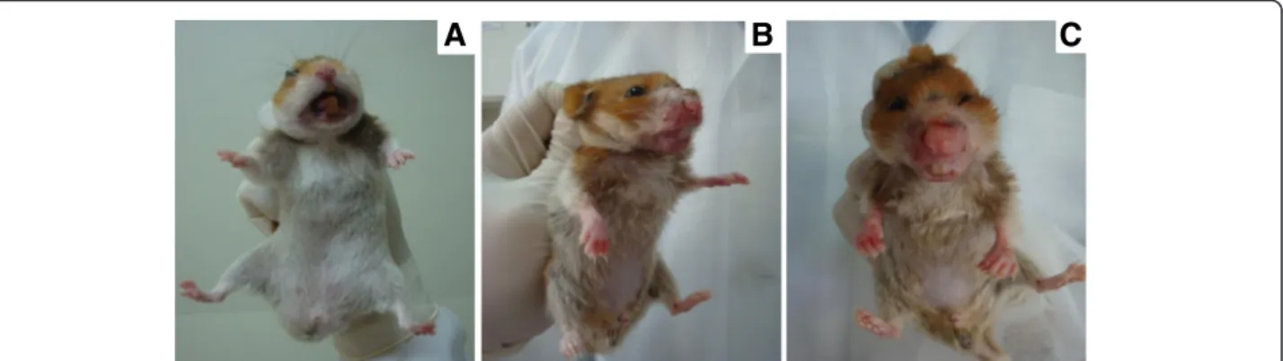
after 6 months, which were more severe at 9 months in hamsters inoculated via the IC route. Hamsters infected with the OP46 strain showed clinical signs suggestive of VL (splenomegaly and ascites) from 3 months after infection regardless of route. Overall, the OP46 strain was associated with more severe clinical signs than PP75 during the monitoring period for all three routes (Table 1). Interestingly, 9 months after IC infection with the OP46 strain, we found desquamation, hair loss, and external mucocutaneous lesions and ulcers lo-calized in the snout, accompanied by edema of the paws in all animals. Furthermore, the animals exhibited severe weight loss, hunched posture, an inability to eat or drink and non-responsiveness to external stimuli (Fig. 1).
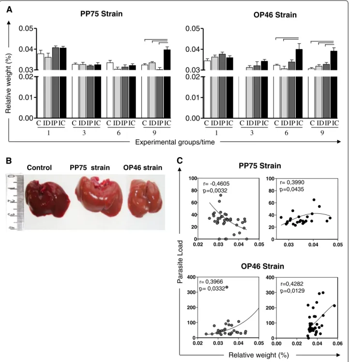
An increase in the relative weight of the liver was ob-served at 9 months in animals infected with the PP75 strain via the IC route compared to the other groups. This increase was also observed in the IC group of ani-mals infected with the OP46 strain beginning in the sixth month and persisting at 9 months (Fig. 2a). Macro-scopically, the livers of animals infected with both strains and by the IC route appeared friable; changed color from red to pale yellowish brown (Fig. 2b). In hamsters in-fected with the PP75 strain, the correlation analysis re-vealed that relative liver weight was negatively correlated with the parasite load in the IP group, but positively cor-related in the IC group. In animals inoculated with the OP46 strain, the correlation analysis revealed that rela-tive liver weight was posirela-tively correlated with its para-site load in both the IP and IC groups (Fig. 2c).
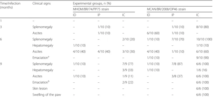
Table 1Clinicopathological analysis of hamsters experimentally infected withL. infantum. Absolute (n) and percentage (%)
Time/infection (months)
Clinical signs Experimental groups, n (%)
MHOM/BR/74/PP75 strain MCAN/BR/2008/OP46 strain
ID IP IC ID IP IC
1 – – – – – – –
3 Splenomegaly – 1/10 (10) – – 1/10 (10) 8/10 (80)
Ascites – 1/10 (10) – 6/10 (60) 1/10 (10) –
6 Splenomegaly – – 2/10 (20) 1/10 (10) 7/10 (70) 10/10 (100)
Hepatomegaly 1/10 (10) – – – – 1/10 (10)
Ascites 4/10 (40) 4/10 (40) 3/10 (30) 4/10 (40) 1/10 (10) 6/10 (60)
Emaciationa – – – 1/10 (10) – 9/10 (90)
9 Splenomegaly 1/10 (10) – 7/9 (77) 1/10 (10) 7/8 (87) 6/6 (100)
Hepatomegaly – – 3/9 (33) 1/10 (10) – 1/6 (16)
Ascites 1/10 (10) – 1/9 (11) – 3/8 (37) 6/6 (100)
Emaciationb – – 2/9 (22) – – 6/6 (100)
Skin lesion – – – – – 6/6 (100)
Swelling of the paw – – – – – 6/6 (100)
Animals were infected with PP75 and OP46 strains ofL. infantumby different routes of inoculation: intradermal (ID), intraperitoneal (IP), and intracardiac (IC)
aWeight loss with preservation of the general condition of the animal
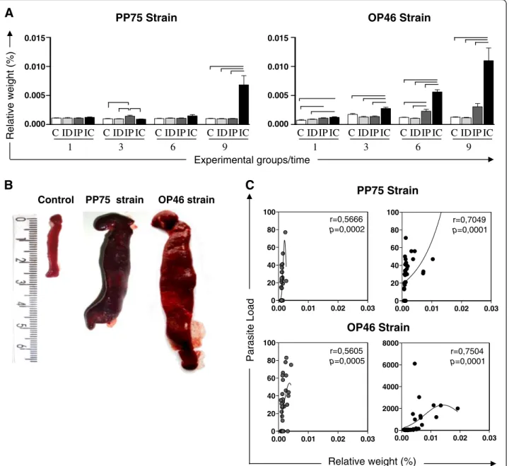
In the analyses of the spleen, hamsters infected with the PP75 strain and inoculated by the IP route had a slight increase in the splenic relative weight compared with the other groups (C, ID and IC) at 3 months. A greater increase in the splenic relative weight was ob-served at 9 months after infection by the IC route com-pared to the C, ID, and IP groups. Animals infected with the OP46 strain via the IP route showed an increase in the relative weight of spleen at 1 month compared to the C group and at 6 months compared to the C and ID groups. The IC route demonstrated a gradual increase in the relative weight of the spleen during the course of in-fection. This increase was observed with regard to the C, ID, and IP groups at 3, 6, and 9 months, and also in rela-tion to the C and ID groups at 1 month (Fig. 3a). The macroscopic splenic changes most frequently observed included an increase in the volume of the organ (spleno-megaly), thickening of edges, rugose and an irregular surface and dark coloration, suggesting hyperemia. It is noteworthy that these alterations were more intense in animals infected with the OP46 strain (Fig. 3b). Further-more, the correlation analysis revealed that splenic rela-tive weight was posirela-tively correlated with the parasite load in the IP and IC groups for both strains (Fig. 3c).
Hamsters infected with the OP46 strain through the intracardiac route exhibit severe anemia with a reduction of red blood cells, hemoglobin, and hematocrit
In hamsters infected with the PP75 strain, a reduction in the hematocrit was observed in all groups (ID, IP, and IC) after 1 month of infection. At 3 months after infec-tion, an increase in the number of platelets was observed in the IP and IC groups compared to the control ani-mals. Reduced counts for erythrocytes, hemoglobin and hematocrit were observed at 6 months. The only differ-ence at 9 months, however, was an increase in the num-ber of erythrocytes in the IP group compared to the C and ID groups.
In animals infected with the OP46 strain, severe anemia was observed in the IC group from the sixth month of infection, with a reduction of red blood cells, hemoglobin and hematocrit in comparison with the other groups (Table 2).
With regard to the white blood cells, data analysis re-vealed statistical differences in the animals infected with the PP75 strain at 3 months, with a reduction in the number of eosinophils in animals inoculated by the IP route in relation to the C group. Moreover, at 9 months a significant de-crease in the monocyte population was observed in the IC group compared with the control group. In contrast, after 1 month of infection with the OP46 strain by the ID and IP routes, there was an increased number of monocytes in re-lation to the C group. After 3 months of infection an in-crease in the lymphocyte population was observed in the group inoculated via the IP route compared to group C. At this same time, animals infected via the IC route presented with leucopenia compared to the C, ID, and IP groups, which was characterized by a remarkable reduction in the monocyte population compared to the C group. After 6 months of infection, the IC group showed some alter-ations: a reduction in the total number of neutrophils was observed compared to the ID group; the number of eosino-phils in relation to the C and ID groups was diminished; the number of lymphocytes was reduced in relation to the C, ID, and IP groups; and the number of monocytes de-clined in relation to the C and IP groups. In a similar man-ner, at 9 months after infection with the OP46 strain, hamsters inoculated by the IP and IC routes were more likely to have leucopenia compared to the C and ID groups, which was characterized by a decrease in the total neutro-phils, eosinoneutro-phils, and monocytes (data not shown).
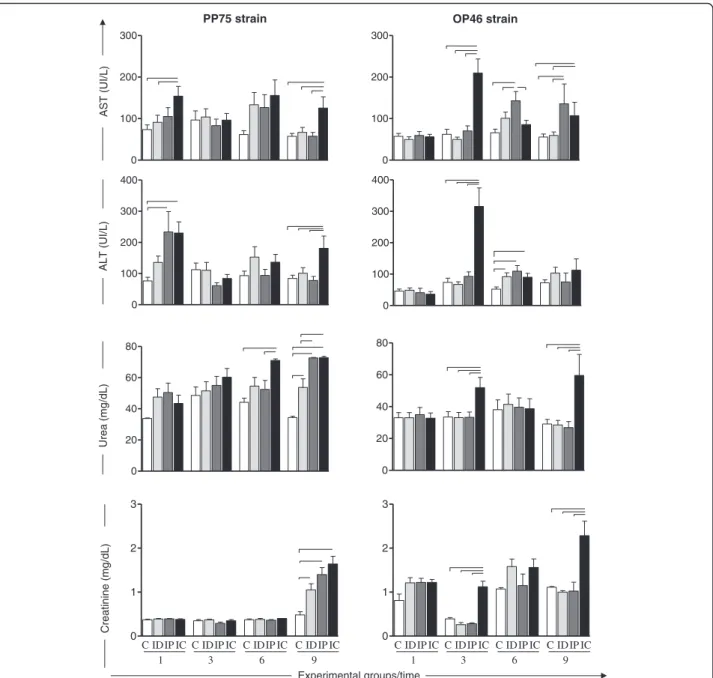
Hamsters demonstrated consistent biochemical changes which present themselves as strong clinical markers of visceral leishmaniasis
The biochemical evaluations are shown in Fig. 4 and consist of the following tests: liver function tests,
A
B
C
A
B
C
Fig. 1Macroscopic changes observed in hamsters 9 months after infection with the OP46 strain. Inanormal hamster,b-csevere cachexia, hair
r= -0,4605
p=0,0032
r=0,4282
p=0,0129
Relative weight (%)
Parasite Load
OP46 Strain PP75 Strain
OP46 Strain PP75 Strain
ID IP IC
1
C ID IP IC
3
C ID IP IC
6
C ID IP IC
9
C ID IP IC
1
C ID IP IC
3
C ID IP IC
6
C ID IP IC
9 C
Relative weight
(%)
Experimental groups/time
A
B
C
Control PP75 strain OP46 strain
r= 0,3990
p=0,0435
r= 0,3966
p= 0,0332 0.00
0.01 0.02 0.03 0.04 0.05
0.00 0.01 0.02 0.03 0.04 0.05
0.02 0.03 0.04 0.05 0
20 40 60 80 100
0.03 0.04 0.05
0 20 40 60 80 100
0.02 0.03 0.04 0.05 0
100 200 300 400
0.00 0.02 0.04 0.06 0
100 200 300 400
Fig. 2Relative weight of the liver of uninfected hamsters as a control group (C,n= 10 animals/time; white) and hamsters experimentally infected
including the measurement of AST and ALT, and renal function tests based on urea and creatinine levels.
In animals infected with the PP75 strain, an increase in the serum AST level was observed in the IC group in relation to the C and ID groups at 1 month as well as at 9 months in comparison to all other groups (C, ID, and IP). Furthermore, an increase in the serum ALT level
was observed in the IC group at 1 month compared to the C group and at 9 months in relation to all other groups (C, ID, and IP) (Fig. 4).
In hamsters infected with the OP46 strain, an increase in the serum AST level was observed in the IC group compared to all groups at 3 months, and in relation to the C and ID groups at 9 months after infection.
OP46 Strain PP75 Strain
Relative weight (%)
r=0,5666
p=0,0002
r=0,7049
p=0,0001
r=0,5605
p=0,0005
r=0,7504
p=0,0001
Relative weight (%)
Parasite Load
ID IP IC
1
C ID IP IC
3
C ID IP IC
6
C ID IP IC
9 C
0.015
0.010
0.005
0.000
0.015
0.010
0.005
0.000
ID IP IC
1
C ID IP IC
3
C ID IP IC
6
C ID IP IC
9 C
A
B
C
Experimental groups/time
OP46 Strain PP75 Strain
Control PP75 strain OP46 strain
0.00 0.01 0.02 0.03 0
20 40 60 80 100
0.00 0.01 0.02 0.03 0
20 40 60 80 100
0.00 0.01 0.02 0.03 0
20 40 60 80 100
0.00 0.01 0.02 0.03 0
2000 4000 6000 8000
Fig. 3Relative weight of the spleen in uninfected hamsters as a control group (C,n= 10 animals/time; white) and hamsters experimentally infected
Similarly, the IP group showed an increase in the AST level in relation to all groups at 3 months after infection and at 9 months in relation to the C and ID groups. Serum ALT levels were observed to be increased in the IC group compared to all groups at 3 months. Further-more, an increase of ALT was observed in the ID, IP, and IC groups compared with the control group at 6 months of follow-up (Fig. 4).
Regarding renal function, increased levels of urea were observed in the IC group compared to the C and IP groups at 6 months after infection with the PP75 strain. Moreover, after 9 months of infection, increased levels of urea were observed in the three experimental groups (ID, IP, and IC) compared to C group. Also, at the last follow up, an increased serum level of urea in the IP and IC groups in relation to the ID group was detected. In the animals infected with the OP46 strain, increased levels of urea were observed in the IC group compared to all other groups (C, ID, and IP) at 3 and 9 months (Fig. 4). The animals infected with the PP75 strain
presented increased serum creatinine levels in all in-fected groups (ID, IP, and IC) compared to the control group at 9 months (Fig. 4). Furthermore, animals in-fected with the OP46 strain also presented an increased serum creatinine level in the IC group compared to the others (C, ID and IP) at 3 and 9 months after infection (Fig. 4).
Discussion
In this study we investigated the clinical and laboratory changes in male golden hamsters infected with L. infan-tum in order to improve our understanding of the pathogenesis studies and clinical aspects of VL. Previous studies by Moreira et al. [7] demonstrated progressive disease in hamsters infected with L. infantum, which was closely related to the presence of high parasitism in the spleen and liver. Herein we evaluated the clinical signs and changes in hematological and biochemical pa-rameters in hamsters that were experimentally infected by routes (ID, IP, and IC) with strains of L. infantum
Table 2Erythrogram of control hamsters and hamsters experimentally infected with strains PP75 and OP46 ofL. infantum
Hematological variables by group
Time/months
1 3 6 9 1 3 6 9
C group
Erythrocytes (106/mm3) 7.2 ± 0.3 7.2 ± 1.1 8.5 ± 1.2 8.4 ± 1.0 7.4 ± 0.4 7.0 ± 0.3 7.1 ± 0.2 6.7 ± 0.3
Hemoglobin (g/dL) 13.9 ± 0.6 13.5 ± 0.8 17.6 ± 2.1 17.5 ± 2.3 15.0 ± 1.0 13.3 ± 0.9 13.3 ± 0.5 13.2 ± 0.7
Hematocrit (%) 35.5 ± 3.6 34.6 ± 7.2 39.5 ± 5.5 38.4 ± 6.7 32.6 ± 1.5 30.5 ± 2.3 32.5 ± 2.3 33.2 ± 2.0
Platelets (106/mm3) 165.8 ± 37.4 142.6 ± 33.8 154.9 ± 143 127.4 ± 12.5 139.9 ± 20.8 136.4 ± 16.9 164.7 ± 15.5 174.0 ± 20.5
Strain/months after infection
PP75 strain OP46 strain
1 3 6 9 1 3 6 9
ID group
Erythrocytes (106/mm3) 6.5 ± 0.4 7.2 ± 0.2 6.5 ± 0.4a 8.5 ± 0.7 7.4 ± 0.3 6.8 ± 0.3 6.7 ± 0.8 6.9 ± 0.3
Hemoglobin (g/dL) 13.7 ± 1.1 13.6 ± 0.5 12.7 ± 1.0a 19.7 ± 4.5 15.2 ± 0.6 13.3 ± 0.7 12.6 ± 1.6 13.7 ± 0.3
Hematocrit (%) 29.3 ± 2.1a 36.3 ± 3.0 31.1 ± 2.7a 38.3 ± 3.9 33.9 ± 1.5 31.2 ± 3.0 33.6 ± 3.6 34.0 ± 2.0
Platelets (106/mm3) 123.8 ± 16.2 168.70 ± 25.26 138.3 ± 19.2 108.1 ± 18.0 156.8 ± 27.5 141.1 ± 24.4 185.5 ± 24.4 189.4 ± 27.3
IP group
Erythrocytes (106/mm3) 6.1 ± 0.1 6.8 ± 0.7 6.7 ± 0.2a 9.8 ± 0.5a,b 7.5 ± 1.0 7.1 ± 0.4 7.0 ± 0.5 6.2 ± 1.0
Hemoglobin (g/dL) 13.4 ± 0.5 13.1 ± 1.2 13.4 ± 0.4a 21.2 ± 1.3 14.4 ± 0.6 13.6 ± 1.2 12.8 ± 1.1 12.0 ± 2.0
Hematocrit (%) 28.9 ± 1.5a 38.7 ± 3.2 31.2 ± 2.7a 43.9 ± 2.2 32.0 ± 2.6 31.4 ± 3.2 36.1 ± 2.6 31.0 ± 4.2
Platelets (106/mm3) 134.1 ± 45.3 221.6 ± 75.0a 132.2 ± 23.4 126.3 ± 14.5 169.3 ± 35.4 145.6 ± 32.1 184.4 ± 32.7 174.6 ± 87.4
IC group
Erythrocytes (106/mm3) 6.6 ± 0.4 7.2 ± 0.7 6.8 ± 0.3a 9.2 ± 1.0 7.1 ± 0.4 6.7 ± 0.3 4.3 ± 1.4a,b,c 4.7 ± 1.9a,b, c
Hemoglobin (g/dL) 13.7 ± 0.8 13.8 ± 1.2 13.6 ± 0.7a 20.0 ± 2.4 14.5 ± 0.9 12.3 ± 1.6 7.0 ± 1.8a,b,c 10.1 ± 4.2a,b
Hematocrit (%) 30.1 ± 1.8a 35.0 ± 3.6 31.5 ± 2.4a 41.5 ± 4.3 31.6 ± 2.1 29.0 ± 4.4 21.0 ± 6.9a,b,c 25.0 ± 9.4a,b
Platelets (106/mm3) 134.1 ± 21.5 144.6 ± 22.3c 135.4 ± 13.5 108.5 ± 17.5 156.5 ± 17.0 110.9 ± 29.8 329.4 ± 153.5a,b,c 291.3 ± 136.4a,b,c
Values (mean ± standard deviation) erytrogram of uninfected hamsters as a control group (C) and infected animals with strains PP75 and OP46 ofL. infantumby
different routes of inoculation: intradermal (ID), intraperitoneal (IP), intracardiac (IC). Significant differences (P< 0.05) are represented by lettersa, b, crelated to the
(MHOM/BR/74/PP75 and MCAN/BR/2008/OP46) that exhibit distinct degrees of virulence and pathogenicity.
The clinical signs observed in our study have also been reported by several other researchers [14, 23, 37–39]. Wilson et al. [40] documented that the clin-ical symptoms of VL in hamsters only occurred 10 months after ID inoculation of L. donovani. How-ever, other studies found that progressive VL was achieved after intradermal infection with 105 para-sites, which is the route closer to natural transmission
by sand fly bites [22]. The hamsters showed clinical signs similar to those observed in symptomatic HVL, including splenomegaly and severe weight loss. In contrast, the frequency of clinical signs when the ID route was used with both strains was relatively low during our study. However, clinical signs were more evident at 9 months after infection with the PP75 strain inoculated by the IC route, but with low fre-quency. With the OP46 strain, the clinical signs were more frequent and were exacerbated in the IC group.
0 100 200 300
0 100 200 300 400
0 20 40 60 80
0 1 2 3
0 100 200 300
0 100 200 300 400
0 20 40 60 80
0 1 2 3
ID IP IC 1
C ID IP IC 3
C ID IP IC 6
C ID IP IC 9
C ID IP IC
1
C ID IP IC 3
C ID IP IC 6
C ID IP IC 9 C
Experimental groups/time
OP46 strain PP75 strain
AST
(UI/L)
ALT (UI/L)
Urea
(mg
/dL)
Creatinine
(mg
/dL)
Fig. 4Activity of AST and ALT; urea and creatinine in the blood plasma of uninfected hamsters as a control group (C,n= 10 animals/time; white)
In other words, our results demonstrated an increased pathogenicity of this strain.
At 9 months after infection with the OP46 strain by the IC route, an accentuated weight loss (cachexia) was observed, as well as mucocutaneous lesions accompan-ied by localized ulcers in the snout and edema in the paws. We speculate that the cachexia may have arisen due to parasites spreading to the liver and spleen [7] as well as the secondary mucocutaneous lesions. Moreover, these data emphasize the capacity of the IC route to cause a severe injury in hamsters, including cutaneous lesion and edema of the lower limbs. These data corrob-orate those reported by Nieto et al. [23] in hamsters in-fected with promastigotes of L. infantum by the IC route. This result clearly demonstrates that classical clin-ical signs observed and reported in human and canine VL also occur in experimental model hamsters. There-fore, we can infer that the route of the inoculum and the strain used are vital conditions for generating progres-sive VL in the hamster model.
The classical hepatomegaly was an uncommon finding in hamsters infected with either strains used in the present work. However, we reported an increased rela-tive liver weight in both groups (PP75 and OP46 strain) when infection occurred by the IC route, and the weight was closely correlated with an increased parasite load. Our results indicate hepatomegaly not from the in-creased size of the liver, but with regard to the weight loss. Hepatomegaly is not necessarily always observed during the disease, which has also been documented by other authors [41, 42]. These differences, as well as the other changes, are probably inherent to the complexity of the effects caused by the parasite and the host im-mune response. Macroscopically, the liver of animals in-oculated via the IC route appeared friable; containing white foci distributed across the liver surface and chan-ged color from red to pale yellowish brown. These same macroscopic changes were also described by other au-thors for hamsters experimentally infected withL. infan-tum and L. donovani using the IC route for the inoculums [42, 43]. At 9 months, the spleen size in ani-mals inoculated via the IC route was exorbitant com-pared to the control group for both strains. We hypothesized that this splenomegaly was due to the high parasitism displayed by these animals [7].
Knowledge of the biochemical and hematological param-eters in hamsters experimentally infected withL. infantum
is extremely limited. Although hematological and biochem-ical profiles have limited applications for a VL diagnosis, they can be very important in evaluating the clinical prog-nosis status, as well as in elucidating the pathogenesis to define therapeutic approaches [8, 44–47]. Blood abnormal-ities are frequently observed in both humans and animal models for VL [48–50]. Leucopenia, neutropenia, and
eosiponenia are frequently found in HVL [51–53]. Accord-ing to Reis et al. [44] the impaired hematological picture in CVL is associated with severe clinical manifestations and demonstrated by leucopenia, which is characterized by lym-phopenia, eosinopenia and monocytopenia. Herein, the changes in the hematological parameters of hamsters in-fected with the PP75 strain were not expressive; we de-tected monocytopenia in the last month of infection only in the IC group. However, the hamsters infected with the OP46 strain through the IC and IP routes presented intense monocytopenia accompanied by leucopenia after the third month of infection. The leucopenia observed in the IC and IP groups at 6 and 9 months after experimental infection, showed a typical LV immunosuppression marked by a de-crease of neutrophils, eosinophils, lymphocytes and/or monocytes. Thus, in the context of our study, the classical hematological changes observed in human and canine VL have been reproduced. The results also confirm that the hematological changes in the IP and IC groups, accompan-ied by the presence of clinical signs suggestive of active VL (especially splenomegaly), are associated with high tissue parasitism (spleen and liver) previously observed by Mor-eira et al. [7]. These findings resulted from the complex interaction related to the potential virulence and pathogen-icity of each of the two strains used.
Normocytic normochromic anemia is a common find-ing in patients with the chronic form of the human dis-ease [52] and also for dogs with CVL [44]. In our study the main changes in the red blood cells of hamsters in-fected with the PP75 strain occurred at 1 and 6 months after infection in the three experimental groups (ID, IP, and IC). Moreover, in animals infected with the OP46 strain, these changes were observed from the sixth month, and were restricted to the IC group only, in which severe anemia, marked by the reduction of eryth-rocytes, hematocrit and hemoglobin was observed. Anemia was also demonstrated by Rica-Capela et al. [38] in 70–80 % of hamsters experimentally infected with dif-ferent forms (promastigotes and amastigotes) of L. infantum.
points were observed for both strains (PP75 and OP46). Our results provide advanced understanding of the role of these biomarkers in the hepatic func-tion during ongoing experimental hamster infecfunc-tion.
Evaluation of renal function consisted of determining serum levels of urea and creatinine. Our results revealed a renal alteration characterized by an increase in urea and creatinine in both strains by different routes of inoculation and at different time points. The most common change in renal physiology in VL is glomerulonephritis, while amyl-oidosis is less common [57]. Studies conducted by Rica-Capela et al. [38] showed a large amount of amyloid in the liver and spleen of hamsters experimentally infected with amastigotes and promastigotes ofL. infantum. These data suggest kidney injury in the animals, perhaps due to the de-position of immune complexes in lymphoid organs.
Therefore, these findings represent an advance in the knowledge of the involvement of experimental infection in hamster model with L. infantum. Data indicate that the infectivity capacity of the parasite, the inoculum routes and the dose used as well as the biological form of the parasite (amastigote and promastigote) can influ-ence the course of the natural history of experimental VL. These aspects make it difficult to select an ideal ex-perimental model that is capable of reproducing the dis-ease in its visceral form exhibiting various clinical manifestations and evolving from acute to chronic form [7, 14, 21]. However, here we showed that the route of infection and the strain used was crucial to influence the biochemical and hematological parameters evaluated in this study. Thus the IC route proved to be more aggres-sive than other routes used and OP46 more virulent and a more pathogenic strain compared to the reference PP75 strain.
Conclusion
Taken together, the results obtained from this experi-mental infection of male golden hamsters infected with
L. infantum having distinct virulence and pathogenicity profiles show that it is possible to reproduce a disease with differing degrees of severity. Many alterations and typical biomarkers, such as hematological/biochemical factors and pathological and clinical signs frequently ob-served in human and/or canine VL, were also detected in this hamster model. Thus, the results presented in this paper indicate that the golden hamster (M. auratus) constitutes an excellent model for analysis of experimen-tal infections caused by different strains of L. infantum
inoculated by different routes, reinforcing its use in pathogenesis studies of VL.
Competing interests
The authors declare that they have no competing interests.
Authors’contributions
ABR, RCG, JVS, CMC and NDM planned the study design. NDM performed laboratory work. JMOC, MTR, BMR, PMAV, HGK and NDM have made substantial contributions to analysis and interpretation of data. BMR, WCV and NDM provided substantial improvement of the manuscript and RCG and ABR provided scientific supervision of the study. All authors approved the final version of the manuscript.
Acknowledgments
CMC, RCG, JV-S, BMR and ABR thank Conselho Nacional de Desenvolvimento Científico e Tecnológico, Brazil, WCV and NDM by CAPES/PNPD for fellowships. The authors are also grateful for Universidade Federal de Ouro Preto (UFOP), the use of facilities at Centro de Ciência Animal (Universidade Federal de Ouro Preto). We are also grateful for the specific grants (FAPEMIG, CNPq, and CAPES) from these financial agencies.
Author details
1
Laboratório de Imunopatologia, Núcleo de Pesquisas em Ciências Biológicas/NUPEB, Universidade Federal de Ouro Preto, Ouro Preto, Minas Gerais, Brasil.2Laboratório de Pesquisas Clínicas, Departamento de Análises Clínicas, Escola de Farmácia, Universidade Federal de Ouro Preto, Ouro Preto, Minas Gerais, Brasil.3Laboratório de Morfopatologia, Departamento de Ciências Biológicas, Universidade Federal de Ouro Preto, Ouro Preto, Minas Gerais, Brasil.4Laboratório de Biologia das Interações Celulares,
Departamento de Morfologia, Universidade Federal de Minas Gerais, Belo Horizonte, Minas Gerais, Brasil.
Received: 23 December 2015 Accepted: 21 March 2016
References
1. Carvalho EM, Teixeira RS, Johnson Jr WD. Cell-mediated immunity in American visceral leishmaniasis: reversible immunosuppression during acute infection. Infect Immun. 1981;33(2):498–500.
2. Carvalho EM, Badaro R, Reed SG, Jones TC, Johnson Jr WD. Absence of gamma interferon and interleukin 2 production during active visceral leishmaniasis. J Clin Invest. 1985;76(6):2066–9.
3. van Griensven J, Balasegaram M, Meheus F, Alvar J, Lynen L, Boelaert M. Combination therapy for visceral leishmaniasis. Lancet Infect Dis. 2010;10(3):184–94.
4. Wilson ME, Sandor M, Blum AM, Young BM, Metwali A, Elliott D, RG, Weinstock JV. Local suppression of IFN-gamma in hepatic granulomas correlates with tissue-specific replication ofLeishmania chagasi. J Immunol. 1996;156(6):2231–9.
5. Smelt SC, Engwerda CR, McCrossen M, Kaye PM. Destruction of follicular dendritic cells during chronic visceral leishmaniasis. J Immunol. 1997;158(8):3813–21.
6. Tropia de Abreu R, Carvalho M, Carneiro CM, Giunchetti RC, Teixeira-Carvalho A, Martins-Filho OA, Coura-Vital W, Correa-Oliveira R, Reis AB. Influence of clinical status and parasite load on erythropoiesis and leucopoiesis in dogs naturally infected withLeishmania(Leishmania)
chagasi. PLoS One. 2011;6(5):e18873.
7. Moreira N, Vitoriano-Souza J, Roatt BM, Vieira PM, Ker HG, de Oliveira Cardoso JM, Giunchetti RC, Carneiro CM, de Lana M, Reis AB. Parasite burden in hamsters infected with two different strains ofLeishmania
(Leishmania)infantum:“Leishman Donovan units”versus real-time PCR. PLoS One. 2012;7(10), e47907.
8. Nicolato Rde C, de Abreu RT, Roatt BM, Aguiar-Soares RD, Reis LE, Carvalho M, Carneiro CM, Giunchetti RC, Bouillet LE, Lemos DS et al. Clinical forms of canine visceral leishmaniasis in naturallyLeishmania infantum-infected dogs and related myelogram and hemogram changes. PLoS One. 2013;8(12), e82947.
9. De Oliveira CI, TM Gomes R, Barral A, Brodskyn C. Mortality due to visceral leishmaniasis: clinical and laboratory characteristics. Drug Discov Today. 2004;1:81–6.
10. Moreno J, Alvar J. Canine leishmaniasis: epidemiological risk and the experimental model. Trends Parasitol. 2002;18(9):399–405.
12. Miralles GD, Stoeckle MY, McDermott DF, Finkelman FD, Murray HW. Th1 and Th2 cell-associated cytokines in experimental visceral leishmaniasis. Infect Immun. 1994;62(3):1058–63.
13. Engwerda CR, Handwerger BS, Fox BS. Aged T cells are hyporesponsive to costimulation mediated by CD28. J Immunol. 1994;152(8):3740–7. 14. Melby PC, Chandrasekar B, Zhao W, Coe JE. The hamster as a model of
human visceral leishmaniasis: progressive disease and impaired generation of nitric oxide in the face of a prominent Th1-like cytokine response. J Immunol. 2001;166(3):1912–20.
15. Engwerda CR, Ato M, Cotterell SE, Mynott TL, Tschannerl A, Gorak-Stolinska PM, Kaye PM. A role for tumor necrosis factor-alpha in remodeling the splenic marginal zone duringLeishmania donovaniinfection. Am J Pathol. 2002;161(2):429–37.
16. Engwerda CR, Ato M, Kaye PM. Macrophages, pathology and parasite persistence in experimental visceral leishmaniasis. Trends Parasitol. 2004;20(11):524–30.
17. Nieto CG, Vinuelas J, Blanco A, Garcia-Alonso M, Verdugo SG, Navarrete I. Detection ofLeishmania infantumamastigotes in canine choroid plexus. Vet Rec. 1996;139(14):346–7.
18. Vinuelas J, Garcia-Alonso M, Ferrando L, Navarrete I, Molano I, Miron C, Carcelen J, Alonso C, Nieto CG. Meningeal leishmaniosis induced byLeishmania infantumin naturally infected dogs. Vet Parasitol. 2001;101(1):23–7.
19. Abreu-Silva AL, Calabrese KS, Tedesco RC, Mortara RA, Goncalves da Costa SC. Central nervous system involvement in experimental infection with
Leishmania(Leishmania)amazonensis. Am J Trop Med Hyg. 2003;68(6):661–5. 20. Rolao N, Melo C, Campino L. Influence of the inoculation route in BALB/c
mice infected byLeishmania infantum. Acta Trop. 2004;90(1):123–6. 21. Carrion J, Nieto A, Iborra S, Iniesta V, Soto M, Folgueira C, Abanades DR,
Requena JM, Alonso C. Immunohistological features of visceral leishmaniasis in BALB/c mice. Parasite Immunol. 2006;28(5):173–83.
22. Gomes R, Teixeira C, Teixeira MJ, Oliveira F, Menezes MJ, Silva C, de Oliveira CI, Miranda JC, Elnaiem DE, Kamhawi S et al. Immunity to a salivary protein of a sand fly vector protects against the fatal outcome of visceral leishmaniasis in a hamster model. Proc Natl Acad Sci U S A. 2008;105(22):7845–50.
23. Nieto A, Dominguez-Bernal G, Orden JA, De La Fuente R, Madrid-Elena N, Carrion J. Mechanisms of resistance and susceptibility to experimental visceral leishmaniosis: BALB/c mouse versus Syrian hamster model. Vet Res. 2011;42:39.
24. Mookerjee A, Sen PC, Ghose AC. Immunosuppression in hamsters with progressive visceral leishmaniasis is associated with an impairment of protein kinase C activity in their lymphocytes that can be partially reversed by okadaic acid or anti-transforming growth factor beta antibody. Infect Immun. 2003;71(5):2439–46.
25. Goto H, Lindoso JA. Immunity and immunosuppression in experimental visceral leishmaniasis. Braz J Med Biol Res. 2004;37(4):615–23. 26. Osorio EY, Zhao W, Espitia C, Saldarriaga O, Hawel L, Byus CV, Travi BL,
Melby PC. Progressive visceral leishmaniasis is driven by dominant parasite-induced STAT6 activation and STAT6-dependent host arginase 1 expression. PLoS Pathog. 2012;8(1), e1002417.
27. Selvapandiyan A, Dey R, Nylen S, Duncan R, Sacks D, Nakhasi HL. Intracellular replication-deficientLeishmania donovaniinduces long lasting protective immunity against visceral leishmaniasis. J Immunol.
2009;183(3):1813–20.
28. Samant M, Gupta R, Kumari S, Misra P, Khare P, Kushawaha PK, Travi BL, Melby PC. Immunization with the DNA-encoding N-terminal domain of proteophosphoglycan ofLeishmania donovanigenerates Th1-type immunoprotective response against experimental visceral leishmaniasis. J Immunol. 2009;183(1):470–9.
29. Banerjee S, Ghosh J, Sen S, Guha R, Dhar R, Ghosh M, Datta S, Raychaudhury B, Naskar K, Haldar AK et al. Designing therapies against experimental visceral leishmaniasis by modulating the membrane fluidity of antigen-presenting cells. Infect Immun. 2009;77(6):2330–42.
30. Kushawaha PK, Gupta R, Sundar S, Sahasrabuddhe AA, Dube A. Elongation factor-2, a Th1 stimulatory protein ofLeishmania donovani, generates strong IFN-gamma and IL-12 response in curedLeishmania-infected patients/ hamsters and protects hamsters againstLeishmaniachallenge. J Immunol. 2011;187(12):6417–27.
31. Prajapati VK, Awasthi K, Gautam S, Yadav TP, Rai M, Srivastava ON, Sundar S. Targeted killing ofLeishmania donovaniin vivo and in vitro with amphotericin B attached to functionalized carbon nanotubes. J Antimicrob Chemother. 2011;66(4):874–9.
32. Selvapandiyan A, Dey R, Gannavaram S, Lakhal-Naouar I, Duncan R, Salotra P, Nakhasi HL. Immunity to visceral leishmaniasis using genetically defined live-attenuated parasites. J Trop Med. 2012;2012:631460.
33. Anuradha, Pal R, Katiyar JC. Sex-influenced population kinetics ofLeishmania donovaniin hamsters. Indian J Exp Biol. 1990;28(9):876–9.
34. Singh N, Samant M, Gupta SK, Kumar A, Dube A. Age-influenced population kinetics and immunological responses ofLeishmania donovaniin hamsters. Parasitol Res. 2007;101(4):919–24.
35. Travi BL, Osorio Y, Melby PC, Chandrasekar B, Arteaga L, Saravia NG. Gender is a major determinant of the clinical evolution and immune response in hamsters infected withLeishmaniaspp. Infect Immun. 2002;70(5):2288–96. 36. Evans TG, Smith D, Pearson RD. Humoral factors and nonspecific immune
suppression in Syrian hamsters infected withLeishmania donovani. J Parasitol. 1990;76(2):212–7.
37. Requena JM, Soto M, Doria MD, Alonso C. Immune and clinical parameters associated withLeishmania infantuminfection in the golden hamster model. Vet Immunol Immunopathol. 2000;76(3–4):269–81. 38. Rica-Capela MJ, Cortes S, Leandro C, Peleteiro MC, Santos-Gomes G,
Campino L. Immunological and histopathological studies in a rodent model infected withLeishmania infantumpromastigotes or amastigotes. Parasitol Res. 2003;89(3):163–9.
39. Dea-Ayuela MA, Rama-Iniguez S, Alunda JM, Bolas-Fernandez F. Setting new immunobiological parameters in the hamster model of visceral
leishmaniasis for in vivo testing of antileishmanial compounds. Vet Res Commun. 2007;31(6):703–17.
40. Wilson ME, Innes DJ, Sousa AD, Pearson RD. Early histopathology of experimental infection withLeishmania donovaniin hamsters. J Parasitol. 1987;73(1):55–63.
41. Melo F, Amaral M, Oliveira P, Lima W, Andrade M, Michalick M, Raso P, Tafuri W, Tafuri W. Diffuse intralobular liver fibrosis in dogs naturally infected with
Leishmania(Leishmania)chagasi. Am J Trop Med Hyg. 2008;79(2):198–204. 42. Aslan H, Dey R, Meneses C, Castrovinci P, Jeronimo SM, Oliva G, Fischer L, Duncan RC, Nakhasi HL, Valenzuela JG et al. A new model of progressive visceral leishmaniasis in hamsters by natural transmission via bites of vector sand flies. J Infect Dis. 2013;207(8):1328–38.
43. Vianna VL, Takiya CM, de Brito-Gitirana L. Histopathologic analysis of hamster hepatocytes submitted to experimental infection withLeishmania donovani. Parasitol Res. 2002;88(9):829–36.
44. Reis AB, Martins-Filho OA, Teixeira-Carvalho A, Carvalho MG, Mayrink W, Franca-Silva JC, Giunchetti RC, Genaro O, Correa-Oliveira R. Parasite density and impaired biochemical/hematological status are associated with severe clinical aspects of canine visceral leishmaniasis. Res Vet Sci. 2006;81(1):68–75. 45. da Costa-Val AP, Cavalcanti RR, de Figueiredo GN, Michalick MS,
Alexander B, Williams P, Melo MN. Canine visceral leishmaniasis: relationships between clinical status, humoral immune response, haematology andLutzomyia(Lutzomyia)longipalpisinfectivity. Vet J. 2007;174(3):636–43.
46. Reis AB, Martins-Filho OA, Teixeira-Carvalho A, Giunchetti RC, Carneiro CM, Mayrink W, Tafuri WL, Correa-Oliveira R. Systemic and compartmentalized immune response in canine visceral leishmaniasis. Vet Immunol Immunopathol. 2009;128(1–3):87–95.
47. Oliveira JMF, Fernandes AC, Dorval MEC, Alves TP, Fernandes TD, Oshiro ET, Oliveira ALL. Mortality due to visceral leishmaniasis: clinical and laboratory characteristics. Rev Soc Bras Med Trop. 2010;43(2):188-93.
48. Caldas AJ, Costa J, Aquino D, Silva AA, Barral-Netto M, Barral A. Are there differences in clinical and laboratory parameters between children and adults with American visceral leishmaniasis? Acta Trop. 2006;97(3):252–8. 49. Fernandez-Guerrero ML, Robles P, Rivas P, Mojer F, Muniz G, de Gorgolas M.
Visceral leishmaniasis in immunocompromised patients with and without AIDS: a comparison of clinical features and prognosis. Acta Trop. 2004;90(1):11–6.
50. Moreno J, Nieto J, Masina S, Canavate C, Cruz I, Chicharro C, arrillo E, Napp S, Reymond C, Kaye PM et al. Immunization with H1, HASPB1 and MML
Leishmaniaproteins in a vaccine trial against experimental canine leishmaniasis. Vaccine. 2007;25(29):5290–300.
51. Campos Jr D. Clinical and epidemiological features of Kala-Azar in children. Pediatr (Rio J). 1995;71:261–5.
52. Pearson RD, Sousa AQ. Clinical spectrum of leishmaniasis. Clin Infect Dis. 1996;22(1):1–13.
54. Nieto CG, Navarrete I, Habela MA, Serrano F, Redondo E. Pathological changes in kidneys of dogs with naturalLeishmaniainfection. Vet Parasitol. 1992;45(1–2):33–47.
55. Solano-Gallego L, Koutinas A, Miro G, Cardoso L, Pennisi MG, Ferrer L, Bourdeau P, Oliva G, Baneth G. Directions for the diagnosis, clinical staging, treatment and prevention of canine leishmaniosis. Vet Parasitol.
2009;165(1–2):1–18.
56. Solano-Gallego L, Miro G, Koutinas A, Cardoso L, Pennisi MG, Ferrer L, Bourdeau P, Oliva G, Baneth G, The LeishVet G. LeishVet guidelines for the practical management of canine leishmaniosis. Parasit Vectors. 2011;4:86. 57. Zatelli A, Borgarelli M, Santilli R, Bonfanti U, Nigrisoli E, Zanatta R, Tarducci A,
Guarraci A. Glomerular lesions in dogs infected withLeishmaniaorganisms. Am J Vet Res. 2003;64(5):558–61.
• We accept pre-submission inquiries
• Our selector tool helps you to find the most relevant journal
• We provide round the clock customer support
• Convenient online submission
• Thorough peer review
• Inclusion in PubMed and all major indexing services
• Maximum visibility for your research
Submit your manuscript at www.biomedcentral.com/submit