R E S E A R C H
Open Access
LBSapSal-vaccinated dogs exhibit increased
circulating T-lymphocyte subsets (CD4
+
and
CD8
+
) as well as a reduction of parasitism after
challenge with
Leishmania infantum
plus salivary
gland of
Lutzomyia longipalpis
Rodrigo Dian de Oliveira Aguiar-Soares
1, Bruno Mendes Roatt
1, Henrique Gama Ker
1, Nádia das Dores Moreira
1,
Fernando Augusto Siqueira Mathias
1, Jamille Mirelle de Oliveira Cardoso
1, Nelder Figueiredo Gontijo
2,
Oscar Bruna-Romero
3, Andréa Teixeira-Carvalho
4, Olindo Assis Martins-Filho
4, Rodrigo Corrêa-Oliveira
5,
Rodolfo Cordeiro Giunchetti
6and Alexandre Barbosa Reis
1,7,8*Abstract
Background:The development of a protective vaccine against canine visceral leishmaniasis (CVL) is an alternative approach for interrupting the domestic cycle ofLeishmania infantum. Given the importance of sand fly salivary proteins as potent immunogens obligatorily co-deposited during transmission ofLeishmaniaparasites, their inclusion in an anti-Leishmaniavaccine has been investigated in the last few decades. In this context, we previously immunized dogs with a vaccine composed ofL. braziliensisantigens plus saponin as the adjuvant and sand fly salivary gland extract (LBSapSal vaccine). This vaccine elicited an increase in both anti-saliva and anti-LeishmaniaIgG isotypes, higher counts of specific circulating CD8+T cells, and high NO production.
Methods:We investigated the immunogenicity and protective effect of LBSapSal vaccination after intradermal challenge with 1 × 10⁷late-log-phaseL. infantumpromastigotes in the presence of sand fly saliva ofLutzomyia longipalpis.The dogs were followed for up to 885 days after challenge.
Results:The LBSapSal vaccine presents extensive antigenic diversity with persistent humoral and cellular immune responses, indicating resistance against CVL is triggered by high levels of total IgG and its subtypes (IgG1 and IgG2); expansion of circulating CD5+, CD4+, and CD8+T lymphocytes and isLeishmania-specific; and reduction of splenic parasite load.
Conclusions:These results encourage further study of vaccine strategies addressingLeishmaniaantigens in combination with proteins present in the saliva of the vector.
Keywords:LBSapSal vaccine, Canine visceral leishmaniasis, Immunogenicity, Experimental challenge,Leishmania infantum, Saliva ofLutzomyia longipalpis
* Correspondence:alexreisufop@gmail.com
1Laboratório de Imunopatologia, Núcleo de Pesquisas em Ciências Biológicas/NUPEB, Instituto de Ciências Exatas e Biológicas, Universidade Federal de Ouro Preto, Ouro Preto, Minas Gerais, Brasil
7Departamento de Análises Clínicas, Escola de Farmácia, Universidade Federal de Ouro Preto, Ouro Preto, Minas Gerais, Brasil
Full list of author information is available at the end of the article
Background
Dogs are the most important domestic reservoir of Leish-mania infantum, the protozoan parasite causing visceral leishmaniasis (VL), and represent the major source of con-tagion for the vector, increasing the risk for human infec-tion [1]. Treatment of infected dogs is not an effective strategy because relapses are frequent, and dogs quickly be-come infectious again [2]. In this context, a vaccine would be an important tool in the control of canine visceral leish-maniasis (CVL) and would also dramatically decrease the infection pressure ofL. infantumfor humans [3].
Significant efforts are being made by several groups to develop a vaccine against CVL [4-18]. Given their wide spectrum of antigenicity, cost, and safety, the first gen-eration vaccines that composed of crude antigens also represent an excellent tool for immunoprophylaxis [10,11,13-15,19]. In phase I and II clinical trials, Mayrink
et al. [10], demonstrated enhanced lymphocyte prolifera-tion and significant protecprolifera-tion (90%) against experimental infection with L. infantum in dogs that had received ultrasound-disrupted, merthiolated promastigotes of L. braziliensis with Bacillus Calmete-Guerin (BCG). Strong cellular proliferation in response to soluble Leishmania
antigens has also been reported in dogs vaccinated with autoclavedL. majorpromastigotes plus BCG as the adju-vant [11]. Moreover, in a double-blind randomized efficacy field trial, a single dose of a vaccine composed of alum-precipitated autoclaved L. major vaccine against CVL mixed with BCG was shown to be safe and decreased the incidence of the CVL from 12% to 3.7%, which is equiva-lent to a 69.3% efficacy rate [20].
In the last few decades, the incorporation of salivary proteins of phlebotomines has been widely used in experi-mental challenge studies, or in seeking potential targets for vaccine development against Leishmania infection; such proteins have even been a part of vaccine compos-ition as an adjuvant or co-adjuvant [14,21-29]. Gomes
et al. [28] showed that hamsters immunized with DNA plasmid coding LJM19 from Lutzomyia longipalpis were protected against a challenge with L. infantum plus Lu. longipalpissalivary gland sonicate [28]. Collin et al. [29], working with dogs immunized with saliva recombinant proteins ofLu. longipalpis(LJL143 and LJM17) and chal-lenged with uninfected or infected sandflies, observed a cellular immune response at the site of the bite charac-terized by lymphocytic infiltration and expression of interferon-γ or interleukin-12 [29]. These results suggest
that the use of Lu. longipalpis saliva proteins could be a good strategy in developing an anti-CVL vaccine in dogs. In this context, previous studies in dogs conducted by our group used a first generation vaccine composed ofL. brazi-liensis antigens plus saponin as an adjuvant and sand fly salivary gland extract (SGE) (LBSapSal vaccine). The immunization elicited increases in the saliva and
anti-Leishmania IgG isotypes, higher counts of circulating and specific T CD8+, and high NO production after immunization [14]. The current study included an analysis of the immuno-genicity and a parasitological investigation of dogs immu-nized with LBSapSal vaccine. The dogs were evaluated for up to 885 days after challenge by intradermal inoculation usingL. infantumpromastigotes plusLu. longipalpisSGE.
Methods
The study protocol was approved by the Ethical Committee for the Use of Experimental Animals at the Universidade Federal de Ouro Preto, Minas Gerais, Brazil.
Sand flies and salivary gland extracts
Closed colonies of Lu. longipalpis were maintained at 25°C and 60%–80% relative humidity according to a published protocol [30]. Sand fly SGE was prepared using the method of Cavalcante et al.[31] in which the acini of salivary glands of 4-day-old, mated, but non–
blood-fed female sand flies were dissected in 0.8% un-buffered saline, broken by sonication for 10 s, and cen-trifuged at 10,000 ×g for 2 min. The supernatant was collected and stored at−70°C prior use.
Study animals, vaccination, and experimental challenge
In this study, we used the LBSapSal vaccine as previously described by Giunchetti et al. [14]. Twenty male and fe-male mongrel dogs were born and reared in the kennels of the Animal Science Center, Universidade Federal de Ouro Preto, Ouro Preto, Minas Gerais, Brazil. The dogs were treated by 7 months of age with an anthelmintic and vac-cinated against rabies (Tecpar, Curitiba-PR, Brazil), canine distemper, type 2 adenovirus, coronavirus, parainfluenza, parvovirus, and leptospira (Vanguard® HTLP 5/CV-L; Pfi-zer Animal Health, New York, NY, USA). The absence of specific anti-Leishmania antibodies was confirmed by indirect fluorescence immunoassay and enzyme-linked immunosorbent assay (ELISA) tests. Ouro Preto city is considered a non-endemic area for visceral leishmaniasis in Brazil. Besides negative serology, other additional effect-ive approaches were performed aiming to rule out Leish-mania infection such as spraying the kennels of UFOP with pyrethroid insecticide and protecting all extension of the kennels with an appropriate and security stainless steel wire mesh to block the access of phlebotomines.
received 600μg ofL. braziliensispromastigote protein plus
SGE (as above) in 1 mL sterile 0.9% saline; and (iv) the LBSapSal group (n= 5) that received 600μg ofL. brazilien-sis promastigote protein plus 1 mg of saponin together with SGE in 1 mL of sterile 0.9% saline. Each group re-ceived three subcutaneous injections in the right flank at intervals of 4 weeks. Three and a half months (105 days) after the last vaccine dose, the dogs were challenged via intradermal injection in the right ear, with 1 × 10⁷ late-log-phase L. infantum promastigotes (MHOM/BR/1972/ BH46) plus SGE obtained from two acini ofLu. longipalpis
salivary glands [32-34]. Dogs were followed for 885 days after challenge. Evaluation of the humoral and cellular im-mune response was performed before challenge (Tbc; i.e. 85 days before experimental challenge) and 20, 90, 274, 435, 541 and 885 days after challenge (dac). The dogs were euthanized in 885 dac and the spleen was collected to evaluate the parasite load.
Blood sample collection
Peripheral blood (5 mL) was collected from the jugular vein of each dog and transferred to tubes containing EDTA as anticoagulant. The absolute count of lympho-cytes in each sample was obtained using a BC-2800 VET auto hematology analyzer (Mindray, China). Blood sam-ples were stored at room temperature for up to 12 h prior to processing.
Humoral immune response
The humoral immune response was evaluated by using soluble lysate of L. infantumantigen (MHOM/BR/1972/ BH46) (SLiA) according to conventional enzyme-linked immunosorbent assay (ELISA) as previously described by Reiset al. [35] and Giunchettiet al. [13]. Briefly the SLiA was coated onto 96-well microplates (MaxiSorp™, Nalge Nunc Intl., Rochester, NY, USA) at a concentration of 10μg/well, the serum samples were added at 1:80 dilution,
the wells were washed, and peroxidase-conjugated goat anti-dog IgG1 or sheep anti-dog IgG and IgG2 (Bethyl Laboratories Inc., Montgomery, TX, USA) were added at dilutions of 1:1000, 1:8000, and 1:16,000, respectively. The wells were then washed, substrate and chromogen (o-phenylenediamine; Sigma–Aldrich Co., St. Louis, MO, USA) were added, and the absorbance was read at 492 nm on a Multiskan® MCC 340 (Labsystems, Helsinki, Finland) automatic microplate reader.
In vitroassays
For in vitro evaluation, peripheral blood mononuclear cells (PBMCs) were isolated from 20 mL of heparinised blood under 10 mL of Ficoll–Hypaque density gradient (Histopaque® 1.077; Sigma Chemical Co.), and centri-fuged at 450 ×g for 40 min at room temperature. The PBMCs were resuspended in Gibco RPMI1640 medium,
homogenized, washed twice with RPMI 1640, centri-fuged at 450 ×g for 10 min at room temperature, ho-mogenized, and resuspended in RPMI 1640 at 107cells/ mL. Briefly, for the mitogenic stimulus assays, 25μL
ali-quots of PBMCs (2.5 × 10⁵cells/well) were added to trip-licate wells together with 25 μL of phytohemagglutinin
(2.5μg/mL; Sigma-Aldrich Chemie GmbH, Taufkirchen,
Germany). Incubations were carried out in a humidified 5% CO2atmosphere at 37°C for 3 days (mitogenic-stimu-lated cultures) or 5 days (antigenic-stimu(mitogenic-stimu-lated cultures). To investigate the immunophenotypic features, PBMCs were cultured in 48-well flat-bottomed tissue culture plates (Costar, Cambridge, MA, USA) containing 650 μL
of supplemented RPMI medium/well. Fifty microliters of PBMCs (5.0 × 10⁵cells/well) was added to triplicate wells together with 100 μL of vaccine soluble antigen (VSA)
(25 μg/mL) or 100 μL of SLcA (25 μg/mL). Incubation
was carried out in a humidified 5% CO2 atmosphere at 37°C for 5 days, after which the PBMCs were removed for immunophenotyping according to Giunchettiet al. [13].
Immunophenotyping and flow cytometry
Unlabeled canine monoclonal antibodies (mAbs) against CD5 (rat-IgG2a: clone YKIX322.3), CD4 (rat-IgG2a: clone YKIX302.9), and CD8 (rat-IgG1: clone YCATE55.9), all purchased from Serotec (USA), were used in an indirect immunofluorescence procedure in which pooled normal rat serum (diluted 1:6000) was employed as the isotypic control, and fluorescein isothiocyanate (FITC)-labelled IgG sheep anti-rat polyclonal antibody was used as the secondary antibody.
Briefly, microplate assays for immunophenotyping ca-nine whole blood leukocytes in both fresh blood samples and PBMCs obtained afterin vitrostimulation were car-ried out according to methods previously described by Giunchetti et al. [13]. The results were expressed as the percentage of positive cells within the gated lymphocytes for cell surface markers presenting bimodal distribution (CD5, CD4, and CD8).
DNA Manipulations, Primers and Real Time PCR of spleen
extension at 60°C for 1 min. Standard curves were prepared for each run using known quantities of pGEM®T plasmids (Promega®, Madison, WI, USA) containing inserts of inter-est [37]. To verify the integrity of the samples, the same procedure was carried out for the GAPDH gene (115-bp fragment GenBank accession number AB038240). Reac-tions were processed and analyzed in an ABI Prism 7500-Sequence Detection System (Applied Biosystems, USA). The results were expressed as the number of amastigotes per milligram of spleen.
Statistical analysis
Statistical analyses were performed using Prism 5.0 soft-ware package (Prism Softsoft-ware, Irvine, CA, USA). Normality
of the data was detected using a Kolmogorov–Smirnoff test. One-way analysis of variance and Tukey post-tests were used to determine the differences between groups. In all cases, the differences were considered significant when p
values were < 0.05.
Results
LBSapSal elicited lasting production of anti–L. infantum
immunoglobulin isotypes (IgG, IgG1, and IgG2) afterL.
infantumexperimental challenge
Significant (p< 0.05) increases in the serum levels of anti-Leishmania total IgG at the time before challenge (Tbc; i.e., 85 days before experimental challenge) and 20, 90, 274, 541, and 885 days after challenge (dac) were
OD 492 nm
Days after challenge (dac)
0 0.25 0.5
0,08
Tbc 20 90 274 435 541 885
IgG 1
a,b,c a,b,c a,b,c
a,b a,b
a,b,c 0 0.25 0.5
0,11
Total IgG
a,b,c a,b,c
a,b,c a,b,c
a,b,c a,b,c
Ratio IgG2/IgG
1
IgG2/IgG1
Ratio IgG2/IgG
1
*
*
*
*
*
*
IgG 2
0 0.25 0.5
0,1
a,b,c a,b,c
a,b,c a,b,c
a,b,c
Tbc 20 90 274 435 541 885
A
C
B
D
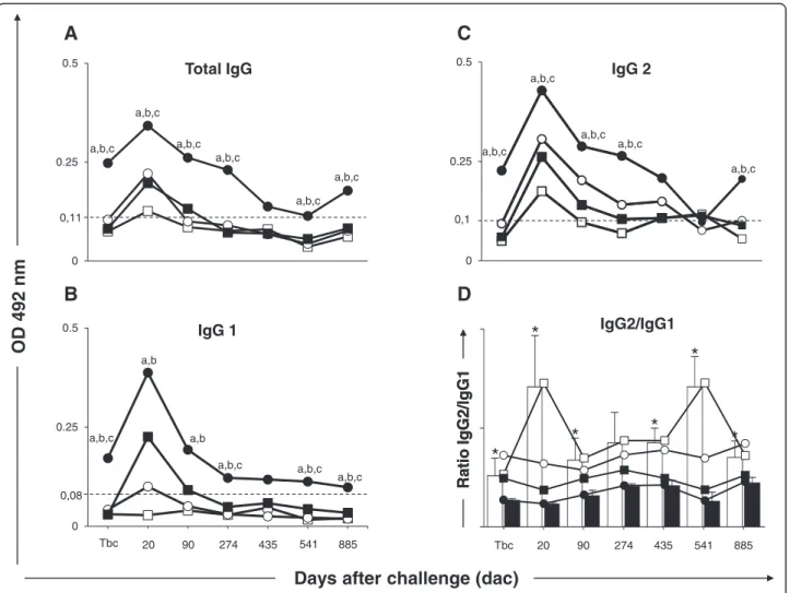
Figure 1Anti-Leishmaniareactivity in serum from dogs submitted to different vaccination protocols before and after intradermal
challenge withL. infantumplus SGE: C (control; ); Sal (SGE; ); LBSal (killedL. braziliensisvaccine plus SGE; ); LBSapSal (killedL.
braziliensisvaccine plus saponin plus SGE; ). IgG2/IgG1 ratio: C (control; ) and LBSapSal (killedL. braziliensisvaccine plus saponin
plus SGE; ). (A)anti–L. infantumtotal IgG;(B)anti–L. infantumIgG1;(C)anti–L. infantumIgG2;(D)IgG2/IgG1 ratio: C (control; white square)
observed in the LBSapSal group compared with the C, Sal, and LBSal groups (Figure 1A). Also, the LBSapSal group elicited higher levels (p< 0.05) of IgG1 at Tbc and 274, 541, and 885 dac compared with the other groups (C, Sal, and LBSal). Moreover, the LBSapSal group showed higher levels (p< 0.05) of IgG1 at 20 and 90 dac compared with C and Sal groups (Figure 1B). Higher levels (p< 0.05) of anti-Leishmania IgG2 were observed in the LBSapSal group at Tbc and 20, 90, 274, and 885 dac compared with other groups (C, Sal, and LBSal) (Figure 1C).
Further analysis demonstrated that the IgG2/IgG1 ra-tio was lower (p< 0.05) in the LBSapSal group compared with C (Tbc and 20, 90, 435, 541, and 885 dac), Sal (Tbc and 20, 90, 435, 541, and 885 dac), and LBSal (Tbc) groups (Figure 1D).
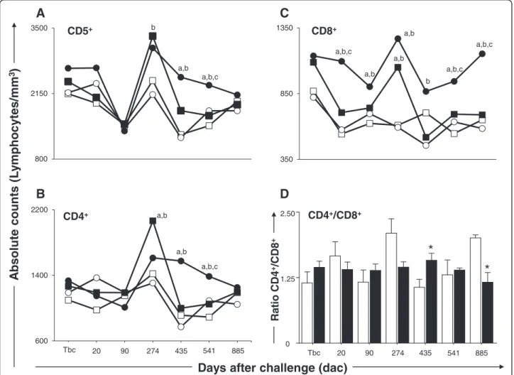
Increased numbers of circulating T lymphocytes and their subsets were displayed in LBSapSal-vaccinated dogs after
L. infantumexperimental challenge
In order to evaluate the cellular immunophenotype pro-file, we enumerated the frequency of T lymphocytes (CD5+) and their major subpopulations (CD4+and CD8+) (Figure 2). Our results revealed an increase (p< 0.05) in the number of circulating CD5+ T lymphocytes in dogs vaccinated with LBSapSal group at 435 dac compared with C and Sal groups and at 541 dac when compared with C, Sal, and LBSal groups (Figure 2A).
Analyses of the data showed an increase (p< 0.05) of circulating CD4+ T lymphocytes in the LBSapSal group compared with the C and Sal groups at 435 dac and the C, Sal, and LBSal groups at 541 dac (Figure 2B). In the same way, higher (p< 0.05) CD8+ T-cell counts were
A
b
solu
te counts
(L
y
m
phoc
ytes/
mm
3
)
Days after challenge (dac)
800 2150
3500 CD5+
a,b b
a,b,c
600 1400 2200
CD4+ a,b
a,b a,b,c
Tbc 20 90 274 435 541 885
350 850
1350 CD8+
a,b,c
b a,b a,b
a,b a,b,c a,b,c
2.50
1.25
0
CD4+/CD8+
*
*
Ra
tio CD4
+/CD8
+
Rati
o CD4
+/C
D
8
+
Tbc 20 90 274 435 541 885
B
A
D
C
Figure 2Cellular profile of circulating lymphocytes in dogs submitted to different vaccination protocols before and after challenge
withL. infantumplus SGE.C (control; ); Sal (SGE; ); LBSal (killedL. braziliensisvaccine plus SGE; ); LBSapSal (killedL. braziliensisvaccine plus saponin plus SGE; ): thex-axis displays the times at which the assays were conducted (Tbc: time before challenge withL. infantum; and 20, 90, 274, 435, 541, and 885 days after challenge [dac] withL. infantum), and they-axis represents the mean values of(A)CD5+,(B)CD4+,(C)
CD8+cells, and(D)CD4+/CD8+ratio, the white and black bars represented the control and LBSapSal groups, respectively. Significant differences
observed in dogs vaccinated with LBSapSal at 20 dac com-pared with the C, Sal, and LBSal groups, at 90 and 274 dac compared with the C and Sal groups, at 435 dac compared with the Sal group, and at 541 and 885 dac in comparison with all other groups (Figure 2C). Further analysis demon-strated that the CD4+/CD8+ratio was higher (p< 0.05) in the LBSapSal group when compared with the C group at 541dac. On the other hand, at 885 dac, the CD4+/CD8+ ratio was lower (p< 0.05) in the LBSapSal group in com-parison with the C group (Figure 2D).
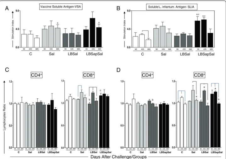
In vitrocell proliferation upon antigenic stimuli is
persistently increased in LBSapSal-vaccinated dogs after
L. infantumexperimental challenge
To explore thein vitrocell proliferation (PBMCs) we used two different antigenic stimuli: VSA (L. braziliensis) in order to evaluate the memory lymphoproliferative immune response against antigens of the vaccine components, and
SLiA to investigate possible lymphoproliferative homology with the etiological agent of VL (L. infantum) (Figure 3). Comparative analysis of the different treatment groups showed a significantly augmented (p< 0.05) stimulation index at 885 dac in the LBSapSal dogs compared with the C (885 dac) groups with VSA stimuli (Figure 3A). In addition, the LBSapSal group exhibited a higher (p< 0.05) lymphoproliferative index at 90 and 435 dac compared with the C and LBSal dogs after SLiA stimuli (Figure 3B).
Higher frequencies of CD4+and CD8+T lymphocytes in antigen-stimulated cultures related to major phenotypic changes in LBSapSal-vaccinated dogs afterL. infantum
experimental challenge
In order to evaluate whether the phenotypic profile of PBMCs in vaccinated/challenged dogs was influenced by VSA or SLiA stimulation, as well as to characterize these cells, we conducted an analysis of the phenotypic features
St
im
ul
at
io
n Index
St
im
ul
at
io
n Index
0.0 4.5 9.0
90 435 885 90 435 885 90 435 885 90 435 885
C Sal LBSal LBSapSal
Soluble L. infantum Antigen - SLiA
a,c a,c
0.0 4.5 9.0
C
90 435 885 90 435 885 90 435 885 90 435 885
Sal LBSal LBSapSal
a
Vaccine Soluble Antigen -VSA
a
Ly
mphocytes
Ratio
Days After Challenge/Groups
0.0 0.5 1.0 1.5
0.0 0.5 1.0 1.5
CD4+ CD8+
b c
0.0 0.5 1.0 1.5
0.0 0.5 1.0 1.5
a
CD4+ CD8+
b
a
a
b
C
90 435 885 90 435 88590 435 885 90 435 885
Sal LBSal LBSapSal C
90 435 885 90 435 885 90 435 88590 435 885
Sal LBSal LBSapSal C
90 435 885 90 435 885 90 435 88590 435 885
Sal LBSal LBSapSal C
90 435 885 90 435 885 90 435 88590 435 885
Sal LBSal LBSapSal
A
C
B
D
Figure 3Cell proliferation response of PBMCs after stimulation with (A) vaccine soluble antigen (VSA) and (B) solubleL. infantum
antigen (SLiA).The lower panels show the immunophenotypic profile ofin vitroPBMCs following stimulation with(C)VSA and(D)SLiA
determined at 90, 435, and 885 dac (days after challenge withL. infantumplus SGE) for vaccinated groups: C (control; ); Sal (SGE; ); LBSal (killedLeishmania braziliensisvaccine plus SGE; ); LBSapSal (killedL. braziliensisvaccine plus saponin plus SGE; ). The results are expressed as the ratio of mean frequencies of CD4+and CD8+cells in the stimulated cultures over non-stimulated cultures (SC/CC). Significant differences
of PBMCs of vaccinated/challenged dogs (Figure 3). In the presence of VSA (Figure 3C), a significant increase (p< 0.05) in the stimulated cell/non-stimulated cell ra-tio of CD8+ T cells was observed in the LBSapSal dogs compared with the LBSal group at 90 dac. When we evaluated the SLiA stimulus (Figure 3D), a significant increase (p< 0.05) in the ratio of CD8+ T cells was ob-served in the LBSapSal dogs at 90 and 435 dac com-pared with 885 dac. In addition, the LBSapSal group showed a higher (p< 0.05) ratio of CD8+T lymphocytes compared with the C dogs at 435 dac. In contrast, the LBSapSal group displayed a significant decrease (p< 0.05) in the CD8+ T-cell ratio compared with the Sal dogs at 885 dac.
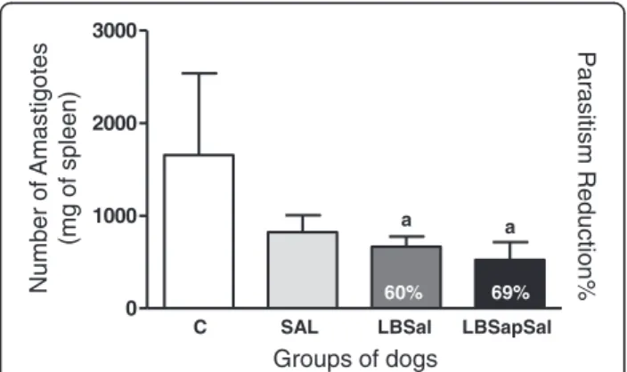
Splenic parasite burden is decreased in LBSapSal-vaccinated dogs afterL. infantumexperimental challenge
The main parasitological features presented by all animals are summarized in Figure 4. As shown in Figure 4, lower (p< 0.05) numbers of amastigotes were observed in the LBSapSal and LBSal groups compared with C dogs. These data are associated with a parasite burden reduction of 60% in LBSal dogs, and 69% in the LBSapSal group com-pared with C animals. These results indicate the high pro-tective potential of the LBSapSal vaccine even for an extended period after challenge (885 dac).
Discussion
The addition of sand fly saliva extract in vector-based vaccines can enhance the ability of the host to control or block the parasitic infection [38-41]. In this sense, our study in dogs evaluated the immunogenicity and efficacy of a vaccine composed of L. braziliensiscrude antigens, saponin as an adjuvant, and sand fly salivary gland ex-tract (LBSapSal vaccine) 885 days after intradermal in-oculation usingL. infantumand SGE ofLu. longipalpis.
In a previous study, our group [14] demonstrated that immunization with LBSapSal vaccine can induce high levels of total IgG, IgG1, and IgG2 anti-Leishmania anti-bodies. Interestingly, the present study demonstrated a significant increase of these immunoglobulins 885 days after experimental challenge. It has been proposed that the serum reactivity observed in vaccinated dogs indi-cates the antigen recognition of L. infantum, suggesting the establishment of immunogenic events [13,14,32]. However, it is still not clear which subclass of immuno-globulin would be associated with a CVL resistance pat-tern. The increases in IgG1 and IgG2 subclasses appear to characterize a mixed profile immune response, as pre-viously described in dogs vaccinated with crude antigens [6,13,14,42,43]. Moreover, there is controversy concerning the association between IgG1 or IgG2 with a profile related to resistance or susceptibility to infection in CVL [44].
Immunophenotyping of canine leukocytes by flow cy-tometry has been used in an attempt to establish pat-terns associated with the cellular profile linked to resistance or susceptibility to infection by L. infantum
[3,45-48], beyond the immunogenic profile in anti-CVL vaccine trials [13,14]. Our results revealed an increase in the number of circulating CD5+ T lymphocytes in dogs vaccinated with LBSal and LBSapSal. This increase was associated with high levels of CD4+ T- and CD8+ T-lymphocyte subpopulations. Interestingly, the LBSapSal vaccine induced a persistent increase in CD8+ T lym-phocytes throughout the period after challenge. In immunophenotypic CVL studies, increases in CD5+ T lymphocytes and subsets of CD4+and CD8+ T lympho-cytes were reported and related to a profile associated with possible resistance against infection by L. infantum
[3,49-51]. In a Brazilian endemic area, vaccination of dogs using Leishmune® was associated with increased frequency of CD8+ T lymphocytes and an absence of clinical signs 18 months after immunization [8]. The in-crease in CD8+ T circulating lymphocytes in dogs vacci-nated against CVL is considered to be an important biomarker of resistance against infection by Leishmania
[13,14]. Thus, the expansion of CD5+ T lymphocytes and the sustained increase of CD4+ and CD8+ T subsets observed in LBSapSal vaccinated dogs seem to reflect the attempt of the immune system to eradicate the para-site after intradermal experimental challenge. This large 0
1000 2000 3000
Parasitism
Reduction%
a a
60% 69%
C SAL LBSal LBSapSal
Number
of
Amastigotes
(mg
of
spleen
)
Groups of dogs
Figure 4Quantification of parasite burden in spleen samples at
885 dac (days after challenge withL. infantumplus SGE) for
vaccinated groups: C (control; ); Sal (SGE; ); LBSal (killedL.
braziliensisvaccine plus SGE; ); LBSapSal (killedL. braziliensis
vaccine plus saponin plus SGE; ).They-axis displays the
antigenic repertoire in the composition of LBSapSal vac-cine probably induces responsive memory T lympho-cytes, which expand in the peripheral blood inducing a transient increase in CD5 and CD4 lymphocytes, due to exposure to parasite in an attempt of the dogs' immune system to control a possible increase or spread of the L. infantumparasite to the organs.
To determine whether the LBSapSal vaccine would be able to activate PBMCs underin vitroantigenic stimula-tion with VSA and SLiA, we measured the stimulastimula-tion index at 90, 435, and 885 dac in cells derived from im-munized dogs. The PBMCs of the Sal and LBSapSal groups were able to recognize and respond to VSA, showing increased stimulation index levels at 885dac compared to the C group. This stimulation observed in PBMC of Sal-vaccinated dogs indicates a nonspecific stimulation and the use of SGE in the experimental chal-lenge could stimulate the immune cells and occasional oscillations occuring in these animals. Moreover, when we stimulated PBMCs with SLiA, an increase in the stimulation index was observed in the LBSapSal group at 90 and 435 dac compared to the C dogs. Interestingly, studies in dogs immunized with a bivalent vaccine com-posed of crude antigens of Leishmaniastrains (L. ama-zonensisand L. braziliensis) associated with BCG as the adjuvant showed greater lymphoproliferative capacity in response to stimulation with antigens of L. infantum
[15]. Furthermore, the ability to stimulate lymphoprolif-erative activity using antigens of Leishmania has been associated with an immune profile related to resistance to CVL [47,49,50], supporting the hypothesis that the LBSapSal vaccine induces a specific immune response against the causative agent of CVL.
The analysis seeking to identify the phenotypic profile of PBMC afterin vitrostimulation (VSA or SLiA) showed an increase in the stimulated ratio of CD8+T lymphocytes in the LBSal and LBSapSal groups in the presence of SLiA compared to the C group. In fact, this result reflects the expansion of circulating CD8+T lymphocytes observed in
ex vivo analysis in the LBSapSal group. In addition, Reis
et al.[3] described high numbers of CD8+ T lymphocytes from the spleens of asymptomatic dogs in the presence of
Leishmaniaantigen compared with symptomatic dogs, in-dicating the importance of these cells to control the L. infantuminfection in dogs. In this sense, both in humans and dogs, asymptomatic infection has been linked to higher Leishmania-specific CD8+ T lymphocytes [52,53]. The data from the in vitro immunophenotypic profile demonstrates a greater ability of the LBSapSal vaccine to control parasites in dogs [3,47] and reflect improved con-trol of tissue parasitism observed in vaccinated dogs. Thus, the LBSapSal-vaccinated dogs showed a Leishmania -spe-cific memory cell consistent with the ability to eliminate parasites after the intradermal experimental challenge.
In the context of the differences between experimental and natural challenges, such as the number of parasites, the life cycle stage (amastigote or promastigote), route of infection, and presence or absence of vector saliva, are im-portant variables that must be taken into account in experi-mental challenges in the canine model. The experiexperi-mental challenge using an intravenous route with high numbers of parasites (107–108promastigotes) seems to be the most ef-ficient challenge model [37]. However, this type of chal-lenge can hide the real experimental vaccine efficacy by suppressing a protective response that could be present in vaccinated dogs [35,54-56]. Thus, an experimental model of infection that most closely resemblesLeishmanianatural transmission is increasingly recommended and desired by many investigators; however, the use of infected sandflies is not easy and remains an obstacle in the dog model. In this work, we used SGE associated with the experimental chal-lenge in an attempt to mimic a sand fly feeding on blood and inoculating an animal with saliva andLeishmania pro-mastigotes. However, our results showed that all dogs remained asymptomatic throughout the follow-up (885 dac), indicating that the use of SGE was not sufficient to replicate the bite environment and enhance experimental infection [21-25].
In this study we selected the spleen as the target tissue for detection of the parasite because it is naturally one of the major sites for the parasites [46]. Further, the real-time PCR technique, used by several authors in order to diagnose and screen the evolution of VL in quantifying the parasite burden, offers high sensitivity, accuracy, and reproducibility [36,57,58]. In our work, we applied the real-time PCR technique to quantify the number of amastigotes per milligram of spleen. Interestingly, we found that the LBSal group (60%) and LBSapSal dogs (69%) had a higher proportion of parasite reduction when compared with the control animals. This reduction in parasite load may be associated with the immune re-sponse induced by salivary components presenting in both vaccines. Some studies show that Lu. longipalpis
salivary proteins induce an immune response associated with protection in dogs [14,29]. Furthermore, in a ham-ster model, salivary protein of a sand fly vector protects against the fatal outcome of visceral leishmaniasis [28]. Thus, the protection obtained in the present study con-firms the capacity of this prototype vaccine (LBSapSal) to limit parasite replication even long after challenge (885 days).
Conclusions
and CD8+T lymphocytes and Leishmania-specific; reduc-tion of parasite load in spleen. The results presented in this work encourage further study of vaccine strategies address-ing Leishmania antigens in combination with proteins present in the saliva of the vector.
Competing interest
The authors have no conflicts of interest.
Authors’contributions
All authors read and approved the final version of the manuscript.
Acknowledgments
The study was supported by Fundação de Amparo à Pesquisa do Estado de Minas Gerais, Brazil (FAPEMIG grant: 02020–09, 02473–10); PAPES V/FIOCRUZ/
CNPq (grant: 403485/2008-8), PDTIS/FIOCRUZ, and CAPES. The authors are grateful for the use of the facilities at Animal Center Science/UFOP, Rede Mineira de Bioterismo (FAPEMIG). The authors also thank Agrohealth Corporation and Chemitec Agro-Veterinária LTDA for support with the provision of medications. NFG, OBR, ATC, OAMF, RCO, RCG, and ABR are grateful to CNPq for fellowships (PQ).
Author details
1
Laboratório de Imunopatologia, Núcleo de Pesquisas em Ciências Biológicas/NUPEB, Instituto de Ciências Exatas e Biológicas, Universidade Federal de Ouro Preto, Ouro Preto, Minas Gerais, Brasil.2Laboratório de Fisiologia de Insetos Hematófagos, Departamento de Parasitologia, Instituto de Ciências Biológicas, Universidade Federal de Minas Gerais, Belo Horizonte, Minas Gerais, Brasil.3Departamento de Microbiologia, Instituto de Ciências Biológicas, Universidade Federal de Minas Gerais, Belo Horizonte, Minas Gerais, Brasil.4Laboratório de Biomarcadores de Diagnóstico e Monitoração, Centro de Pesquisas René Rachou, Fundação Oswaldo Cruz-FIOCRUZ, Belo Horizonte, Minas Gerais, Brasil.5Laboratório de Imunologia Celular e Molecular, Centro de Pesquisas René Rachou, Fundação Oswaldo Cruz-FIOCRUZ, Belo Horizonte, Minas Gerais, Brasil.6Laboratório de Biologia das Interações Celulares, Departamento de Morfologia, Universidade Federal de Minas Gerais, Belo Horizonte, Minas Gerais, Brasil.7Departamento de Análises Clínicas, Escola de Farmácia, Universidade Federal de Ouro Preto, Ouro Preto, Minas Gerais, Brasil.8Instituto Nacional de Ciência e Tecnologia em Doenças Tropicais/INCT-DT, Salvador, Bahia, Brasil.
Received: 25 July 2013 Accepted: 18 January 2014 Published: 7 February 2014
References
1. Coura-Vital W, Marques MJ, Veloso VM, Roatt BM, Aguiar-Soares RD, Reis LE, Braga SL, Morais MH, Reis AB, Carneiro M:Prevalence and factors associated withLeishmania infantuminfection of dogs from an urban area of Brazil as identified by molecular methods.PLoS Negl Trop Dis2011,5(8):e1291. 2. Alvar J, Molina R, San Andres M, Tesouro M, Nieto J, Vitutia M, González F,
San Andrés MD, Boggio J, Rodriguez F,et al:Canine leishmaniasis: clinical, parasitological and entomological follow-up after chemotherapy.Ann Trop Med Parasitol1994,88:371–378.
3. Reis AB, Giunchetti RC, Carrillo E, Martins-Filho OA, Moreno J:Immunity to
Leishmaniaand the rational search for vaccines against canine leishmaniasis.Trends Parasitol2010,26(7):341–349. Review.
4. Lemesre JL, Holzmuller P, Cavaleyra M, Goncalves RB, Hottin G, Papierok G:
Protection against experimental visceral leishmaniasis infection in dogs immunized with purified excreted secreted antigens ofLeishmania infantumpromastigotes.Vaccine2005,23:2825–2840.
5. Lemesre JL, Holzmuller P, Goncalves RB, Bourdoiseau G, Hugnet C, Cavaleyra M, Papierok G:Long-lasting protection against canine visceral leishmaniasis using the LiESAp-MDP vaccine in endemic areas of France: double-blind randomized efficacy field trial.Vaccine2007,25:4223–4234.
6. Fujiwara RT, Vale AM, França da Silva JC, da Costa RT, Quetz Jda S, Martins Filho OA, Reis AB, Corrêa Oliveira R, Machado-Coelho GL, Bueno LL, Bethony JM, Frank G, Nascimento E, Genaro O, Mayrink W, Reed S, Campos-Neto A:Immunogenicity in dogs of three recombinant antigens (TSA, LeIF and LmSTI1) potential vaccine candidates for canine visceral leishmaniasis.Vet Res2005,36:827–838.
7. Borja-Cabrera GP, Correia Pontes NN, da Silva VO, de Souza Paraguai E, Santos WR, Gomes EM, Luz KG, Palatnik M, Palatnik de Sousa CB:Long lasting protection against canine kala-azar using the FML-QuilA saponin vaccine in an endemic area of Brazil (Sao Goncalo do Amarante, RN).
Vaccine2002,20:3277–3284.
8. Borja-Cabrera GP, Santos FN, Bauer FS, Parra LE, Menz I, Morgado AA, Soares IS, Batista LM, Palatnik-de-Sousa CB:Immunogenicity assay of the Leishmune vaccine against canine visceral leishmaniasis in Brazil.Vaccine2008,
26:4991–4997.
9. Fernandes AP, Costa MM, Coelho EA, Michalick MS, de Freitas E, Melo MN, Luiz Tafuri W, Resende Dde M, Hermont V, Abrantes Cde F, Gazzinelli RT:
Protective immunity against challenge withLeishmania (Leishmania) chagasiin beagle dogs vaccinated with recombinant A2 protein.Vaccine 2008,26:5888–5895.
10. Mayrink W, Genaro O, Silva JC, da Costa RT, Tafuri WL, Toledo VP, da Silva AR, Reis AB, Williams P, da Costa PW:Phase I and II open clinical trials of a vaccine againstLeishmania chagasiinfections in dogs.Mem Inst Oswaldo Cruz1996,91:695–697.
11. Lasri S, Sahibi H, Sadak A, Jaffe CL, Rhalem A:A Immune responses in vaccinated dogs with autoclavedLeishmania majorpromastigotes.Vet Res1999,30:441–449.
12. Panaro MA, Acquafredda A, Lisi S, Lofrumento DD, Mitolo V, Sisto M, Fasanella A, Trotta T, Bertani F, Consenti B, Brandonisio O:Nitric oxide production by macrophages of dogs vaccinated with killedLeishmania infantum
promastigotes.Comp Immunol Microbiol Infect Dis2001,24:187–195.
13. Giunchetti RC, Correa-Oliveira R, Martins-Filho OA, Teixeira-Carvalho A, Roatt BM, de Oliveira Aguiar-Soares RD, de Souza JV, das Dores Moreira N, Malaquias LC, Mota e Castro LL, de Lana M, Reis AB:Immunogenicity of a killedLeishmania
vaccine with saponin adjuvant in dogs.Vaccine2007,25:7674–7686.
14. Giunchetti RC, Correa-Oliveira R, Martins-Filho OA, Teixeira-Carvalho A, Roatt BM, de Oliveira Aguiar-Soares RD, Coura-Vital W, de Abreu RT, Malaquias LC, Gontijo NF, Brodskyn C, de Oliveira CI, Costa DJ, de Lana M, Reis AB:A killed
Leishmaniavaccine with sand fly saliva extract and saponin adjuvant displays immunogenicity in dogs.Vaccine2008,26:623–638.
15. Giunchetti RC, Reis AB, da Silveira-Lemos D, Martins-Filho OA, Correa-Oliveira R, Bethony J, Vale AM, da Silva Quetz J, Bueno LL, França-Silva JC, Nascimento E, Mayrink W, Fujiwara RT:Antigenicity of a whole parasite vaccine as promising candidate against canine leishmaniasis.Res Vet Sci 2008,85:106–112.
16. Carcelen J, Iniesta V, Fernandez-Cotrina J, Serrano F, Parejo JC, Corraliza I, Gallardo-Soler A, Marañón F, Soto M, Alonso C, Gómez-Nieto C:The chimerical multi-component Q protein fromLeishmaniain the absence of adjuvant protects dogs against an experimentalLeishmania infantum
infection.Vaccine2009,27:5964–5973.
17. Fiuza JA, Santiago Hda C, Selvapandiyan A, Gannavaram S, Ricci ND, Bueno LL, Bartholomeu DC, Correa-Oliveira R, Nakhasi HL, Fujiwara RT:Induction of immunogenicity by live attenuatedLeishmania donovanicentrin deleted parasites in dogs.Vaccine2013,31(14):1785–1792.
18. Moreno J, Vouldoukis I, Martin V, McGahie D, Cuisinier AM, Gueguen S:Use of a LiESP/QA-21 vaccine (CaniLeish) stimulates an appropriate Th1-dominated cell-mediated immune response in dogs.PLoS Negl Trop Dis 2012,6(6):e1683.
19. Ravindran R, Ali N:Progress in vaccine research and possible effector mechanisms in visceral leishmaniasis.Curr Mol Med2004,4(6):697–709.
20. Mohebali M, Khamesipour A, Mobedi I, Zarei Z, Hashemi-Fesharki R: Double-blind randomized efficacy field trial of alum precipitated autoclaved Leishmania major vaccine mixed with BCG against canine visceral leishmaniasis in Meshkin-Shahr district, I.R. Iran.Vaccine2004,22:4097–4100.
21. Titus RG, Ribeiro JM:Salivary gland lysates from the sand flyLutzomyia longipalpisenhanceLeishmaniainfectivity.Science1988,239(4845):1306–1308.
22. Samuelson J, Lerner E, Tesh R, Titus R:A mouse model ofLeishmania braziliensis braziliensisinfection produced by coinjection with sand fly saliva.J Exp Med1991,173(1):49–54.
23. Warburg A, Saraiva E, Lanzaro GC, Titus RG, Neva F:Saliva ofLutzomyia longipalpissibling species differs in its composition and capacity to enhance leishmaniasis.Philos Trans R Soc B1994,345(1312):223–230.
24. Lima HC, Titus RG:Effects of sand fly vector saliva on development of cutaneous lesions and the immune response toLeishmania braziliensisin BALB/c mice.Infect Immun1996,64(12):5442–5445.
EnhancedLeishmania braziliensisinfection following pre-exposure to sandfly saliva.PLoS Negl Trop Dis2007,1:e84.
26. Belkaid Y, Kamhawi S, Modi G, Valenzuela J, Noben-Trauth N, Rowton E, Ribeiro J, Sacks DL:Development of a natural model of cutaneous leishmaniasis: powerful effects of vector saliva and saliva pre-exposure on the long-term outcome ofLeishmania majorinfection in the mouse ear dermis.J Exp Med1998,188(10):1941–1953.
27. Kamhawi S, Belkaid Y, Modi G, Rowton E, Sacks D:Protection against cutaneous leishmaniasis resulting from bites of uninfected sand flies.
Science2000,290(5495):1351–1354.
28. Gomes R, Teixeira C, Teixeira MJ, Oliveira F, Menezes MJ, Silva C, de Oliveira CI, Miranda JC, Elnaiem DE, Kamhawi S, Valenzuela JG, Brodskyn CI:Immunity to a salivary protein of a sand fly vector protects against the fatal outcome of visceral leishmaniasis in a hamster model.Proc Natl Acad Sci U S A2008,
105:7845–7850.
29. Collin N, Gomes R, Teixeira C, Cheng L, Laughinghouse A, Ward JM, Elnaiem DE, Fischer L, Valenzuela JG, Kamhawi S:Sand Fly Salivary Proteins Induce Strong Cellular Immunity in a Natural Reservoir of Visceral Leishmaniasis with Adverse Consequences forLeishmania.PLoS Pathog2009,5(5):e1000441. 30. Modi GB, Tesh RB:A simple technique for mass rearingLutzomyia
longipalpisandPhlebotomus papatasi(Diptera: Psychodidae) in the laboratory.J Med Entomol1983,20(5):568–569.
31. Cavalcante RR, Pereira MH, Gontijo NF:Anti-complement activity in the saliva of phlebotomine sand flies and other haematophagous insects.
Parasitology2003,127(1):87–93.
32. Roatt BM, Aguiar-Soares RD, Vitoriano-Souza J, Coura-Vital W, Braga SL, Corrêa-Oliveira R, Martins-Filho OA, Teixeira-Carvalho A, de Lana M, Figueiredo Gontijo N, Marques MJ, Giunchetti RC, Reis AB:Performance of LBSap vaccine after intradermal challenge withL. infantumand saliva of
Lu. longipalpis: immunogenicity and parasitological evaluation.PLoS One 2012,7(11):e49780.
33. Costa DJ, Carvalho RM, Abbehusen M, Teixeira C, Pitombo M, Trigo J, Nascimento F, Amorim L, Abreu-Silva AL, do Socorro Pires Cruz M, Miranda JC, Fukutani K, de Oliveira CI, Barral A, Barral-Netto M, Brodskyn C:Experimental infection of dogs withLeishmaniaand saliva as a model to study canine visceral leishmaniasis.PLoS One2013,8(4):e60535.
34. Paranhos M, dos Santos WC, Sherlock I, Oliveira GG, de Carvalho LC:
Development of eosinophilia in dogs intradermically inoculated with sand fly saliva andLeishmania (Leishmania) chagasistationary-phase promastigotes.Mem Inst Oswaldo Cruz1993,88(2):249–251.
35. Reis AB, Martins-Filho OA, Teixeira-Carvalho A, Carvalho MG, Mayrink W, França-Silva JC, Giunchetti RC, Genaro O, Corrêa-Oliveira R:Parasite density and impaired biochemical/hematological status are associated with severe clinical aspects of canine visceral leishmaniasis.Res Vet Sci2006,
81:68–75.
36. Bretagne S, Durand R, Olivi M, Garin JF, Sulahian A, Rivollet D, Vidaud M, Deniau M:Real-time PCR as a new tool for quantifyingLeishmania infantum
in liver in infected mice.Clin Diagn Lab Immunol2001,8:828–831.
37. Moreira ND, Vitoriano-Souza J, Roatt BM, Vieira PMA, Ker HG, de Oliveira Cardoso JM, Giunchetti RC, Carneiro CM, de Lana M, Reis AB:Parasite burden in hamsters infected with two different strains ofLeishmania
(Leishmania)infantum:“Leishman Donovan Units”versus real-time PCR.
PLoS One2012,10:1371. journal.pone.0047907.
38. Titus RG, Bishop JV, Mejia JS:The immunomodulatory factors of arthropod saliva and the potential for these factors to serve as vaccine targets to prevent pathogen transmission.Parasite Immunol2006,28(4):131–141.
39. Brodskyn C, de Oliveira CI, Barral A, Barral-Netto M:Vaccines in leishmaniasis: advances in the last five years.Expert Rev Vaccines2003,2(5):705–717.
40. Kamhawi S:The biological and immunomodulatory properties of sand fly saliva and its role in the establishment ofLeishmaniainfections.Microbes Infect2000,2(14):1765–1773.
41. Mejia JS, Bishop JV, Titus RG:Is it possible to develop panarthropod vaccines?Trends Parasitol2006,22(8):367–370.
42. Araújo MS, de Andrade RA, Sathler-Avelar R, Magalhães CP, Carvalho AT, Andrade MC, Campolina SS, Mello MN, Vianna LR, Mayrink W, Reis AB, Malaquias LC, Rocha LM, Martins-Filho OA:Immunological changes in canine peripheral blood leukocytes triggered by immunization with first or second generation vaccines against canine visceral leishmaniasis.Vet Immunol Immunopathol2011,141(1-2):64–75.
43. Araujo MS, de Andrade RA, Sathler-Avelar R, Teixeira-Carvalho A, Andrade MC, Vianna LR, Mayrink W, Reis AB, Malaquias LC, Mello MN, Martins-Filho OA:
T-cell-derived cytokines, nitric oxide production by peripheral blood monocytes and seric anti-Leishmania(Leishmania)chagasiIgG subclass patterns following immunization against canine visceral leishmaniasis using Leishvaccine and Leishmune.Vaccine2009,27:1008–1017.
44. Day MJ:Immunoglobulin G subclass distribution in canine leishmaniosis: a review and analysis of pitfalls in interpretation.Vet Parasitol2007,147:2–8.
45. Araujo MS, de Andrade RA, Vianna LR, Mayrink W, Reis AB, Sathler-Avelar R, Teixeira-Carvalho A, Andrade MC, Mello MN, Martins-Filho OA:Despite Leishvaccine and Leishmune trigger distinct immune profiles, their ability to activate phagocytes and CD8+ T-cells support their high-quality immunogenic potential against canine visceral leishmaniasis.
Vaccine2008,26:2211–2224.
46. Reis AB, Teixeira-Carvalho A, Giunchetti RC, Guerra LL, Carvalho MG, Mayrink W, Genaro O, Corrêa-Oliveira R, Martins-Filho OA:Phenotypic features of circulating leucocytes as immunological markers for clinical status and bone marrow parasite density in dogs naturally infected byLeishmania chagasi.Clin Exp Immunol2006,146:303–311.
47. Reis AB, Martins-Filho OA, Teixeira-Carvalho A, Giunchetti RC, Carneiro CM, Mayrink W, Tafuri WL, Corrêa-Oliveira R:Systemic and compartmentalized immune response in canine visceral leishmaniasis.Vet Immunol Immunopathol2009,128:87–95.
48. Giunchetti RC, Martins-Filho OA, Carneiro CM, Mayrink W, Marques MJ, Tafuri WL, Correa-Oliveira R, Reis AB:Histopathology, parasite density and cell phenotypes of the popliteal lymph node in canine visceral leishmaniasis.Vet Immunol Immunopathol2008,121:23–33.
49. Pinelli E, Boog CJ, Rutten VP, Van DB, Bernadina WE, Ruitenberg EJ:A canine CD8+ cytotoxic T-cell line specific forLeishmania infantum
infected macrophages.Tissue Antigens1994,43:189–192.
50. Pinelli E, Killick-Kendrick R, Wagenaar J, Bernadina W, Del Real G, Ruitenberg J:
Cellular and humoral immune responses in dogs experimentally and naturally infected withLeishmania infantum.Infect Immun1994,62:229–235.
51. Pinelli E:Cytokines in canine visceral leishmaniasis. InCytokines in veterinary medicine.Edited by Schijns HM. Netherlands: Utrecht University; 1997:217–247.
52. Pinelli E, Gonzalo RM, Boog CJ, Rutten VP, Gebhard D, Del Real G, Ruitenberg J:Leishmania infantum-specific T cell lines derived from asymptomatic dogs that lyse infected macrophages in a major histocompatibility complex-restricted manner.Eur J Immunol1995,
25:1594–1600.
53. Mary C, Auriault V, Faugere B, Dessein AJ:Control ofLeishmania infantum
infection is associated with CD8(+) and gamma interferon- and
interleukin-5-producing CD4(+) antigen-specific T cells.Infect Immun 1999,67:5559–5566.
54. Poot J, Rogers ME, Bates PA, Vermeulen A:Detailed analysis of an experimental challenge model forLeishmania infantum(Jpc strain) in dogs.Vet Parasitol2005,130(1–2):41–53.
55. Peters NC, Egen JG, Secundino N, Debrabant A, Kimblin N, Kamhawi S, Lawyer P, Fay MP, Germain RN, Sacks D:In vivoimaging reveals an essential role for neutrophils in leishmaniasis transmitted by sand flies.
Science2008,321:970–974.
56. Peters NC, Kimblin N, Secundino N, Kamhawi S, Lawyer P, Sacks DL:Vector transmission ofLeishmaniaabrogates vaccine-induced protective immunity.PLoS Pathog2009,5:e1000484.
57. Manna L, Reale S, Viola E, Vitale F, Foglia Manzillo V, Pavone LM, Caracappa S, Gravino AE:Leishmania DNA load and cytokine expression levels in asymptomatic naturally infected dogs.Vet Parasitol2006,142:271–280.
58. Alves CF, de Amorim IF, Moura EP, Ribeiro RR, Michalick MS, Kalapothakis E, Bruna-Romero O, Tafuri WL, Teixeira MM, Melo MN:Expression of IFN-gamma, TNF-alpha, IL-10 and TGF-beta in lymph nodes associates with parasite load and clinical form of disease in dogs naturally infected with
Leishmania(Leishmania)chagasi.Vet Immunol Immunopathol2009,
128:343–349.
doi:10.1186/1756-3305-7-61
Cite this article as:Aguiar-Soareset al.:LBSapSal-vaccinated dogs exhibit increased circulating T-lymphocyte subsets (CD4+and CD8+) as well as
a reduction of parasitism after challenge withLeishmania infantumplus