Expression levels of candidate genes for intramuscular fat deposition
in two Banna mini-pig inbred lines divergently selected for fatness traits
Su-Mei Zhao
1#, Wei-Zhen Li
2#, Hong-Bin Pan
1, Ying Huang
1, Ming-Hua Yang
1, Hong-Jiang Wei
1and Shi-Zheng Gao
11
Yunnan Key Laboratory of Animal Nutrition and Feed Science, Yunnan Agricultural University,
Kunming, China.
2
College of Animal Science and Technology, Yunnan Agricultural University, Kunming, China.
Abstract
Intramuscular fat (IMF) content plays an important role in meat quality. Many genes involved in lipid and energy me-tabolism were identified as candidate genes for IMF deposition, since genetic polymorphisms within these genes were associated with IMF content. However, there is less information on the expression levels of these genes in the muscle tissue. This study aimed at investigating the expression levels of sterol regulating element binding protein-1c (SREBP-1c), diacylglycerol acyltransferase (DGAT-1), heart-fatty acids binding protein (H-FABP), leptin receptor (LEPR) and melanocortin 4 receptor (MC4R) genes and proteins in two divergent Banna mini-pig inbred lines (BMIL). A similar growth performance was found in both the fat and the lean BMIL. The fat meat and IMF content in the fat BMIL were significantly higher than in the lean BMIL, but the lean meat content was lower. The serum triacylglycerol (TAG) and free fatty acid (FFA) contents were significantly higher in the fat than in the lean BMIL. The expression levels of SREBP-1c, DGAT-1 and H-FABP genes and proteins in fat BMIL were increased compared to the lean BMIL. However, the expression levels of LEPR and MC4R genes and proteins were lower.
Keywords: intramuscular fat (IMF), expression levels, Banna mini-pig inbred line (BMIL).
Received: December 19, 2011; Accepted: June 28, 2012.
Introduction
In pig production, intramuscular fat (IMF) content is an important determinant of meat quality characteristics such as tenderness, juiciness and flavor (Fernandezet al., 1999). Because of the importance of IMF on the economics of pig production, it is important to elucidate the molecular mechanisms underlying intramuscular fat deposition (Woodet al., 2008). Particularly, in the Chinese local pigs, the IMF content is higher than in other commercial pig breeds (Zhao et al., 2009), which makes them an ideal model for elucidating the mechanism of IMF deposition.
During the past decades, many efforts have been made to appoint possible candidate genes for IMF deposi-tion (Schwabet al., 2009). Sterol regulating element bind-ing protein-1c (SREBP-1c), acyl CoA:diacylglycerol acyl-transferase (DGAT-1) (Mourot and Kouba, 1998; Dinget al., 2000; Chenet al., 2008; Switonskiet al., 2010) and the intracellular heart fatty acid binding protein (H-FABP) af-fect the IMF content in pigs (Gerbenset al., 1997, 1998,
1999, 2000; Glatzet al., 2003; Gardanet al., 2007; Liet al., 2007; Liet al., 2010; Switonskiet al., 2010). Genetic vari-ants of the leptin receptor (LEPR) and the melanocortin 4 receptor (MC4R) genes were reported to be associated with IMF deposition (Mackowskiet al., 2005; Oviloet al., 2005; Bruunet al., 2006; Jokubkaet al., 2006; Kimet al., 2006; Piórkowskaet al., 2010; Fanet al., 2009, 2010; Liet al., 2010; Switonskiet al., 2010). Although LEPR and MC4R are mainly studied in hypothalamus, some reports indicated that they could be expressed in muscle tissue as well (Stinckenset al., 2009; Liet al., 2010; Larkinaet al., 2011; Tyraet al., 2011). Global genomics analyses also led to the hypothesis that IMF deposition could be affected by both lipid metabolism and energy metabolism (Liuet al., 2009; Cánovaset al., 2010). However, most of the studies only focused on lipogenic genes, ignoring energy metabolism genes (Mourot and Kouba, 1998; Zhaoet al., 2009). More-over, these reports usually used different pig breeds as fat and lean models, which were fed diets according to their in-dividual nutritional standard. These studies could not rule out the possibility that the different expressions of genes might be due to breed characteristics rather than to actual variations in IMF deposition.
Send correspondence to Shi-Zheng Gao. Yunnan Key Laboratory of Animal Nutrition and Feed Science, Yunnan Agricultural Univer-sity, Kunming 650201, China. E-mail: [email protected]. #These authors contributed equally to this work and should both be regarded as the first authors.
The Banna Mini-pig Inbred Line (BMIL) was estab-lished in China in 1980 (Huoet al., 2012). The original an-cestors were a sow and her son. The further reproduction occurred by full-sibling or parent-offspring mating. Pro-gressive inbreeding generated the fat and the lean BMIL. The body weight of mature pigs reaches about 45.00 ±
1.50 kg. They are ideal experimental models for a better un-derstanding of the genetic mechanisms leading to divergent fat deposition phenotypes, because they have the same or a very similar background genotype and were fed the same diets in the same environment (Guoet al., 2008). The aim of this study was to compare the expression levels of the SREBP-1c, DGAT-1, H-FABP, LEPR and MC4R genes and proteins in fat and lean BMIL and to explain the phenotypic variations of IMF deposition in these two diver-gent BMILs.
Materials and Methods
The animal experiments and protocols used in this study were approved by the Yunnan Agricultural Univer-sity Institutional Animal Care and Use Committee. The fat and lean BMIL were fed in the Yunnan Key Laboratory of BMIL, Yunnan Agricultural University, China.
Fat and lean BMIL animals of the 25th generation were selected, with an inbreeding coefficient of 99.56%. A total of twelve healthy pigs were fed ad libitum the same corn-soybean diets (Table 1) in the same environmental
conditions for 1.5-1.6 ± 0.45 years. Their body weights were determined every week, and their growth perfor-mances were calculated. Then the pigs were slaughtered by exsanguination after electrical stunning, and their hot car-cass weight was measured. The fat-free lean and fat con-tents were calculated from the left side of the carcass, after a 20 h chill at 2 °C. The average backfat thickness of the first, tenth, last-rib and last-lumbar sites were calculated. Longissimus muscle samples from the last ribs were col-lected from each one of the animals, snap-frozen in liquid nitrogen, and stored at -80 °C prior to analysis.
IMF content
The IMF content, expressed as weight percentage of dry muscle tissue, was evaluated 24 h after slaughter, using the Soxhlet petroleum-ether extraction method.
Serum parameters
Blood samples were centrifuged at 600 x g for 15 min. Serum was obtained and stored at -20 °C for posterior anal-yses. Serum FFA and TG concentrations were determined using the respective commercial kit assays (Nanjing Jiancheng Biochemical Reagent Co., Nanjing, China) ac-cording to the protocols provided by the manufacturer.
RNA extraction and Reverse transcription (RT)
Total RNA was extracted from the muscle tissues us-ing Trizol reagent accordus-ing to the manufacturer’s protocol (Takala, Japan). Total RNA concentration was then quanti-fied by measuring the absorbance at 260 nm in an Eppen-dorf Biospectrophotometer (EppenEppen-dorf AG, Hamburg, Germany). The absorption ratios (260/280 nm) ranged from 1.8 to 2.0. The integrity of the RNA was verified by 1.4% agarose-formaldehyde gel electrophoresis. Total RNA (2mg) was then reverse-transcribed by incubation at 42 °C for 1 h in a 25 mL mixture consisting of MMLV (Promega Co., Shanghai, China), RNase inhibitor (Pro-mega), and oligo dT. The reaction was terminated by heat-ing to 95 °C for 5 min and quick coolheat-ing oni.e.cDNA was stored at -20 °C for PCR amplification.
Real-Time Quantitative PCR (RT-qPCR)
RT-qPCR was performed to quantify the mRNA ex-pression abundance of genes SREBP1c, H-FABP, DGAT1, LEPR and MC4R. ß-actin was used as internal control. The 25mL PCR mixture contained 12.5mL of 2iQ SYBR Green Supermix, 0.5mL (10 mM) of each primer (Table 2), and 1mL of cDNA. Mixtures were incubated in an iCyler iQ Real-time Detection system (Bio-Rad Laboratories, Hercu-les, CA, USA). A melting curve analysis was performed to ensure that only a single PCR product was amplified. Con-trol reactions were set for each sample. PCR amplification efficiencies were between 91% and 103%. The amount of specific target was calculated according to the following
Table 1- Composition of diets.
Percentage (%)
Corn 68.98
Wheat bran 22.53
Soybean meal 5.62
Fish meal 0.50
Limestone 0.97
Monocalcium phosphate 0.10
Salt 0.30
Premix1 1.00
Calculated nutritional composition
Crude protein 10.42
Total Lysine 0.69
Digestive energy (MJ/kg) 13.11
Calcium 0.46
Total phosphorus 0.37
Available phosphorus 0.14
1
formula: ratio = 2-DCTtarget (sample - calibrator)/2-DCT ß-actin (sample -
cali-brator)
. All primers used were designed by Primer Premier 5 and synthesized by Shanghai Shenggong Biological Com-pany (Shanghai, China).
Western blotting
Muscle samples were collected and homogenized on ice in 700mL buffer A [50 mM Tris-HCl (pH 7.5), 50 mM NaF, 5 mM sodium pyrophosphate, 1 mM EDTA, 1 mM DTT, 0.1 mM phenylmethylsulfonyl fluoride, 10% glyc-erol] containing 1% Triton X-100, 5 mM aprotinin, leu-peptin and pepstatin. The lysates were centrifuged at 6000 g for 20 min at 4 °C to remove insoluble material. Thereafter, supernatant extracts were collected and protein concentra-tion determined, using the method described by Bradford (1976). Then the extracts were frozen at -80 °C until the western blot analyses were performed.
To measure SREBP-1c, DGAT-1, H-FABP, LEPR and MC4R protein expression, 50mg of total whole-cell protein extract were separated by sodium dodecyl sulfate-polyacrylamide gel electrophoresis (12% resolving gel), transferred to a nitrocellulose membrane and probed over-night, with rabbit polyclonal anti- SREBP-1c, anti-DGAT-1, anti-H-FABP, anti-LEPR, and anti-MC4R antibodies (Sigma, USA) at 1:500, 1:500, 1:1000, 1:1000 and 1:2000 dilutions, respectively. The membranes were then probed with a goat anti-rabbit IgG-horseradish peroxidase conju-gate (1:20,000 dilution) (Sigma, USA) for 1 h at room tem-perature. Blots were developed using the SuperSignal West Pico Chemiluminescent Substrate system (Bio-Rad, Her-cules, CA, USA) and imaged on X-film, for image analysis and densitometry. Signal intensity was quantified using the Quantity One 1-D analysis software (Bio-Rad, Hercules, CA, USA).
Statistical analysis
Data regarding growth performance, sera parameters and mRNA expression were analyzed using pairedt-tests with the Statistical Packages for Social Science (SPSS) 12.0 software. All data were presented as mean±S.E.M. Differences were considered statistically significant if p < 0.05.
Results
Growth performance, carcass traits and serum parameters
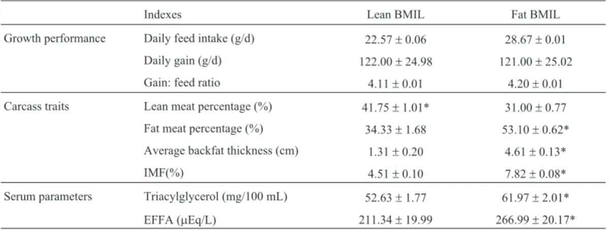
Growth performance, carcass traits and serum param-eters are presented in Table 3. There was no significant dif-ference in growth performance between the fat and lean BMIL (p > 0.05). A significant reduction in fat percentage was found in the fat BMIL compared to the lean BMIL (p < 0.05). A significant increase of lean meat was observed in the fat BMIL (p < 0.05). The serum concentrations of TG and FFA as well as the IMF content were significantly higher in the fat BMIL than in the lean BMIL (p < 0.05).
Expression levels of genes and proteins
Figures 1 and 2 show the expression levels of the SREBP1c, H-FABP, DGAT1, LEPR and MC4R genes and proteins in muscle tissue of fat and lean BMIL animals. The expression levels of the H-FABP, SREBP-1c and DGAT1 genes and proteins were significantly higher in the fat BMIL than in the lean BMIL (p < 0.05). The mRNA and protein abundance of MC4R and LEPR was lower in the fat BMIL compared to the lean BMIL. The difference in LEPR gene expression between the fat and lean BMIL was signif-icant (p < 0.05).
Table 2- Specific primers used for real-time quantitative PCR (RT-qPCR)
Gene name Sequence Product size Position Accession number
b-actin F: 5’-GCCGCACCACTGGCATTGTC-3’ 399 bp Exon 2 DQ845171
R: 5’-AGGTAGTTTCGTGGATGCCGCAG-3’ Exon 3
SREBP-1c F: 5’-GCGACGGTGCCTCTGGTAGT-3’ 218 bp Exon 1 AY496867
R: 5’-CGCAAGACGGCGGATTTA-3’ Exon 2
H-FABP F: 5’-CCTGGAAGCTAGTGGACAGC-3’ 227 bp Exon 1 AJ416019
R: 5’-TGCCTCTTTCTCGTAAGTGC-3’ Exon 2
DGAT-1 F: 5’- AAGGACGGACACGACGATG-3’ 289 bp Exon 1 AY093657
R: 5’- GGAACGCAGTCACAGCAAAG-3’ Exon 5
LEPR F: 5’-GTGATAACTGCATTTGACTTGGC-3’ 285 bp Exon 1 NM001024587
R: 5’-CTGCAATGTTGTCTGCATGTACAG-3’ Exon 1
MC4R F: 5’-TGGAGAAAATCGCTGAGGCTACC-3’ 632 bp Exon1 NM214173
Discussion
In this study, the growth performance of fat and lean BMIL animals showed no significant difference. However,
an increase in fat meat as well as in IMF content and re-duced lean meat content in the fat BMIL compared to the lean BMIL was found. As a rule, animal obesity models
Figure 1- mRNA expression levels of SREBP-1c, H-FABP, DGAT1, LEPR and MC4R (A, B, C, D and E, respectively) in the fat and lean BMIL. Means ±SE with asterisk indicate a significant difference between the two groups (p < 0.05).
Table 3- Growth performance, carcass traits and serum parameters
Indexes Lean BMIL Fat BMIL
Growth performance Daily feed intake (g/d) 22.57±0.06 28.67±0.01
Daily gain (g/d) 122.00±24.98 121.00±25.02
Gain: feed ratio 4.11±0.01 4.20±0.01
Carcass traits Lean meat percentage (%) 41.75±1.01* 31.00±0.77
Fat meat percentage (%) 34.33±1.68 53.10±0.62*
Average backfat thickness (cm) 1.31±0.20 4.61±0.13*
IMF(%) 4.51±0.10 7.82±0.08*
Serum parameters Triacylglycerol (mg/100 mL) 52.63±1.77 61.97±2.01*
EFFA (mEq/L) 211.34±19.99 266.99±20.17*
weigh more than their lean counterparts. However, a simi-lar feed intake was also reported in the lean Yorkshire and fat Ossabaw pigs (Wangsnesset al., 1980). This may sug-gest that the different fat deposition in the fat and lean BMIL pigs does not result from their feed intake, but rather from their genotypes for fatness traits.
TAG, the major component of IMF in muscles, is stored within intramuscular adipocytes (Gao and Zhao, 2009). It could be synthesized using fatty acids which were
de novosynthesized or originated from feed nutrients. Hau-seret al. (1997) reported that the plasma of fat pigs had a higher percentage of triglycerides than the plasma of lean pigs. Our results also showed that the fat BMIL had higher serum TAG and FFA concentrations as well as IMF content compared with the lean BMIL. These data indicate that more TAG or FFA from feed nutrients may be allotted to the muscle tissue of fat BMIL, leading to a higher IMF de-position than in the lean BMIL.
In general, an increase in IMF content is mainly due to an increase in TG contents (Fernandezet al., 1999).
Con-sequently, the TG metabolism in muscle tissue should be a target for identification of genes involved in IMF deposi-tion, especially the TAG synthesis metabolism. SREBP-1c can stimulate the transcription of genes encoding acetyl-CoA carboxylase (ACC) (Zhuanget al., 2003) and fatty acid synthase (FAS) enzymes (Maganaet al., 2000). H-FABP plays a role in transporting the fatty acids, and has been regarded as a candidate gene for IMF deposition in pigs (Gerbens et al., 1998; Glatz et al., 2003; Chmur-zynska, 2006; Damonet al., 2006; Gardan et al., 2007). DGAT are microsomal enzymes which catalyze the final step of the triglyceride synthesis pathway (Yen et al., 2008). Overexpression of DGAT enzymes in transgenic mice is sufficient to increase cellular TAG storage in skele-tal muscle (Koliwadet al., 2010). Our results show that the expression levels of the SREBP-1c, H-FABP and DGAT-1 genes and proteins in the fat BMIL pigs were higher than in the lean BMIL pigs. However, the difference in DGAT-1 protein expression was not significant. The expression of the DGAT-1 gene and protein was positively correlated
(Coleman and Lee, 2004). The results suggest that, in the fat BMIL, more fatty acids would be synthesizedde novo
and transported to store TAG.
Intramuscular TG is a major form of energy storage and represents a significant fuel for cellular metabolism (Gao and Zhao, 2009). MC4R and LEPR play an important role in the control of energy homeostasis. Many reports have shown that the genetic variants of MC4R and LEPR are associated with porcine fat deposition (Mackowskiet al., 2005; Oviloet al., 2005; Bruunet al., 2006; Jokubkaet al., 2006; Kimet al., 2006; Fanet al., 2009, 2010; Liet al., 2010; Piórkowskaet al., 2010; Switonskiet al., 2010). The LEPR gene mediates the regulation of leptin effects (Halaas and Friedman, 1997; Houseknecht and Porto-carrero, 1998;Liet al., 2010). The MC4R gene has been most closely linked to controlling the energy balance in ro-dents. The interaction between melanocortins and their re-ceptors in the hypothalamus is one of the main neuro-endocrine pathways controlling energy balance (Yeoet al., 2000; Wardlaw, 2001). However, some studies have indi-cated that these genes could also be expressed in the muscle tissue (Stinckenset al., 2009; Liet al., 2010; Larkinaet al., 2011; Tyraet al., 2011). Our data show that the mRNA and protein abundance of LEPR and MC4R in the fat BMIL was lower than in the lean BMIL. However, the difference in MC4R protein expression was not significant. This may be due to the fact that the main function of MC4R is to regu-late the food intake (Kimet al., 2006). Since in the present study the feed intake by the fat and the lean BMIL pigs was similar,this may suggestthat the IMF deposition could be affected by energy metabolism-related genes.
In conclusion, fat and lean BMIL with divergent phe-notypes for IMF content did not result from feeding differ-ences, but rather were influenced by feed nutrients TAG and FFA and the expression levels of genes and proteins which participate in the TAG synthesis process and the en-ergy metabolism.
Acknowledgments
This work was supported by the National Key Pro-gram of Transgenic Project of China (No. 2009ZX08009-140B), the National Key Foundation Re-search Development Project of China (973 Project, number 2007CB116201), the National Natural Science Foundation of China (No.30660132 and 31060331) and the Yunnan Natural Science Foundation of China (No. 2009CD056). The authors would like to thank Dr. Marinus F.W. te Pas for editing the manuscript.
References
Bradford MM (1976) A rapid and sensitive method for the quan-titation of microgram quantities of protein utilizing the prin-ciple of protein-dye binding. Anal Biochem 72:248-254. Bruun CS, Jørgensen CB, Nielsen VH, Andersson L and
Fredholm M (2006) Evaluation of the porcine melanocortin
4 receptor (MC4R) gene as a positional candidate for a fat-ness QTL in a cross between Landrace and Hampshire. Anim Genet 37:359-362.
Cánovas A, Quintanilla R, Amills M and Pena RN (2010) Muscle transcriptomic profiles in pigs with divergent phenotypes for fatness traits. BMC Genomics 11:372-387.
Chen J, Yang XJ, Xia D, Chen J, Wegner J, Jiang Z and Zhao RQ (2008) Sterol regulatory element binding transcription fac-tor 1 expression and genetic polymorphism significantly af-fect intramuscular fat deposition in the longissimus muscle of Erhualian and Sutai pigs. J Anim Sci 86:57-63.
Chmurzynska A (2006) The multigene family of fatty acidbinding proteins (FABPs): Function, structure and polymorphism. J Appl Genet 47:39-48.
Coleman RA and Lee DP (2004) Enzymes of triacylglycerol syn-thesis and their regulation. Prog Lipid Res 43:134-176. Damon M, Louveau I, Lefaucheur L, Lebret B, Vincent A, Leroy
P, Sanchez MP, Herpin P and Gondret F (2006) Number of intramuscular adipocytes and fatty acid binding protein-4 content are significant indicators of intramuscular fat level in crossbred Large White x Duroc pigs. J Anim Sci 84:1083-1092.
Ding ST, Schinckel AP, Weber TE and Mersmann HJ (2000) Ex-pression of porcine transcription factors and genes related to fatty acid metabolism in different tissues and genetic popu-lations. J Anim Sci 78:2127-2134.
Fan B, Lkhagvadorj S, Cai W, Young J, Smith RM, Dekkers JC, Huff-Lonergan E, Lonergan SM and Rothschild MF (2010) Identification of genetic markers associated with residual feed intake and meat quality traits in the pig. Meat Sci 84:645-650.
Fan B, Onteru SK, Plastow GS and Rothschild MF (2009) De-tailed characterization of the porcine MC4R gene in relation to fatness and growth. Anim Genet 40:401-409.
Fernandez X, Monin G, Talmant A, Mourot J and Lebret B (1999) Influence of intramuscular fat content on the quality of pig meat-1. Composition of the lipid fraction and sensory char-acteristics of m. longissimus lumborum. Meat Sci 53:59-65. Gao SZ and Zhao SM (2009) Physiology, affecting factors and
strategies for control of pig meat intramuscular fat. Recent Pat Food Nutr Agric 1:59-74.
Gardan D, Louveau I and Gondret F (2007) Adipocyte- and heart-type fatty acid binding proteins are both expressed in subcutaneous and intramuscular porcine (Sus scrofa) adi-pocytes. Comp Biochem Physiol B 148:14-19.
Gerbens F, Rettenberger G, Lenstra JA, Veerkamp JH and te Pas MFW (1997) Characterization, chromosomal localization, and genetic variation of the porcine heart fatty acid-binding protein. Mamm Genome 8:328-331.
Gerbens F, Jansen A, van Erp AJM, Harders F, Meuwissen THE, Rettenberger G, Veerkamp JH and te Pas MF (1998) The adipocyte fatty acid-binding protein locus: Characterization and association with intramuscular fat content in pigs. Mamm Genome 9:1022-1026.
Gerbens F, van Erp AJM, Harders FL, Verburg FJ, Meuwissen THE, Veerkamp JH and te Pas MFW (1999) Effect of ge-netic variants of the heart fatty acid-binding protein gene on intramuscular fat and performance traits in pigs. J Anim Sci 77:846-852.
Glatz JF, Schaap FG and Binas B (2003) Cytoplasmic fatty acid-binding protein facilitates fatty acid utilization by skeletal muscle. Acta Physiol Scand 178:367-371.
Guo X, Xia X, Tang R, Zhou J, Zhao H and Wang K (2008) De-velopment of a real-time PCR method for Firmicutes and Bacteroidetes in faeces and its application to quantify intes-tinal population of obese and lean pigs. Lett Appl Microbiol 47:367-373.
Halaas JL and Friedman JM (1997) Leptin and its receptor. J Endocrinol 155:215-216.
Hauser N, Mourot J, De Clercq L, Genart C and Remacle C (1997) The cellularity of developing adipose tissues in Pietrain and Meishan pigs. Reprod Nutr Dev 37:617-625.
Houseknecht KL and Portocarrero CP (1998) Leptin and its recep-tors: Regulators of whole body energy homeostasis. Domest Anim Endocrinol 15:457-475.
Huo J, Huo H, Wang P, Zeng Y and Xiao H (2012) The Associa-tion ofMC1Rgene with coat color of Banna Mini-Pig Inbred Line (BMIL). J Anim Vet Adv 1:503-508.
Jokubka R, Maak S, Kerziene S and Swalve HH (2006) Associa-tion of a melanocortin 4 receptor (MC4R) polymorphism with performance traits in Lithuanian White pigs. J Anim Breed Genet 123:17-22.
Kim KS, Lee JJ, Shin HY, Choi BH, Lee CK, Kim JJ, Cho BW and Kim TH (2006) Association of melanocortin 4 receptor (MC4R) and high mobility group AT-hook 1 (HMGA1) polymorphisms with pig growth and fat deposition traits. Anim Genet 37:419-421.
Koliwad SK, Streeper RS, Monetti M, Cornelissen I, Liana C, Terayama K, Naylor S, Rao M, Hubbard B and Farese RV (2010) DGAT1-dependent triacylglycerol storage by macrophages protects mice from diet-induced insulin resis-tance and inflammation. J Clin Invest 120:756-767. Larkina TA, Sazanova AL, Fomichev KA, Barkova Olu,
Saza-nova AA, Malwski T and Jaszczak K (2011) Expression pro-filing of candidate genes for abdominal fat mass in domestic chickenGallus gallus. Genetika 47:1140-1144.
Li B, Zerby HN and Lee K (2007) Heart fatty acid binding protein is upregulated during porcine adipocyte development. J Anim Sci 85:1651-1659.
Li X, Kim SW, Choi JS, Lee YM, Lee CK, Choi BH, Kim TH, Choi YI, Kim JJ and Kim KS (2010) Investigation of porcine FABP3 and LEPR gene polymorphisms and mRNA expres-sion for variation in intramuscular fat content. Mol Biol Rep 37:3931-3939.
Liu J, Damon M, Gutton N, Guisle I, Ecolan P, Vincent A, Cherel P and Gondret F (2009) Differentially-expressed genes in pig longissimus muscles with contrasting levels of fat, as identified by combined transcriptomic, reverse transcription PCR, and proteomic analyses. J Agric Food Chem 57:3808-3817.
Mackowski M, Szymoniak K, Szydlowski M, Kamyczek M, Eckert R, Rozycki M and Switonski M (2005) Missense mu-tations in exon 4 of the porcine LEPR gene encoding extra-cellular domain and their association with fatness traits. Anim Genet 36:135-137.
Magana MM, Koo SH, Towle HC and Osborne TF (2000) Differ-ent sterol regulatory elemDiffer-ent-binding protein-1 isoforms uti-lize distinct co-regulatory factors to activate the promoter for fatty acid synthase. J Biol Chem 275:4726-4733.
Mourot J and Kouba M (1998) Lipogenic enzyme activities in muscles of growing Large White and Meishan pigs. Livest Prod Sci 55:127-133.
Ovilo C, Fernández A, Noguera JL, Barragán C, Letón R, Rodrí-guez C, Mercadé A, Alves E, Folch JM and Varona L (2005) Fine mapping of porcine chromosome 6 QTL and LEPR ef-fects on body composition in multiple generations of an Ibe-rian by Landrace intercross. Genet Res 85:57-67.
Piórkowska K, Tyra M, Rogoz M, Ropka-Molik K, Oczkowicz M and Rózycki M (2010) Association of the melanocortin-4 re-ceptor (MC4R) with feed intake, growth, fatness and carcass composition in pigs raised in Poland. Meat Sci 85:297-301. Schwab CR, Mote BE, Du ZQ, Amoako R, Baas TJ and Rothschild MF (2009) An evaluation of four candidate genes for use in selection programmes aimed at increased in-tramuscular fat in Duroc swine. J Anim Breed Genet 126:228-236.
Stinckens A, Luyten T, Van den Maagdenberg K, Janssens S, De Smet S, Georges M and Buys N (2009) Interactions between genes involved in growth and muscularity in pigs: IGF-2, myostatin, ryanodine receptor 1, and melanocortin-4 recep-tor. Domest Anim Endocrinol 37:227-235.
Switonski M, Stachowiak M, Cieslak J, Bartz M and Grzes M (2010) Genetics of fat tissue accumulation in pigs: A com-parative approach. J Appl Genet 51:153-168.
Tyra M, Ropka-Molik K, Eckert R, Piorkowska K and Oczkowicz M (2011) H-FABP and LEPR gene expression profile in skeletal muscles and liver during ontogenesis in various breeds of pigs. Domest Anim Endocrinol 40:147-154. Wangsness PJ, James LG and Sherritt GW (1980) Feeding
behav-ior of lean and obese pigs. Physiol Behav 24:407-410. Wardlaw SL (2001) Obesity as a neuroendocrine disease: Lessons
to be learned from proopiomelanocortin and melanocortin receptor mutations in mice and men. J Clin Endocrinol Metabol 86:1442-1446.
Wood JD, Enser M, Fisher AV, Nute GR, Sheard PR, Richardson RI, Hughes SI and Whittington FM (2008) Fat depositon, fatty acid composition and meat quality: A review. Meat Sci 78:343-358.
Yen CL, Stone SJ, Koliwad S, Harris C and Farese RV (2008) Thematic review series: Glycerolipids. DGAT enzymes and triacylglycerol biosynthesis. J Lipid Res 49:2283-2301. Yeo GS, Farooqi IS, Challis BG, Jackson RS and O’Rahilly S
(2000) The role of melanocortin signalling in the control of body weight: Evidence from human and murine genetic models. QJM 2000 93:7-14.
Zhao SM, Liu LY, Zhang X, Ge CR, Liu YG and Gao SZ (2009) Effects of monoclonal antibody on fat tissue development, carcass composition, growth performance and fat metabo-lism of pigs by subcutaneous injection. Livest Sci 122:8-15. Zhao SM, Ren LJ, Chen L, Zhang X, Cheng ML, Li WZ, Zhang
YY and Gao SZ (2009) Differential expression of lipid me-tabolism related genes in porcine muscle tissue leading to different intramuscular fat deposition. Lipids 44:1029-1037. Zhuang Y, Yin L and Hillgartner FB (2003) SREBP-1 integrates the actions of thyroid hormone, insulin, cAMP, and medium chain fatty acids on ACC alpha transcription in hepatocytes. J Lipid Res 44:356-368.
Associate Editor: Alexandre Rodrigues Caetano