Glycaemic Control in Skeletal Muscle Cells
Stephen A. Myers1*., Alex Nield1., Guat-Siew Chew2
, Mark A. Myers1
1Collaborative Research Network and the School of Health Sciences, University of Ballarat, Mount Helen Campus, Victoria, Australia,2School of Health Sciences, University of Ballarat, Mount Helen Campus, Victoria, Australia
Abstract
Dysfunctional zinc signaling is implicated in disease processes including cardiovascular disease, Alzheimer’s disease and diabetes. Of the twenty-four mammalian zinc transporters, ZIP7 has been identified as an important mediator of the ‘zinc wave’ and in cellular signaling. Utilizing siRNA targeting Zip7 mRNA we have identified that Zip7 regulates glucose metabolism in skeletal muscle cells. An siRNA targetingZip7mRNA down regulatedZip7mRNA 4.6-fold (p = 0.0006) when compared to a scramble control. This was concomitant with a reduction in the expression of genes involved in glucose metabolism includingAgl,Dlst,Galm, Gbe1, Idh3g, Pck2,Pgam2, Pgm2, Phkb, Pygm, Tpi1, Gusb and Glut4. Glut4 protein expression was also reduced and insulin-stimulated glycogen synthesis was decreased. This was associated with a reduction in the mRNA expression ofInsr, Irs1andIrs2, and the phosphorylation of Akt. These studies provide a novel role forZip7in glucose metabolism in skeletal muscle and highlight the importance of this transporter in contributing to glycaemic control in this tissue.
Citation:Myers SA, Nield A, Chew G-S, Myers MA (2013) The Zinc Transporter,Slc39a7(Zip7) Is Implicated in Glycaemic Control in Skeletal Muscle Cells. PLoS ONE 8(11): e79316. doi:10.1371/journal.pone.0079316
Editor:Barbara Bardoni, CNRS UMR7275, France
ReceivedApril 25, 2013;AcceptedSeptember 22, 2013;PublishedNovember 12, 2013
Copyright:ß2013 Myers et al. This is an open-access article distributed under the terms of the Creative Commons Attribution License, which permits unrestricted use, distribution, and reproduction in any medium, provided the original author and source are credited.
Funding:This study was funded by a School of Health Sciences Seeding Grant, University of Ballarat, Victoria Australia. The funders had no role in study design, data collection and analysis, decision to publish, or preparation of the manuscript.
Competing Interests:The authors have declared that no competing interests exist.
* E-mail: [email protected]
.These authors contributed equally to this work.
Introduction
Cellular zinc storage, release and distribution are controlled by a family of zinc transporters and metallothioneins. In mammals two families of zinc transporters exist: the zinc efflux (Slc30/ZnT) and the zinc influx (Slc39/ZIP) proteins [1]. ZnT proteins transport zinc out of the cell or into subcellular compartments in the presence of high cytoplasmic zinc. In contrast, ZIP proteins transport zinc into the cell or out of subcellular compartments when cytosolic zinc is low or depleted [2].
There is increasing interest in the importance of zinc transporters in diseases associated with dysfunctional cellular signaling. In particular, a significant role for these transporters in maintaining essential glucose and lipid metabolism has been identified. For example, in myocytes isolated from the femoral muscle of ZnT7 knockout mice, a reduction in insulin signaling pathway activity was observed [3]. The ZnT7 null mice were susceptible to diet-induced glucose intolerance and insulin resistance and this was associated with a decrease in the expression of the insulin receptor, insulin receptor substrate 2 and Akt1 [3]. ZnT3, ZnT5 and ZnT8 gene expression are differentially regulated by glucose in INS-IE cells, and streptozotocin-treated ZnT3 null mice have decreased insulin gene expression and insulin secretion that resulted in hyperglycemia [4]. Moreover, ZnT8 plays a critical role in the synthesis and secretion of insulin and therefore represents a pharmacological target for treating disorders of insulin secretion including diabetes [5].
Zinc mediates its effects through two mechanisms; early zinc signaling (EZS) and late zinc signaling (LZS) [6]. LZS occurs several hours after an extracellular signaling event and depends on changes in the expression of zinc-related molecules such as zinc transporters and metallothioneins [6,7]. In contrast, EZS occurs minutes after an extracellular stimulus and does not involve transcriptional-dependent changes [6,7]. Zinc signaling mecha-nisms are involved in eliciting an increase in intracellular zinc concentrations2the ‘zinc wave’ phenomenon [8]. Thus, in this situation zinc acts as a second messenger that activates pathways associated with cellular signaling. In fact, zinc has been categorized as an insulin-mimetic with several groups examining the role of its mimetic activity on glucose [9–13] and lipid [13,14] metabolism. In this context ZIP7 has been identified as a key zinc transporter implicated in the ‘‘zinc wave’’ and is suggested to be a ‘‘gatekeeper’’ of cytosolic zinc release from the ER [8]. Endog-enous ZIP7 is predominately localized to the Golgi apparatus [15], the ER [16], or both [17] and has been implicated in breast cancer progression [8,17,18]. Studies in tamoxifen-resistant MCF-7 breast cancer cells identified that ZIP7 was responsible for activation of multiple tyrosine kinases that are implicated in the aggressive phenotype of tamoxifen-resistant breast cancer [8,19,20]. Recent evidence in MCF7 cells suggests that ZIP7 is phosphorylated by CK2 and is associated with the regulated release of zinc from intracellular stores to phosphorylate kinases implicated in cell proliferation and migration [8].
ZIP7 may also be implicated in metabolic processes associated with glycaemic control. Here we report evidence for a novel role forZip7in modulating glycaemic control in skeletal muscle cells. We find that the attenuation of Zip7mRNA in C2C12 skeletal muscle cells modulates genes involved in carbohydrate metabolism and glycogen synthesis. These studies demonstrate a previously unprecedented role for Zip7 in regulating glycaemic control in skeletal muscle and provide a platform to further explore the potential of this transporter in skeletal muscle insulin resistance.
Materials and Methods
Cell culture
Proliferating mouse C2C12 myoblasts in all experiments were cultured and maintained in DMEM supplemented with 10% Fetal Bovine Serum and physiological zinc concentrations (20mM
ZnSO4), (Life Technologies, Mulgrave, Victoria, Australia).
Differentiation of myoblasts into post-mitotic, multi-nucleated myotubes was induced by mitogen withdrawal (i.e. DMEM supplemented with 20mM Zn SO4and 2% horse serum for three
days). Assessment of the muscle-specific, contractile and metabolic C2C12 muscle phenotype was assessed by measuring the expression of markers of differentiation and metabolic processes as previously described [21]. The time course experiments on differentiated C2C12 skeletal muscle cells were performed over 60 minutes in the presence of 10 nM insulin, 20mM ZnSO4 and
10mM pyrithione (see Figures S1 and S2). RNA Extraction and cDNA Synthesis
Mouse quadriceps muscle was a kind gift from Dr. Paul Lewandowski, Deakin University, Australia with approval from the Deakin University Animal Welfare Committee (A37/2007). Total RNA was extracted from C12C2 cells and C57Bl/6J mouse quadriceps using TRI-Reagent (Sigma-Aldrich, Castle Hill, NSW, Australia) according to the manufacturer’s protocol. Total RNA was then treated with 2 U of DNase1 for 30 min at 37uC followed by purification of the RNA through an RNeasy purification column system (Qiagen, Chadstone, Victoria, Australia). RNA quantity and quality was measured using a Nanodrop spectro-photometer (Thermo Scientific, Scoresby, Victoria, Australia). A High Capacity cDNA Synthesis kit was used to synthesize cDNA from 2mg of total RNA using random hexamers according to the manufacturer’s instructions (Life Technologies). The cDNA was diluted to 400ml in nuclease-free water and stored at220uC.
Mouse glucose metabolism and zinc transporter arrays
The Mouse Glucose Metabolism RT2Profiler PCR Array was purchased from SA Biosciences, Qiagen; Catalogue No. 330321 PAMM-006ZA. This array profiles the expression of 84 genes involved in the regulation and enzymatic pathways of glucose and glycogen metabolism (Table S1). The Zinc Transporter RT2 Profiler Custom PCR Array (Qiagen) contained the genes for the two zinc transporter families,Slc30a1-10andSlc39a1-14.
cDNA synthesis of RT2mouse glucose metabolism and zinc transporter PCR array
cDNA synthesis using the RT2First Strand Kit was performed as described by the manufacturer (Qiagen). Briefly, potential genomic DNA was eliminated from 500 ng of total RNA using buffer GE at 42uC for 5 min. The RNA was then reverse transcribed in a total of 20ml reaction volume for 15 mins at 42uC then the reaction was stopped by heating the sample at 95uC for 5 min.
Quantitative Real-time PCR
Quantitative PCR (qPCR) was performed on a RealPlex PCR detection system (Eppendorf, North Ryde, New South Wales, Australia) in triplicate on at least three independent RNA preparations. Target cDNA levels were analyzed in 10ml reactions with SensiMix SYBR No-ROX (Bioline, Alexandria, New South Wales, Australia). Primers (GeneWorks, South Australia, Australia) for markers of skeletal muscle cell differentiation and metabolism, Myogenin,Tnni1,Tnni2,Abca1,Fabp3andSrebp-1c(Table S2) have been previously described [21–23]. Other primers (Table S2) for the amplification of target gene sequences were designed using the NCBI Primer Blast Tool http://www.ncbi.nlm.nih.gov/tools/ primer-blast/index.cgi, with the exception ofIrs1(PrimerBank ID: 29825829a1),Irs2(PrimerBank ID: 3661525a1) andInsr (Primer-Bank ID: 6754360a1) which were obtained from the Primer(Primer-Bank Database http://pga.mgh.harvard.edu/primerbank/index.html [24–26]. Note: all primers were rigorously analyzed by BLAST for target gene specificity and designed to be genomic resistant (i.e. at least one primer crossed an exon-exon boundary). qPCR was performed using 4ml of cDNA template (20 ng) and 40 cycles of
95uC for 15 seconds, 57uC for 15 seconds and 72uC for 20 seconds. The relative level of target gene expression was normalized to Gapdh, or eukaryotic elongation factor 2 (Eef2) as described in the results section and associated errors were calculated using the guidelines described by Bookout and Mangelsdorf [27].
Primer design to detect endogenous and exogenous pCMV-Zip7
Primers were designed to specifically target endogenous and exogenous Zip7 mRNA. For the pCMV-Zip7 overexpression plasmid, we placed the forward primer on the Zip7 mRNA sequence and the reverse primer on the plasmid C-myc tag (see Table S2). For the specific amplification of endogenousZip7we designed primers on the 59UTR. This region is omitted on the pCMV-Zip7expression plasmid.
Quantitative Real-time PCR of RT2mouse glucose metabolism and zinc transporter PCR array
The qPCR for the RT2 glucose metabolism and zinc transporter arrays were performed as outlined in the guidelines supplied by the manufacturers (Qiagen). Briefly, cDNA (102ml) from the RT2First Strand Kit was added to 1350ml of 2 x RT2 SYBR Green and 1248ml of H2O. To each well of the glucose
metabolism array 96-well plate, 25ml of sample was added. qPCR was performed on a RealPlex PCR detection system (Eppendorf) using 45 cycles of amplification consisting 95uC for 15 seconds and 60uC for 1 minute.
Transient transfections of siRNA molecules
Zip7 overexpression plasmid and transient transfection in C2C12 skeletal muscle cells
A full-length mouse cDNA Zip7 expression plasmid was obtained from Origene Technologies, Inc (Clone ID MR216531; Rockville, MD). The pCMV control plasmid was created by excision of the full-length Zip7 gene by restriction digest at enzyme sitesSgf1 andMlu1 followed by end-filling and blunt-end ligation. Briefly, 1mg of pCMV-Zip7 plasmid was
digested in the presence of 10X Fast Digest Green Buffer and 1 unit of Sgf1 and Mlu1 restriction enzymes (Fermentas, Thermo Scientific) for 5 min at 37uC. For the end-filling, approximately 1mg of pCMV plasmid DNA was incubated with 0.5 mM dNTPs
and 1 unit of Klenow fragment and incubated at 30uC for 15 minutes. The pCMV control plasmid was circularized in the presence of 2ml of 10X T4 DNA ligase, 100 ng of plasmid vector
and 1ml of T4 DNA ligase and incubated at 4uC for approximately 16 h.
The pCMV-Zip7 and pCMV control plasmid were transiently transfected into C2C12 skeletal muscle cells using Lipofectamine 2000 (Life Technologies) as instructed by the supplier. Briefly, C2C12 skeletal muscle cells were grown to 80% confluence and 4mg of pCMV-Zip7 and the pCMV control vector were mixed
with 5ml of Lipofectamine 2000 and 500ml of optimen. Following a 20 min incubation at RT, the Lipofectamine-DNA reagent was pipetted onto the C2C12 cells contained in a 6-well plate and supplemented with 1.5 ml of differentiation media. The cells were differentiated for 72 hr and subsequent RNA and protein was extracted as described below.
Protein Extraction and Western Blot
Total cellular protein from the scramble control and the
siRNA-Zip7transfected C2C12 cells was isolated by scraping cells with RIPA buffer (contains protease and phosphatase inhibitor cocktail) then the samples were place on ice for 1 hr with constant vortexing every 10 mins. The cells were then sonicated for 5 second pulses for 30 seconds at 50% duty followed by boiling for 10 min. The protein samples were centrifuged at 13,000 rpm for 5 min and the supernatant collected. Total protein concentration was measured using a BCA kit (BIORAD, Gladesville, New South Wales) as outlined by the manufacturer’s instructions.
Total soluble protein (100mg) from the scramble control and siRNA-Zip7transfected C2C12 cell lines was resolved on a 4–15% SDS-PAGE gradient gel (BIORAD) and transferred to a nitrocellulose membrane. The membranes were blocked overnight in 5% skim milk in TBS-Tween 20 followed by an overnight incubation with either Glut4 (Cell Signaling, 1:2000; Catalog No: 2213), Gapdh (1:5,000; Catalog No: sc-48167) (Santa Cruz Biotechnology, Santa Cruz, CA), Akt (Cell Signaling 1:5000 Catalog No:9272), pAkt (Cell Signaling, 1:5000, Catalog No: 4058) antibodies. Following 4615 minute washes the membrane
was incubated with either anti-mouse HRP (Cell Signaling, Catalog No: 7076) for Glut4 (1:5000); anti-Rabbit HRP (Cell Signaling, Catalog No:7074) for Akt and pAkt (1:5000), and anti-goat HRP (Santa Cruz; Catalog No: sc-2020) for Gapdh (1:5000) for 1 hr at RT. Immunoreactive signals were detected using enhanced chemiluminescence SuperSignal West Pico Substrate (Pierce) and visualized by autoradiography or a UVITEC Alliance digital imaging system (Thermo Fisher Scientific, Victoria Australia). Note: to assess protein loading consistency, the membranes were stripped with Restore Plus Western Blot Stripping Buffer (Thermo Fisher) by incubating the membrane in the buffer for 15 min at RT. Membranes were subsequently washes in TBS-Tween and blocked with 5% skim milk before adding the primary antibody.
Glycogen synthesis assay
The glycogen synthesis assay was performed as described by the manufacturer’s (BioVision, Life Research, Scoresby Victoria, Australia). Briefly, C2C12 cells were transfected with
siRNA-Zip7 and the scramble control as described above. Following differentiation, C2C12 cells were treated with 10 nM insulin for 60 minutes. Cell lysates were collected in 200ml of dH2O on ice
and homogenates were boiled for 5 min to inactivate enzymes. Samples were then centrifuged at 13000 rpm for 5 min and the supernatant was collected. Samples were then prepared by performing hydrolysis of glycogen to glucose and then mixed with OxiRed probe to generate colour (lmax= 570 nm). Note: a glucose
control was also performed in the absence of glucoamylase to determine background glucose levels. These were subsequently subtracted from the glycogen readings. The glycogen concentra-tion in the samples was calculated by C = Ay/Sv where Ay is the amount of glycogen (mg) in the sample as determined from a standard curve and Sv is the sample volume (ml).
Statistics
Data obtained from individual qPCR was assessed by a Student’s unpaired t-test on at least three independent biological replicates. Statistical significance was denoted as the average6
standard deviation of the mean. Data was considered statistically significance when thePvalue was#0.05. *P,0.05; **P,0.01 and ***P,0.001. The analysis of the gene arrays was performed with the RT2 Profiler PCR Array Data Analysis Software v3.5 (SA Biosciences).
Results
The Slc39a (Zip) zinc transporters are differentially expressed in C2C12 skeletal muscle cells and mouse quadriceps
To determine the expression levels of theSlc39azinc transporter family in mouse C2C12 skeletal muscle cells and mouse quadriceps we utilized a custom zinc gene array (SABiosciences, Qiagen) with primer sequences that are specific for theSlc39a (Zip)
mouse zinc transporter genes (i.e.Slc39a1-14). Quantitative real-time PCR (qPCR) was performed and the expression of each zinc transporter transcript was measured relative to the ‘housekeeping gene’,Gapdh.
The zinc transportersSlc39a1 and Slc39a7were highly expressed in C2C12 skeletal muscle cells (Figure 1A). Lower levels of expression were observed for Slc39a3, 6, 9, 10, 11, 13 and 14. Minimal or no expression was observed inSlc39a2, 4, 5, 8, and12
(Figure 1A). In mouse quadriceps we observed high levels of expression for all of theSlc39atransporters with the exception of
Slc39a5(Figure 1B).
Slc39a7 (Zip7) is expressed during C2C12 skeletal muscle differentiation
responsible for contraction and the extreme metabolic demands placed on this tissue [21]. During this period of differentiation, we observed that Zip7 mRNA is highly expressed in proliferating myoblasts and was constitutively expressed during skeletal muscle cell differentiation when normalized toEef2(Figure 2A).
In order to assess the differentiation status of the C2C12 cells and to demonstrate that they had acquired a differentiated, contractile and metabolic phenotype, qPCR was performed on the marker genes myogenin (MyoG), a gene that encodes the hierarchical basic helix loop regulator and is specifically required for differentiation [28], the slow twitch (type I) and the fast twitch (type II) isoforms of the contractile protein troponin I (Tnni1and
TnniII), and the metabolic genes Abca1 (ATP-binding cassette proteins), Fabp3(fatty acid binding protein 3) andSrebp1c(sterol regulatory binding element protein). Expression of bothMyoGand the contractile protein genes (type I and II, Tnni1 and Tnni2, respectively) were dramatically increased and confirmed the differentiation of the myoblast C2C12 skeletal cell line to the myotube phenotype (Figure 2B–D). Additionally, genes involved
in lipid metabolism (Abca1andSrebp1c) (Figure 2E and 2G) were also induced while Fabp3 was downregulated during muscle differentiation (Figure 2F) which is consistent with previous studies [22,29–31] and confirms that the muscle cells had acquired the appropriate contractile and metabolic phenotype.
siRNA-Zip7 Expression Represses Endogenous Zip7 mRNA in Skeletal Muscle Cells
To elucidate the biological role ofZip7in the context of glucose metabolism we selectively ablated the expression of this transport-er in C2C12 skeletal muscle cells utilizing a siRNA-Zip7molecule. An siRNA targeting mouseGapdhand a scramble sequence that contains no known homology to the mouse, rat or human genome were utilized as controls. The siRNA-Gapdhwas used to determine the robustness of the transfection and the ability to successfully attenuate a specific target gene that is constitutively expressed. Accordingly, C2C12 cells were transfected with the scramble control, siRNA-Gapdh or the siRNA-Zip7 and subsequently differentiated for three days.
Initially, we aimed to validate the specificity and robustness of the siRNA transfection in C2C12 cells by transfecting an
siRNA-Gapdhto determine transfection efficacy and siRNA specificity. We identified a significant reduction in Gapdh mRNA (4-fold, p = 0.0023) in the siRNA-Gapdh transfected cells compared to the scramble control (Figure 3A). We then transfected C2C12 skeletal muscle cells with an siRNA targeting Zip7 mRNA. Quantitative PCR was then performed to measure the expression levels of endogenousZip7relative toEef2in RNA isolated from the scramble control and Zip7transfected cell lines. We observed a significant reduction in the mRNA levels of Zip7 (4.6-fold, p = 0.0006) when compared to the scramble control (Figure 3B).
To determine that the attenuation of Zip7 was not due to differentialEef2mRNA expression, qPCR was also performed on
Eef2normalized to Gapdh. No change in the level of Eef2 in the
Zip7-siRNA cell lines were observed when normalized to Gapdh
mRNA (Figure 3C). We also tested the relative expression ofZip7
in siRNA-GapdhC2C12 cells. There was no change inZip7mRNA expression in theGapdhreduced C2C12 cell lines (Figure 4D).
SinceZip1was also highly expressed in C2C12 skeletal muscle cells (Figure 1) we decided to selectively reduce the expression of this transporter with an siRNA-Zip1to determine if there were any compensatory changes in Zip7 expression. C2C12 cells were transfected with the scramble control and siRNA-Zip1 and endogenousZip1andZip7mRNA was measured. We successfully attenuated endogenous levels of Zip1mRNA (approximately 3-fold, p = 0.0025) in the C2C12 cell lines (Figure 3E). No change in endogenous expression ofZip7mRNA (p = 0.1040) was observed in the siRNA-Zip1cell lines (Figure 3F).
The attenuation of Zip7 resulted in no change in other zinc transporters
To determine the expression status of the other zinc transporter family members in the presence of theZip7reduced C2C12 cell lines we utilized a custom gene array that contains the primer sequences for theSlc30a/ZnT(1–10) andSlc39a/Zip(1–14) family members. cDNA from the scramble control and the siRNA-Zip7
C2C12 cells were assayed to assess for compensatory changes in the other family members due to reduced Zip7 mRNA. We identified that the reduction of Zip7 had no effect on the expression of the Slc30a/ZnT family members (Figure 4A). In the Slc39a/Zip arrays, reduced expression of Zip7 resulted in a significant attenuation of Zip7 mRNA as expected. We also
Figure 1. mRNA expression of the zinc transporters Slc39a (Zip) relative to the housekeeping gene, Gapdh in mouse C2C12 skeletal muscle cells and mouse quadriceps. A–B): Slc39a1-14
Figure 2. Relative expression of Slc39a7 (Zip7) and markers of skeletal muscle differentiation in C2C12 cell lines.A).Slc39a7(Zip7) expression relative to Eef2, B–D). Markers of skeletal muscle differentiation: myogenin (MyoG) and the troponins 1 and 2 (Tnni1 and Tnni2), respectively. E–G). Markers of metabolism: ATP-binding cassette transporter protein 1 (Abca1), fatty-acid binding protein 3 (Fabp3) and sterol regulatory element binding protein 1c (Srebp-1c), respectively. PMB = proliferating myoblasts; D1–3 = day 1 to day 3 of differentiation of myotubes, respectively. Error bars indicated the6SD from three independent biological samples.
doi:10.1371/journal.pone.0079316.g002
Figure 3. Zip7 mRNA is attenuated by si-RNA-Zip7. Relative expression of Gapdh, Zip7, Zip1 and Eef2 in the scramble control and corresponding siRNA cells, respectively. A).Gapdhrelative toEef2in siRNA-Gapdhcells B).Zip7relative toEef2in siRNA-Zip7cells C).Eef2relative to
Gapdhin siRNA-Zip7cells D).Zip7relative toEef2in siRNA-Gapdhcells E).Zip1relative toEef2in siRNA-Zip1cells, and F).Zip7relative toEef2in
observed a small, but significant reduction in the expression of
Zip13andZip14mRNA (Figure 4B).
To further assess the reduced expression ofZip13andZip14in theZip7reduced C2C12 cells we designed primer pairs specific for
Zip13 and Zip14 to independently test the validity of this observation. We performed qPCR onZip13andZip14expression in the scramble control and siRNA-Zip7 C2C12 cells. No significant changes in the level of expression for these zinc transporters were observed (Figure 4C).
Attenuation of Zip7 mRNA in C2C12 cells is associated with changes in several genes implicated in glucose metabolism
We utilized a Mouse Glucose Metabolism RT2Profiler PCR Array (SABiosciences, Qiagen) that contains profiles for the expression of 84 key genes implicated in the regulation of enzymatic pathways of glucose and glycogen metabolism to assess potential pathways that are modulated by Zip7(Table S1). We observed that the attenuation ofZip7mRNA in C2C12 skeletal muscle cells resulted in changes in several genes implicated in glucose metabolism. These includeAgl(Amylo-1,6-glucosidase, 4 alpha-glucanotransferase, p = 0.002997), Dlst (Dihydrolipoamide S-acetyltransferase, p = 0.035894), Galm (Galactose mutarotase, p = 0.001714), Gbe1 (Glucan-1,4-alpha branching enzyme 1, p = 0.003227),Idh3g(Isocitrate dehydrogenase 3 NAD+gamma, p = 0.015324), Pck2 (Phosphoenolpyruvate carboxykinase 2, p = 0.002191),Pgam2(Phosphoglycerate mutase 2, p = 0.031514),
Pgm2(Phosphoglucomutase 2, p = 0.027981),Phkb(Phosphorylase kinase beta, p = 0.032247),Pygm(Muscle glycogen phosphorylase, p = 0.004097),Tpi1 (Triosephosphate isomerase 1, p = 0.021080) andGusb(Glucuronidase beta, p = 0.013637) (Table 1 and Table S1).
We further validated several of these genes with a focus on glycogen metabolism (Pgm2, Phkb, Pygm and Gbe1) by designing new primer pairs and performing qPCR on the scramble control versus the siRNA-Zip7cDNA. We observed significant downreg-ulation in these genes in concordance with the PCR array data (Figure 5A–D).
We speculated that given genes implicated in glycogen metabolism were affected by reduced Zip7 mRNA levels, that perhaps the glucose transporter,Glut4might be downregulated in the siRNA-Zip7 cells. Glut4 predominately transports glucose across the plasma membrane which is further processed by oxidative (glycolysis) or non-oxidative (glycogenesis) pathways [32]. Accordingly, qPCR was performed for Glut4 mRNA expression in the scramble control and the siRNA-Zip7 C2C12 cells. We observed a significant downregulation of Glut4 in the siRNA-Zip7cells (p = 0.0096) (Figure 6A). We also tested for Glut4 immunoreactive protein in the scramble control and siRNA-Zip7
C2C12 cells. Accordingly we observed a significant reduction in immunoreactive Glut4 in the siRNA-Zip7C2C12 cells compared to the scramble control (Figure 6B). Gapdh was used as a protein loading control and showed that similar amounts of total soluble protein were resolved (Figure 6B).
Reduced Zip7 compromises insulin-induced glycogen synthesis and phosphorylation of AKT in C2C12 skeletal muscle cells
Cellular glucose utilization by Glut4 is responsible for glyco-genesis in muscle [33] and with increasing plasma insulin concentration, glycogen synthase is activated by insulin and glycogen synthesis predominates [34]. Moreover, a core compo-nent of glycogen synthesis is the insulin-induced phosphorylation of AKT in a process that leads to the activation of glycogen synthase [34]. To test the efficacy of insulin to induce phosphor-ylation of Akt and thus confirm the robustness of the C2C12 skeletal muscle cell line to respond to insulin, skeletal muscle cells
Figure 4. Reduced Zip7 expression has minimal influence on the expression of other zinc transporter genes. A). Relative expression of theZnTs (Slc30a1-10) toGapdhin the scramble control versus the siRNA-Zip7. B). Relative expression of theZips (Slc39a1-14) to
Gapdhin the scramble control versus the siRNA-Zip7. C and D). Relative expression ofZip13andZip14, respectively in the siRNA-Zip7cells. Error bars indicated the6SD from three independent biological samples. *P,0.05, ***P,0.001.
were treated with 10 nM insulin over 60 mins mins and subsequent protein was extracted as described in Material and Methods. We observed that 10 nM of insulin activated pAkt after 5 min followed by a robust phosphorylation of Akt over the 60 min time course (Figure S1), and thus confirmed the validity of skeletal muscle cell line to respond to insulin treatment (Figure S1). Similarly, to test whether maintaining the C2C12 line in the presence of 20mM ZnSO4 (see Materials and Methods, Cell
culture) affected the phosphorylation status of AKT we treated cells with ZnSO4 alone and ZnSO4 in the presence of an
ionphore, pyrithione (Figure S2). Accordingly, 10mM of pyr-ithione in the presence of 20mM ZnSO4 induced a rapid
phosphorylation of AKT within 15 minutes and this increased further over 30 and 60 minutes of treatment (Figure S2). We did not observe an increase in AKT phosphorylation in the presence of ZnSO4 alone and thus confirmed that maintaining our cell
culture system in the presence of 20mM ZnSO4had no effect on
AKT phosphorylation.
Given that Zip7 modulates core genes implicated in glucose metabolism, we tested whether glycogen synthesis was compro-mised in the attenuatedZip7skeletal muscle cells. We treated the scramble and siRNA-Zip7C2C12 cells with 10nM insulin over 60 minutes and performed glycogen synthesis. We observed a significant reduction in glycogen synthesis in the siRNA-Zip7
when compared to the scramble control (Figure 6C). As expected, we observed a significant induction of glycogen synthesis on exposure to insulin in the scramble control cells, however this effect was blunted in theZip7-siRNA C2C12 (Figure 6C).
To determine a potential mechanism of action for the reduced glycogen synthesis in the presence of reduced Zip7 mRNA we performed qPCR on the insulin receptor (Insr) and the most predominant isoforms of the insulin receptor substrate molecules that are expressed in skeletal muscle, insulin receptor substrate 1 (Irs1), and insulin receptor substrate 2 (Irs2) [32]. These substrates serve as docking molecules for several SH2-containing proteins and the subsequent activation of downstream signaling molecules that result in the activation of AKT, which mediates many of insulin’s metabolic effects by modulating gluconeogenesis, protein synthesis and glycogen synthesis [33]. Accordingly, the reduced expression ofZip7in the C2C12 skeletal muscle cells resulted in a significant reduction in the expression of the Insr, Irs1 and Irs2
(Figure 7A–C). In order to confirm that the reduction of these key genes was associated with a reduction in signaling we performed immunoblot analysis on phosphorylated Akt (pAkt). We observed a significant reduction in pAkt in theZip7-siRNA compared to the scramble control (Figure 7D).
Table 1.Fold changes in expression of glucose metabolic genes in the siRNA-Zip7 compared to the scramble control.
PATHWAY: GLUCOSE METABOLISM
GLYCOLYSIS T-TEST Fold Up- or Down-Regulation
Gene Symbol Description Gene Name p value* siRNA-Zip7/Scramble
NM_176963 Galm Galactose mutarotase 0.00171 21.71
NM_010368 Gusb Glucuronidase, beta 0.01363 21.15
NM_018870 Pgam2 Phosphoglycerate mutase 2 0.03151 21.6
NM_028132 Pgm2 Phosphoglucomutase 2 0.02798 21.36
NM_009415 Tpi1 Triosephosphate isomerase 1 0.02108 21.24
GLUCONEOGENESIS T-TEST Fold Up- or Down-Regulation
Gene Symbol Description Gene Name p value* siRNA-Zip7/Scramble
NM_028994 Pck2 Phosphoenolpyruvate carboxykinase 2 (mitochondrial) 0.00219 1.82
TCA CYCLE T-TEST Fold Up- or Down-Regulation
Gene Symbol Description Gene Name p value* siRNA-Zip7/Scramble
NM_030225 Dlst Dihydrolipoamide S-succinyltransferase 0.03589 21.14
NM_008323 Idh3g Isocitrate dehydrogenase 3 (NAD+), gamma 0.01532 21.31
PATHWAY: GLYCOGEN METABOLISM
SYNTHESIS T-TEST Fold Up- or Down-Regulation
Gene Symbol Description Gene Name p value* siRNA-Zip7/Scramble
NM_028803 Gbe1 Glucan (1,4-alpha-), branching enzyme 1 0.00322 21.98
DEGRADATION T-TEST Fold Up- or Down-Regulation
Gene Symbol Description Gene Name p value* siRNA-Zip7/Scramble
NM_001081326 Agl Amylo-1,6-glucosidase, 4-alpha-glucanotransferase 0.00299 21.41
NM_011224 Pygm Muscle glycogen phosphorylase 0.00409 21.75
REGULATION T-TEST Fold Up- or Down-Regulation
Gene Symbol Description Gene Name p value* siRNA-Zip7/Scramble
NM_199446 Phkb Phosphorylase kinase beta 0.03224 21.43
*P values,0.05
Overexpression of Zip7 in C2C12 cells induces genes associated with glucose metabolism
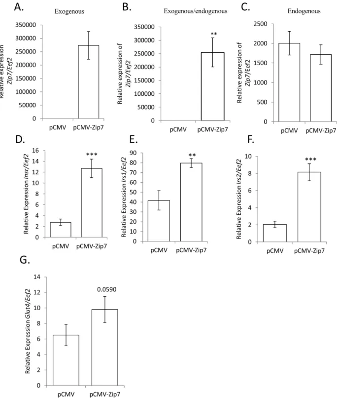
We observed that reduced Zip7 mRNA in C2C12 skeletal muscle cells was associated with changes in genes implicated in glucose metabolism. For example, a significant reduction in the expression ofPgm2, Phkb, Pygm, Gbe1, Glut4,Insr, Irs1andIrs2was observed in the Zip7-siRNA C2C12 cells compared to the scramble control (see Figures 5, 6, 7). Accordingly, to determine if by overexpressingZip7we could observe the converse effect on gene expression, we transiently transfected an overexpressionZip7
plasmid (pCMV-Zip7) into C2C12 skeletal muscle cells and after 72 hours collected RNA for subsequent qPCR analysis. We observed a significant induction in the expression of exogenous
Zip7in the pCMV-Zip7 expressing C2C12 cells compared to the pCMV control (Figure 8A and B). To confirm that the majorZip7
mRNA transcript observed was from the overexpression of the pCMV-Zip7plasmid we performed PCR using primers that were specific for the endogenous form ofZip7mRNA. We observed that Zip7 mRNA was expressed at relatively much lower levels in both the pCMV and pCMV-Zip7 transfected cells (Figure 8C)
Figure 5. Reduced Zip7 expression alters gene expression of key glucose metabolic genes.A–E). Relative expression ofPgm2, Phkb, Pygm
andGeb1mRNA toEef2in the scramble control and the siRNA-Zip7, respectively. Error bars indicated the6SD from three independent biological samples. *P,0.05, **P,0.01.
confirming that the major Zip7 transcript resulted from the overexpression system.
Moreover, we found that the overexpression of Zip7 mRNA induced the expression of the insulin receptor (Insr); insulin receptor substrate 1 (Isr1) and insulin receptor substrate 2 (Isr2) (see Figure 8D–F). This was in contrast to Figure 7 where a reduction in the expression ofZip7mRNA resulted in reduced expression of
Insr, Isr1andIsr2. We also observed an increase inGlut4mRNA in the pCMV-Zip7 overexpression system, however this result did not attain significance (p = 0.0590). Similarly, Glut4 protein levels were not significantly changed in the pCMV-Zip7 overexpression system when compared to the pCMV control (data not shown).
Discussion
Intracellular zinc homeostasis is largely regulated by two families of zinc transporters (ZnTs and ZIPs) that traffic zinc across biological membranes [35,36]. Dysregulation of zinc signaling leads to a number of disease states including cancer [19,37], autoimmune disease [38,39], cardiovascular disease [40,41] and diabetes [42–45]. Of this family, ZIP7 is important in maintaining physiological and cellular zinc homeostasis through its ability to initiate the ‘zinc wave’ and provide cytosolic zinc ions that are involved in cellular signaling processes. Although many zinc transporters respond to fluctuating zinc levels and alter their subcellular localization, ZIP7 is an exception and is restricted constitutively to the membrane of the Golgi apparatus and/or the endoplasmic reticulum [15–17]. Furthermore,Zip7gene expres-sion and intracellular location are not altered in response to changes in intracellular zinc status [3]. Studies in breast cancer cells have elucidated a role for this transporter in cell signaling events [8,20]; however, the role of ZIP7 with respect to the control of the genetic programs associated with carbohydrate metabolism in skeletal muscle has not been addressed. Here we provide the first evidence for a metabolic role forZip7in modulating glycaemic control in skeletal muscle and provide support for further studies in processes associated with insulin resistance in this tissue.
Zip7mRNA is highly expressed in differentiated C2C12 cells and mouse quadriceps. AlthoughSlc39a1was also highly expressed in C2C12 skeletal muscle cells, homozygous knockout ofSlc39a1in mice produces no phenotype when dietary zinc intake is normal [46] suggesting compensatory actions from other family members. To explore compensatory mechanisms from other zinc transport-ers we performed qPCR on all of the family membtransport-ers in the scramble control and the siRNA-Zip7C2C12 skeletal muscle cells. We did not observe major changes in expression of the other members of the zinc transporters which suggest that the attenuation of Zip7 has no other effect on these genes. Of the zinc transporters, it should be emphasized that, in addition to ZIP13, [47] ZIP7 is the only other zinc transporter localized to the Golgi apparatus and not the plasma membrane [15] and compensation is therefore unlikely. Given that ZIP7 is localized exclusively on the Golgi apparatus or the ER [17], and the fact that no compensatory changes in the other transporters were observed in the reduced Zip7 C2C12 cells suggests that this transporter may be unique in its specialized function in transporting zinc from the ER or Golgi into the cytosol [19].
In contrast to theZipexpression profile in C2C12 cells, we also observed moderate levels of expression for all of the Zip
transporters (except forZip5) in mouse quadriceps. Zip7mRNA was more highly expressed in C2C12 cells (approximately 15-fold) when compared to the expression found in quadriceps. It should be noted that quadriceps contain a mix of muscle fibre-types (oxidative type 1 and glycolytic type II) [48]. Similar studies on
Figure 6. Reduced Zip7 expression reduces the expression of Glut4 and decreases glycogen synthesis.A). Relative expression of
Glut4mRNA toEef2in the scramble control and siRNA-Zip7C2C12 cells. B). Western blot for immunoreactive Glut4 and Gapdh in protein lysates from the scramble control and the siRNA-Zip7C2C12 cells. C). Assay for glycogen synthesis in scramble and siRNA-Zip7cells treated with 10 nM insulin for 1 hour. D). Error bars indicated the 6 SD from three independent biological samples forGlut4mRNA, and six independent transient transfections of the siRNA-Zip7 for the glycogen synthesis. **P,0.01, ***P,0.001.
scramble control and siRNA-Zip7transfected C2C12 skeletal muscle cells. Error bars indicate the6SD from three independent biological samples for the mRNA analysis ofInrs, Irs1andIrs2. **P,0.01, ***P,0.001. Western blot analysis for pAkt and Akt was performed three times on six independent and pooled transient transfections of the scramble control and siRNA-Zip7.
doi:10.1371/journal.pone.0079316.g007
Figure 8. The exogenous overexpression ofZip7 mRNA induces the expression of the insulin receptor (Insr), insulin receptor substrate 1 (Irs1), insulin receptor substrate 2 (Isr2) andGlut4mRNA.A. Relative expression of exogenousZip7mRNA. B. Relative expression of endogenous and exogenous Zip7mRNA. C. Relative expression of endogenous expression ofZip7mRNA. D-F. Relative expression ofInsr, Irs1and
Irs2mRNA. G. Relative expression ofGlut4mRNA (not significant P = 0.0590). Error bars indicate the6SD from two independent biological samples for the mRNA analysis that consisted of at least three independent transfections. **P,0.01, ***P,0.001.
other muscle fibre types; soleus (type I), plantaris (type II) and anterior tibialis (type II) also demonstrated differences in the level of expression for the orphan nuclear receptor,Coup-tfIIin these tissues in comparison to C2C12 cells [49]. Moreover, studies on protein arginine methyltransferase 3, 4 and 5 (PRMT3–5) in mouse skeletal muscle tissue and C2C12 cells found high expression of PRMT3–5 in gastrocnemius in comparison to only high expression of PRMT4 (with no or minimal expression of PRMT3 and 5, respectively) [50]. Although these relative expression discrepancies exist between in vitro and in vivo model systems, the C2C12 cell culture model is a well-established and validated system to study the effects of metabolic processes [21,51,52]. For example, data derived from thisin vitromodel with liver X receptor (LXR) and peroxisome proliferating activated receptor (PPAR) agonists and their role in metabolism (e.g. energy expenditure, running endurance, lipid metabolism and cholesterol efflux) has been validated and reproduced in mice [51–55].
Our study revealed that subsets of genes involved in glucose metabolism (Agl, Dlst, Galm, Gbe1,Idh3g, Pck2, Pgam2, Pgm2, Phkb, Pygm, Tpi1, GusbandGlut4) are altered whenZip7expression was reduced. This is further highlighted by the fact that related genes in similar and other pathways (see Table S1) were refractory to the attenuation of Zip7 expression. These data are noteworthy for several reasons. For example, in skeletal muscle, GLUT4 predominately transports glucose across the plasma membrane which is further processed by oxidative (glycolysis) or non-oxidative (glycogenesis) pathways [32]. Thus, the decline in Glut4 protein in theZip7-reduced C2C12 cells would suggest a reduction in glucose transport and subsequent genes associated with oxidative and non-oxidative pathways. Similarly, this is supported by the observation that several genes implicated in glycolysis (Galm, Gusb, Pgam2, Pgm2 and Tpi1) and glycogen synthesis (Gbe1, Agl, Pgm2, PygmandPhkg2) were reduced in theZip7attenuated cells. In skeletal muscle, these genes play critical roles in the oxidative and non-oxidative pathways, respectively. This is further support-ed by the rsupport-eduction in the mRNA of the insulin receptor (Insr), and the insulin receptor substrates 1 and 2 (Isr1 and Isr2) and the subsequent reduction of basal and insulin-mediated glycogen storage in C2C12 myotubes whenZip7expression was attenuated (see Figure 6).
Skeletal muscle is particularly important in maintaining glucose homeostasis because approximately 70-90% of whole body insulin-mediated induction of glucose uptake occurs in muscle where it is incorporated into glycogen for storage [56]. Moreover, in insulin-resistant states, insulin-induced glucose uptake and glycogen synthesis is markedly reduced in skeletal muscle [32,57]. Accordingly, in association with a reduction in genes involved in glycogen metabolism and the fact that there was a reduction in glycogen synthesis, we also observed a significant decrease in the phosphorylation status of AKT in the reduced Zip7 expressing C2C12 cells. This is consistent with recent studies where a siRNA targetingZIP7significantly decreased zinc-induced pAKT after 5 minutes of 20mM zinc treatment in MCF-7 tamoxifen-resistant breast cancer cells [8]. Moreover, in a recent study in aZip9gene knockout chicken DT40 cell model, the levels of phosphorylation of Akt and Erk were significantly reduced [58]. Given the role of Zip7 in facilitating zinc flux into the cytosol [8], and the fact that previous studies have shown that zinc can activate pAKT [8,59], it will be important to determine whether Zip7 in skeletal muscle plays a similar role in mediating zinc flux and signaling events that lead to phosphorylation of AKT and the mobilization of glucose transporters.
Zinc is a well-known inhibitor of protein tyrosine phosphatases (PTPs) [60] with a reported inhibition constant in the nanomolar
range [61]. Zinc inhibits PTP1B, a cytoplasmic phosphatase that interacts with the insulin receptor and catalyzes its dephosphor-ylation resulting in the attenuation of insulin signaling [62]. Based on these results, and the fact that the insulin signaling pathway depends on the status of tyrosine phosphatases and the release of zinc into the cytosol, we hypothesize that reduced expression of
Zip7could lead to a reduction in the cytosolic zinc pool that is available for cellular signaling. For example, in the testes of diabetic mice treated with the zinc chelator, TPEN, a significant down-regulation of Akt-mediated glucose metabolism signaling was observed that was reflected by reduced phosphorylation of Akt and Gsk-3b[63]. Moreover, treatment of 3T3-L1 adipocytes with ZnCl2increased tyrosine phosphorylation of the insulin receptor
beta subunit and enhanced the transport of glucose in the absence of insulin through the PI3-kinase/Akt pathway [53]. Furthermore, in myocytes isolated from the femoral muscle of mice with a ZnT7 knock-out (these mice display low zinc status) there was reduced insulin signaling pathway activity and these mice were insulin resistant. This was also congruent with a reduction in the mRNA expression ofInsr,Irs2andAkt[3].
Based on the observations that Zip7 plays a crucial role in facilitating cytosolic zinc flux [8], and given the role of zinc as a second messenger that activates pathways associated with cellular signaling, these studies now show a new role forZip7in regulating the critical gene programs involved in glucose uptake and glycogen storage in skeletal muscle. In particular, the mRNA down-regulation of Insr, Irs1 and Irs2, in association with reduced phosphorylation of Akt and reduced Glut4 expression, suggests that Zip7 activity may be amenable to manipulation as a novel approach for the treatment of insulin resistance in skeletal muscle.
Supporting Information
Figure S1 A. Western blot analysis for insulin-induced phos-phorylation of AKT in C2C12 skeletal muscle cells. C2C12 skeletal muscle cells were differentiated in 2% horse serum for 3 days and then treated in the absence or presence of 10 nM of insulin for 60 minutes. Total cellular protein was collected and the presence for immunoreactive pAkt and Akt was assessed. This immunoblot is a representation of three independent biologically insulin-treated C2C12 cell preparations. B. Average densitometry quantification of pAkt/Akt. pAkt quantified by densitometry on immunoblots from three independent experiments normalized to total Akt and displayed as the mean 6 SD with significant (**P#0.01,*** P#0.001) changes over time 0.
(TIF)
Figure S2 Western blot analysis for zinc induced phosphoryla-tion of AKT in the absence and presence of 10mM pyrithione in C2C12 skeletal muscle cells. C2C12 skeletal muscle cells were differentiated in 2% horse serum for 3 days and then treated in the presence (+) or absence (-) of 10mM of pyrithione over 60 minutes. Total cellular protein was extracted and the presence for immunoreactive pAKT and AKT was performed by western blot analysis. This immunoblot represents at least three independent biological replicates.
(TIF)
Table S1 Fold changes in expression of glucose metabolic genes in the siRNA-Zip7compared to the scramble control.
(DOC)
Acknowledgments
Thank you to Fahima Ahmady (Biomedical Technician, University of Ballarat) for her help with reagent orders and general laboratory duties.
Author Contributions
Conceived and designed the experiments: SM AN GC MAM. Performed the experiments: SM AN GC. Analyzed the data: SM AN GC MAM. Contributed reagents/materials/analysis tools: SM MAM. Wrote the paper: SM.
References
1. Cousins RJ, Liuzzi JP, Lichten LA (2006) Mammalian Zinc Transport, Trafficking, and Signals. J Biol Chem 281:24085–24089.
2. Gaither LA, Eide DJ (2001) Eukaryotic zinc transporters and their regulation. BioMetals 14:251–270.
3. Huang L, Kirschke CP, Lay YAE, Levy LB, Lamirande DE, et al. (2012) Znt7-null mice are more susceptible to diet-induced glucose intolerance and insulin resistance. J Biol Chem 282:37053–37063.
4. Smidt K, Jessen N, Petersen AB, Larsen A, Magnusson N, et al. (2009) SLC30A3 Responds to Glucose- and Zinc Variations in beta-Cells and Is Critical for Insulin Production and In Vivo Glucose-Metabolism During beta-Cell Stress. PLoS ONE 4:e5684.
5. Chistiakov DA, Voronova NV (2009) Zn2+-transporter-8: A dual role in diabetes. Biofactors 35: 356–363.
6. Fukada T, Yamasaki S, Nishida K, Murakami M, Hirano T (2011) Zinc homeostasis and signaling in health and diseases. J Biol Inorg Chem 16:1123– 1134.
7. Yamasaki S, Hasegawa A, Hojyo S, Ohashi W, Fukada T, et al. (2012) A Novel Role of the L-Type Calcium Channela1D Subunit as a Gatekeeper for Intracellular Zinc Signaling: Zinc Wave. PLoS ONE 7:e39654.
8. Taylor KM, Hiscox S, Nicholson RI, Hogstrand C, Kille P (2012) Protein Kinase CK2 Triggers Cytosolic Zinc Signaling Pathways by Phosphorylation of Zinc Channel ZIP7. Sci Signal 5:ra11.
9. Ilouz R, Kaidanovich O, Gurwitz D, Eldar-Finkelman H (2002) Inhibition of glycogen synthase kinase-3bby bivalent zinc ions: insight into the insulin-mimetic action of zinc. Biochem Biophys Res Commun 295:102–106. 10. Moniz T, Amorim MJ, Ferreira R, Nunes A, Silva A, et al. (2011) Investigation
of the insulin-like properties of zinc(II) complexes of 3-hydroxy-4-pyridinones: Identification of a compound with glucose lowering effect in STZ-induced type I diabetic animals. J Inorg Biochem 105:1675–1682.
11. Simon SF, Taylor CG (2001) Dietary Zinc Supplementation Attenuates Hyperglycemia in db/db Mice. Exp Biol Med 226:43–51.
12. Wijesekara N, Chimienti F, Wheeler MB (2009) Zinc, a regulator of islet function and glucose homeostasis. Diabetes Obes Metab 11:202–214. 13. Yoshikawa Y, Ueda E, Kojima Y, Sakurai H (2004) The action mechanism of
zinc(II) complexes with insulinomimetic activity in rat adipocytes. Life Sci 75:741–751.
14. Coulston L, Dandona P (1980) Insulin-like Effect of Zinc on Adipocytes. Diabetes 29:665–667
15. Huang L, Kirschke CP, Zhang Y, Yu YY (2005) The ZIP7 Gene (Slc39a7) Encodes a Zinc Transporter Involved in Zinc Homeostasis of the Golgi Apparatus. J Biol Chem 280:15456–15463.
16. Taylor KM, Morgan HE, Johnson A, Nicholson RI (2004) Structure-function analysis of HKE4, a member of the new LIV-1 subfamily of zinc transporters. Biochem J 377:131–139.
17. Taylor KM, Morgan HE, Smart K, Zahari NM, Pumford S, et al. (2007) The emerging role of the LIV-1 subfamily of zinc transporters in breast cancer. Mol Med 13:396–406.
18. Lichten LA, Cousins RJ (2009) Mammalian Zinc Transporters: Nutritional and Physiologic Regulation. Annu Rev Nutr 29:153–176.
19. Hogstrand C, Kille P, Nicholson RI, Taylor KM (2009) Zinc transporters and cancer: a potential role for ZIP7 as a hub for tyrosine kinase activation. Trends Mol Med 15:101–111.
20. Taylor KM, Vichova P, Jordan N, Hiscox S, Hendley R, et al. (2008) ZIP7-Mediated Intracellular Zinc Transport Contributes to Aberrant Growth Factor Signaling in Antihormone-Resistant Breast Cancer Cells. Endocrinology 149:4912–4920.
21. Myers SA, Wang SCM, Muscat GEO (2006) The Chicken Ovalbumin Upstream Promoter-Transcription Factors Modulate Genes and Pathways Involved in Skeletal Muscle Cell Metabolism. J Biol Chem 281:24149–24160. 22. Maxwell MA, Cleasby ME, Harding A, Stark A, Cooney GJ, et al. (2005) Nur77
Regulates Lipolysis in Skeletal Muscle Cells: Evidence for cross-talk between the beta-adrenergic and an orphan nuclear hormone receptor pathway. J Biol Chem 280:12573–12584.
23. Lau P, Nixon SJ, Parton RG, Muscat GEO (2004) RORa Regulates the Expression of Genes Involved in Skeletal Cells: Caveolin-3 and CPT-1 are direct targets of ROR. J Biol Chem 279:36828–36840.
24. Spandidos A, Wang X, Wang H, Seed B (2010) PrimerBank: a resourse of human and mouse PCR primer pairs for gene expression detection and quantification. Nulc Acids Res 38:D792–799.
25. Spandidos A, Wang X, WangH, Dragnev S, Thurber T, et al. (2008) A comprehensive collection of experimentally validated primers for Polymerase Chain Reaction quantitation of murine transcript abundance. BMC Genomics 9:6333.
26. Wang X, Seed B (2003) A PCR primer bank for quantitative gene expression analysis. Nulc Acids Res 31:e154; 1–8.
27. Bookout A, Mangelsdorf D (2003) Quantitative real-time PCR protocol for analysis of nuclear receptor signaling pathways. Nucl Recept Signal 1:e012. 28. Muscat GE, Rea S, Downes M (1995) Identification of a regulatory function for
an orphan receptor in muscle: COUP-TF II affects the expression of the myoD gene family during myogenesis. Nucleic Acids Res 23:1311–1318.
29. Shimokawa T, Kato M, Ezaki O, Hashimoto S (1998) Transcriptional regulation of muscle-specific genes during myoblast differentiation. Biochem Biophys Res Commun 246:287–292.
30. Lau P, Nixon SJ, Parton RG, Muscat GE (2004) RORalpha regulates the expression of genes involved in lipid homeostasis in skeletal muscle cells: caveolin-3 and CPT-1 are direct targets of ROR. J Biol Chem 279:36828– 36840.
31. Zhu C, Hu DL, Liu YQ, Zhang QJ, Chen FK, et al. (2011)Fabp3Inhibits Proliferation and Promotes Apoptosis of Embryonic Myocardial Cells. Cell Biochem Biophys. 60:259–266.
32. Peppa M, Koliaki C, Nikolopoulos P, Raptis SA (2010) Skeletal Muscle Insulin Resistance in Endocrine Disease. J Biomed Biotechnol 10.1155/2010/527850. 33. Biddinger SB, Emanueli B (2011) Insulin Resistance in the Metabolic Syndrome. In: Metabolic Basis of Obesity, Ahima RS (ed.), Springer New York. Pp.175– 198.
34. Bajaj M, DeFronzo RA (2003) Metabolic and molecular basis of insulin resistance. J Nucl Cadrdiol 10:311–323.
35. Liuzzi JP, Cousins RJ (2004) Mammalian Zinc Transporters. Annu Rev Nutr 24:151–172.
36. Myers SA, Nield A, Myers M (2012) Zinc Transporters, Mecahnisms of Action and Therapeutic Utility: Implications for Type 2 Diabetes Mellitus. J Nutr Metab doi:10.1155/2012/173712
37. Jayaraman AK, Jayaraman S (2011) Increased level of exogenous zinc induces cytotoxicity and up-regulates the expression of the ZnT-1 zinc transporter gene in pancreatic cancer cells. J Nutr Biochem 22:79–88.
38. Delli AJ, Vaziri-Sani F, Lindblad B, Elding-Larsson H, Carlsson A, et al. (2012) Zinc Transporter 8 Autoantibodies and Their Association With SLC30A8 and HLA-DQ Genes Differ Between Immigrant and Swedish Patients With Newly Diagnosed Type 1 Diabetes in the Better Diabetes Diagnosis Study. Diabetes 10:2556–2564.
39. Kawasaki E, Nakamura K, Kuriya G, Satoh T, Kobayashi M, et al. (2011) Differences in the humoral autoreactivity to zinc transporter 8 between childhood- and adult-onset type 1 diabetes in Japanese patients. Clin Immunol
138:146–153.
40. Patrushev N, Seidel-Rogol B, Salazar G (2012) Angiotensin II Requires Zinc and Downregulation of the Zinc Transporters ZnT3 and ZnT10 to Induce Senescence of Vascular Smooth Muscle Cells. PLoS ONE 7:e33211. 41. Foster M, Samman S (2010) Zinc and redox signaling: perturbations associated
with cardiovascular disease and diabetes mellitus. Antioxid Redox Signal 13:1549–1573.
42. Stadler N, Heeneman S, Voo S, Stanley N, Giles GI, et al. (2012) Reduced metal ion concentrations in atherosclerotic plaques from subjects with Type 2 diabetes mellitus. Atherosclerosis 222:512–518.
43. Ferdousi S, Mia AR (2012) Serum levels of copper and zinc in newly diagnosed type-2 diabetic subjects. Mymensingh Med J 21:475–478.
44. Basaki M, Saeb M, Nazifi S, Shamsaei HA (2012) Zinc, Copper, Iron, and Chromium Concentrations in Young Patients with Type 2 Diabetes Mellitus. Biol Trace Elem Res 148:161–164.
45. Ruchi S, Ashok K (2011) A study of Age Related Decrease in Zinc and Chromium and its Correlations with type 2 Diabetes Mellitus. Res J Chem Environment. 15:75–80.
46. Kambe T, Geiser J, Lahner B, Salt DE, Andrews GK (2008) Slc39a1 to 3 (subfamily II) Zip genes in mice have unique cell-specific functions during adaptation to zinc deficiency. Am J Physiol Regul Integr Comp Physiol. 294:R1474–R1481.
47. Bin BH, Fukada T, Hosaka T, Yamasaki S, Ohashi W, et al. (2011) Biochemical characterization of human ZIP13 protein: a homo-dimerized zinc transporter involved in the spondylocherio dysplastic Ehlers-Danlos syndrome. J Biol Chem 286:40255–40265.
48. Armstrong RB, Phelps RO (1984) Muscle fiber type composition of the rat hindlimb. Am J Anat 171:259–272.
50. Wang SCM, Dowhan DH, Eriksson NA, Muscat GEO (2012) CARM1/ PRMT4 is necessary for the glycogen gene expression programme in skeletal muscle cells. Biochem J 444:323–331.
51. Raichur S, Fitzsimmons RL, Myers SA, Pearen MA, Lau P, et al. (2010) Identification and validation of the pathways and functions regulated by the orphan nuclear receptor, ROR alpha1, in skeletal muscle. Nucleic Acids Res 38:4296–4312.
52. Wang YX, Zhang CL, Yu RT, Cho HK, Nelson MC, et al. (2004) Regulation of muscle fiber type and running endurance by PPARdelta. PLoS Biol 2:e294. 53. Muscat GE, Wagner BL, Hou J, Tangirala RK, Bischoff ED, et al. (2002)
Regulation of cholesterol homeostasis and lipid metabolism in skeletal muscle by liver X receptors. J Biol Chem. 277:40722–40728.
54. Dressel U, Allen TL, Pippal JB, Rohde PR, Lau P, et al. (2003) The peroxisome proliferator-activated receptor beta/delta agonist, GW501516, regulates the expression of genes involved in lipid catabolism and energy uncoupling in skeletal muscle cells. Mol Endocrinol 17:2477–2493.
55. Tanaka T, Yamamoto J, Iwasaki S, Asaba H, Hamura H, et al. (2003) Activation of peroxisome proliferator-activated receptor delta induces fatty acid beta-oxidation in skeletal muscle and attenuates metabolic syndrome. Proc Natl Acad Sci USA 100:15924–15929.
56. Jensen J, Jebens E, Brennesvik EO, Ruzzin J, Soos MA, et al. (2006) Muscle glycogen inharmoniously regulates glycogen synthase activity, glucose uptake,
and proximal insulin signaling. Am J Physiol Endocrinol Metab 290:E154– E162.
57. Petersen KF, Dufour S, Savage DB, Bilz S, Solomon G, et al. (2007) The role of skeletal muscle insulin resistance in the pathogenesis of the metabolic syndrome. Proc Natl Acad Sci USA 104:12587–12594.
58. Taniguchi M, Fukunaka A, Hagihara M, Watanabe K, Kambe T, et al. (2013) Essential role of the zinc transporter ZIP9/SLC39A9 in regulating the activations of Akt and Erk in B-cell receptor signaling pathway in DT40 cells. Plos One 8:e58022.
59. Tang X-h, Shay NF (2001) Zinc Has an Insulin-Like Effect on Glucose Transport Mediated by Phosphoinositol-3-Kinase and Akt in 3T3-L1 Fibro-blasts and Adipocytes. J Nutr 131:1414–1420.
60. Brautigan DL, Bornstein P, Gallis B (1981) Phosphotyrosyl-protein phosphatase. Specific inhibition by Zn. J Biol Chem 256:6519–6522.
61. Maret W, Jacob C, Vallee BL, Fischer EH (1999) Inhibitory sites in enzymes: Zinc removal and reactivation by thionein. Proc Natl Acad Sci USA 96:1936– 1940.
62. Ma Y-m, Tao R-y, Liu Q, Li J, Tian J-y et al. (2011) PTP1B inhibitor improves both insulin resistance and lipid abnormalities in vivo and in vitro. Mol and Cell Biochem 357:65–72.