ISSN 1553-345X
© 2006 Science Publications
Corresponding Author: Chang Won Choi, Department of Biology and Medicinal Science, Pai Chai University, Daejeon, Korea 302-735, Tel: 8242-520-5617
148
Molecular Characterization of
Soybean Mosaic Virus
NIa Protein and its
Processing Event in Bacterial Expression
1
Bong Kum Choi,
1Jae Sung Park,
1Hye Jin Ahn,
1Hye Jung Yum,
1Vasudevan Ayyappan
1
Lakshmi Sumitra Vijayachandran,
1Ji Sue Kim,
1Sei Chang Kim and
1,2Chang Won Choi
1
Department of Biology and Medicinal Science, Pai Chai University, Daejeon, Korea 302-735
2
Biomedicinal Research Center (RRC), Pai Chai University, Daejeon, Korea 302-735
Abstract:Soybean mosaic virus (SMV)-CN18 is an Rsv resistance-breaking (RB) isolate to overcome soybean resistance genes Rsv1, Rsv3 and Rsv4. The aim of this study was to characterize nuclear inclusion protein a (NIa protein) of RB isolate at the molecular level and demonstrate its processing into genome-linked protein (VPg) and NIa-Pro domains in Esherichia coli containing a bacterial expression pET vector inserted with NIa gene. The full-length of NIagene was synthesized by reverse transcription-polymerase chain reaction (RT-PCR) and its 1298 nucleotides (nt) and 432 amino acids (aa) were deduced. The nt and aa sequences of NIa gene of SMV-CN18 shared high identities with the corresponding sequences of the NIa gene of the known SMV isolates, suggesting that the NIa is a highly conserved protein. The NIa-Pro domain contains a highly conserved structural motif for proteolysis, while the VPg domain contains a nuclear localization signal (NLS), a putative NTP-binding site and cellular factor-NTP-binding sites. The phylogenetic tree revealed that less divergence of NIa protein exists among twelve SMV isolates, which can be supported by a low bootstrap value between clades. In addition, the full-length of NIa gene, amplified by RT-PCR, was ligated into pET-28b E. coli expression vector with an N-terminal His6-tag. Optimal conditions for expression were at 1mM treatment of IPTG at 25°C for 5 hr. The released protein from bacterial lysates remained soluble and proved the processing form of the NIa polyprotein. E. coli expression system shows the processed product of 29 kDa VPg in SDS-PAGE confirmed by western blot analysis in both crude extracts and purified elution products, using Ni2+-NTA resin. The present study indicates that the N-terminal region of NIa which is processed and expressed in bacteria.
Key words: Soybean mosaic virus (SMV), Rsv resistance-breaking (RB) isolate, nuclear inclusion protein a (NIa), processing, genome-linked protein (VPg), NIa-Pro, highly conserved protein, E. coli expression
INTRODUCTION
Soybean mosaic virus (SMV), a member of the genus Potyvirus, is a major pathogen of soybean (Glycine max L.). SMV has a positive-sense single-stranded RNA genome of 9588 nucleotides, with VPg on the 5’end and poly(A) track at the 3’end. The genome of potyvirus encodes a single large polyprotein that is proteolytically processed by three virus-encoded proteases[1]. One of the proteases, nuclear inclusion protein a (NIa), possessed structural motifs that showed similarity with cellular serine protease, with the substitution of Ser by a Cys as the active site[2,3]. The NIa is actually a 49 kDa polyprotein consisting of two domains, the genome-linked protein (VPg) domain at the N-terminus and the protease (NIa-Pro) domain at the C-terminus[4,5]. The NIa protease plays an important role in the processing of the remaining two-thirds of polyprotein in cis- and trans-processing by catalyzing
Recently, we have reported twelve emerging Rsv resistance-breaking (RB) isolates of SMV, among them SMV-CN18 has an ability to overcome soybean resistance gene Rsv1, Rsv3 and Rsv4, respectively[25]. In this study, the NIa gene was synthesized by reverse transcription (RT)-PCR using SMV-CN18 genomic RNA as a template, compared its sequences of nucleotides (nt) and amino acids (aa) with those of eleven previous reported SMV isolates and characterized its expression in E. coli to observe the processing event into VPg and NIa-Pro domains.
MATERIALS AND METHODS
Viral strain, purification and RNA extraction: An RB isolate, SMV-CN18, was purified from the infected soybean leaves and its genomic RNA was prepared as described in a previous investigation[25].
RT-PCR amplification of NIa gene and cDNA cloning: The primers used for the amplification of NIa gene were designed based on the conserved nucleotide sequences of known SMV isolates and listed in Table 1. Reverse transcription (RT) reaction was performed on 100 ng of viral RNA in a reaction volume of 20 l containing 50 mM Tris-HCl, pH 8.3, 75 mM KCl, 10 mM DTT, 3 mM MgCl2, 1 mM dNTP, 50 pmol of reverse primer, 20 U of RNase inhibitor (Takara, Japan) and 200 U Molony murine leukaemia virus reverse transcriptase (Promega, USA). For amplification, 20 l of RT mix was added to 80 l of reaction mixture containing 10 mM Tris-HCl, pH 8.3, 50 mM KCl, 1.5~4.5 mM MgCl2, 2.5U Taq polymerase (Takara, Japan), 0.25 mM dNTP, 50 pmol of forward and reverse primers each. The thermocycler (Bio-Rad, Gene Cycler, USA) was programmed for template denaturation at 94ºC for 1 min, primer annealing at 55ºC for 2 min and DNA synthesis at 72ºC for 3 min. A final 7 min extension step at 72ºC was performed at the end of 35 cycles. Ten RB isolates of SMV in a previous study[25] were also subjected to monitor optimal condition for RT-PCR. The purified fragments were directly ligated into pGEM-T Easy Vector System (Promega, USA) and transformed into E. coli JM109 by conventional CaCl2 procedure. Transformants were selected on Luria-Bertani (LB) medium supplemented with 100 g mL-1 ampicillin, X-Gal and IPTG, incubating overnight at 37ºC.
Table 1: RT-PCR primers used for the cloning of Soybean mosaic virus NIa gene
Gene Primer polarity Sequence (5’ 3’)
NIa-reverse 3’ end TCCTTTCTCCCTTGAACTGTCa
NIa-forward 5’ end ATCAACTCAATGAAAGAAGAGb aReverse primer containing stop codon (bold, underline), T represents
a modified base for termination.
bForward primer containing start codon (bold, underline), T represents
a modified base for initiation.
Nucleotide and amino acid sequences analyses and phylogenetic tree: The plasmid containing NIa gene was prepared from the transformed bacterial cells using QIA Plasmid Prep Kit (QIAGEN, Germany) and used for sequencing analysis. After linearization of the plasmid containing entire NIa coding region, nt sequences were determined in both directions by the dideoxynucleotide chain termination method using the ABI Model 337 automatic DNA sequencer. The complete genomic sequences of the SMV-CN18 were deposited as EMBL Database Accession no. AJ619757. For sequence comparisons, NIa gene products of the reference isolates were obtained from the NCBI data library including SMV-G2 (S42280) and -G7-1 (AF241739; then designated G7 in USA)[26], SMV-G7d (AY216987)[27], SMV-N (NC002634)[28], SMV-G5b (AY294044: then designated G5 in Korea) and -G7H (AY294045)[29], SMV-HH5 (AJ310200: then designated Huanghuai) and -HZ (AJ312439: then designated Severe)[30], as well as web-published isolates SMV-G72 (AY216010), -Aa (AB100442) and -Aa15-M2 (AB100443). Distance matrices for complete NIa sequences were calculated from the multiple sequence alignments by the DNAMAN version 5.2.9 (Lynnon Biosoft, Quebec, Canada). Phylogenetic analysis was performed using the generated matrices as an input in DNAMAN to build an unrooted tree and the statistical significance of branching was estimated by bootstrap resampled data sets based on 1000 replications.
150 Total proteins were analyzed by 10% SDS-PAGE followed by Western blotting to detect 6X His-tagged protein using Ni2+-NTA conjugated antibody against 6X His (Qiagen, USA). The membrane was developed by conventional method with nitrobluetetrazolium (NBT)/ 5-bromo-4-chloro-3-indolylphosphate (BCIP) (MBI Fermentas, Canada) in 100 mM Tris, 100 mM NaCl and 5 mM MgCl2, pH 9.5.
RESULTS AND DISCUSSION
RT-PCR amplification of NIa gene: Successful amplification of NIa gene from SMV-CN18 was performed by using forward and reverse primers in a range (1.5-4.5 mM) of magnesium concentrations, among which 2.5 mM of MgCl2 was the optimal condition (Fig. 1a). Under the optimal RT-PCR condition, we detected an array of amplification products of expected size 1,298 bp fragments of NIa gene that encoded 433 aa with a predicted molecular mass of 49 kDa from soybean leaves inoculated with ten SMV RB isolates (Fig. 1b).
Fig. 1: (a) Amplified products of SMV-CN18 NIa gene by RT-PCR under various MgCl2 concentration. Lanes 1 (1.5 mM), 2 (2.5 mM), 3 (3.5 mM) and 4 (4.5 mM). (b) Amplified products of NIa gene of SMV isolates at 2.5 mM MgCl2 by RT-PCR. Lanes 1 (CN1), 2 (CN2), 3 (CN3), 4 (CN9), 5 (CN12), 6 (CN13), 7 (CN15), 8 (CN17), 9 (CN21) and 10 (CN31). M represents 1kb DNA ladder
NIa sequence comparisons and phylogenetic analysis: For the investigation of sequence diversity of NIa, the nt and aa sequences of SMV-CN18 were aligned with the corresponding sequences of the NIa gene of the known SMV isolates (data not shown). The highest nt sequence identity was 98% with SMV-HH5, while the lowest nt sequence identity was 92% with that of SMV-N. In aa similarities SMV-CN18 shows the value of 98% with G2 and G5b, respectively and 99% with the remaining nine isolates, suggesting that the NIa is a highly conserved protein among the isolates (Table 2). In addition, it possessed structural motifs for proteolysis that conserved among potyviruses[8,31]. Presumed codons among SMV isolates for the catalytic triad are composed of H32, D78 and C150. On the other hand, the VPg contains a nuclear localization signal (NLS) that is conserved among SMV isolates by the aa residues from 41 to 50 (KKGKGKGSTR). This NLS is
quite similar to the NLS (KKGKTKGKTH) in Potato virus A (PVA) VPg[24] and the NLS (NKGKRKGTTR) in Tobacco etch virus (TEV) VPg[32]. SMV VPg also contains a conserved 7 aa residues (A38YTKKGK44) which has been proposed as a putative NTP-binding site in the VPg of PVA, its deletion reduced nucleotide-binding capacity and debilitated uridylylation reaction[33]. Recently, a cellular factor called ‘PVIP’ that interacts with VPg of potyviruses has been identified in some plants. Two domains controlled the interactions with PVIP, suggesting that PVIP plays a role in assistant factor to support potyvirus movement in plants[34]. Sequence comparison of SMV and other four potyviruses, Turnip mosaic virus (TuMV), Lettuce mosaic virus (LMV), PVA and TEV, were aligned for the two domains. It shows that four residues in the first domain (VP aa 1 to 16) and six residues in the second domain (VP aa 40 to 64) are identical with those of all four viruses (Fig. 2).
Fig. 2: Amino acid sequences of the determinant between VPg and PVIP interaction. SMV VPg sequences were aligned using DNAMAN version 5.2.9 software, with corresponding region from other potyviruses obtained from the NCBI data library. Only regions 1 to 16 and 40 (41 or 42) to 64 (65 or 66) are shown. Identical (star), related (:) and unmatched (triangle) amino acid sequences are indicated
Table 2: Sequence identities in nucleotides and amino acids of NIa gene between CN18 and known isolates of Soybean mosaic virus
Strain/ isolate CN18 Accession number
G2 93/98a S42280
G5b 93/98 AY294044
G7-1 96/99 AF241739
G7-2 97/99 AY216010
G7d 97/99 AY216987
G7H 96/99 AY294045
Aa 96/99 AB100442
Aa15-M2 96/99 AB100443
HH5 98/99 AJ310200
HZ 97/99 AJ312439
N 92/99 NC002634
aNumbers in bold type at right corner refer amino acid sequence
identities and numbers in plain type at left corner refer to nucleotide sequence identities.
twenty-five SMV isolates, all those SMV isolates can be grouped into three major types and seven subtypes by similarity clustering[25]. On the other hand, the less divergence of NIa protein existing among twelve SMV isolates, which can be supported by low bootstrap values between clades, suggests that the phylogenetic tree is essentially meaningless. Based on the result, it is logical to hypothesize that the recombination event in the NIa coding region rarely occurred between SMV isolates.
Fig. 3: Phylogenetic analysis for NIa protein amino acid sequences of 12 SMV isolates. The tree (unrooted) was constructed by the DNAMAN version 5.2.9. The grouping occurred after bootstrapping the data (only values > 10% are shown) and the numbers on the branches indicate bootstrap percentages based on 1000 resamplings
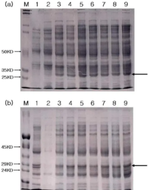
Fig. 4: 10% SDS-PAGE of the expressed NIa protein under various concentrations of IPTG at 37ºC for 5 h. (a) the supernatant of culture lysate after IPTG treatment, Lanes 1 (wild-type control, 1 mM IPTG treatment), 2 (sample without IPTG treatment) and 3-7 (samples with 0.01 mM, 0.05 mM, 0.1 mM, 0.5 mM and 1mM IPTG treatment, respectively). (b) Lane 1 represents a wild-type control treated with 1 mM IPTG. Lanes 2-6 are samples treated with 1 mM, 2 mM, 3 mM, 4 mM and 5 mM IPTG, respectively. M represents protein Mr marker and unlabelled arrow indicates the VPg
Fig. 5: Time course of NIa protein expression under 1 mM IPTG treatment at 25°C. (a) Culture supernatants and (b) pellets. Lanes 1 (wild type control, culture for 5 h) and 2 (no IPTG treated control, culture for 5 h). Lanes 3-9 represent samples treated with IPTG for 1 h, 3 h, 5 h, 7 h, 9 h, 12 h and 24 h, respectively. M represents protein Mr marker and unlabelled arrow indicates the VPg
Fig. 6: Western blot analysis of the processed NIa protein. The recombinant VPg was detected about size of 29KDa protein using Ni2+-NTA conjugated antibody against 6X His. Lane 1 is crude extract, and lanes 2-7 represent the eluted protein samples during purification using Ni2+-NTA resin
152 without His-tag could be released from the processed NIa, and only VPg could be detected by antibody specific to His-tag. Theoretically, the size of native NIa, NIa-Pro and VPg are 49, 27 and 23 kDa, respectively, from the cDNA sequences. Taking intoaccount the N-terminal His-tag, the size of NIa, NIa-Pro and VPg were estimated as 55, 27 and 29 kDa, respectively. The position of band close to the expected size of protein was found at 29 kDa on SDS-PAGE gel (Fig. 5a, b, lanes 3-9), and its migration was corresponding with the predicted VPg. The recombinant protein containing His residues is easily purified by metal-chelation chromatography[35]. To confirm the processing in the purified proteins from E. coli, we performed Western blotting using an Ni2+-NTA conjugated antibody against 6X His. We did not find the unprocessed polyprotein to bind Ni2+ affinity column, but detected the 29 kDa protein corresponding to VPg (Fig. 6). Our results indicate that the anti-His antibodies recognized VPg but failed to recognize unprocessed polyprotein which further processed VPg and NIa-Pro. Our results were corresponding to the previous results that the VPg alone without NIa-Pro was detected when the entire coding region of NIa protein of TuMV was expressed in E. coli[36]. The NIa-Pro of TEV was also expressed in E. coli as a recombinant protein with His-tag, but the expression of unprocessed polyprotein was unsuccessful[37]. The full-length of TuMV NIa gene was expressed as an unprocessed polyprotein (49 kDa) in E. coli by site-directed mutagenesis to block the processing as previously described by Ménard et al.[38]. According to our results and previous reports above mentioned, the unprocessed NIa polyprotein seems to exist for a brief time during its expression in E. coli.
ACKNOWLEDGEMENTS
This work was supported by a grant from Ministry of Commerce, Industry and Energy (MOICE) through the Research Center for Bio-Medicinal Resources (Bio-Med RRC) at Pai Chai University.
REFERENCES
1. Dougherty, W.G. and B.L. Semler, 1993. Expression of virus-encoded proteinases: Functional and structural similarities with cellular enzymes. Microbiol. Rev., 57: 781-822.
2. Gorbalenya, A.E., A.P. Donchenko, E.V. Koonin and V.M. Blinov, 1989. Cysteine proteases of positive strand RNA viruses and chymotrypsin-like serine proteases: A distinct super-family with a common structural fold. FEBS Lett., 243: 103-114. 3. Bazan, J.F. and R.J. Fletterick, 1990. Structural and
catalytic models of trypsin-like viral proteases. Semin. Virol., 1: 311-322.
4. Murphy, J.F., R.E. Rhoads, A.G. Hunt and J.G. Shaw, 1990. The VPg of tobacco etch virus RNA is the 49-kDa proteinase or the N-terminal 24-kDa part of the proteinase. Virology, 178: 285-288. 5. Dougherty, W.G. and T.D. Parks, 1991.
Post-translational processing of the tobacco etch virus 49-kDa small nuclear inclusion polyprotein: identification of an internal cleavage site and delimitation of VPg and proteinase domains. Virology, 183: 449-456.
6. Carrington, J.C. and W.G. Dougherty, 1988a. Small nuclear inclusion protein encoded by a plant potyvirus genome is a protease. J. Virol., 61: 2540-2548.
7. Carrington, J.C. and W.G. Dougherty, 1988b. A viral cleavage site cassette: Identification of amino acid sequences required for tobacco etch virus polyprotein processing. Proc. Natl. Acad. Sci. USA., 85: 3391-3395.
8. Garcia, J.A., S. Lain, M.T. Cervera, J.L. Riechmann and M.T. Martin, 1990. Mutational analysis of plum pox potyvirus polyprotein processing by the NIa protease in Escherichia coli. J. Gen. Virol., 71: 2773-2779.
9. Joseph, J. and H.S. Savithri, 2000. Mutational analysis of the NIa protease from pepper vein banding potyvirus. Arch. Virol., 145: 2493-2502. 10. Wimmer, E., 1982. Genome-linked proteins of
viruses. Cell, 28: 199-201.
11. Murphy, J.F., P.G. Klein, A.G. Hunt and J.G. Shaw, 1996. Replacement of the tyrosine residue that links a potyviral VPg to the viral RNA is lethal. Virology, 220: 535-538.
12. Yambao, M. LM., C. Masuta, K. Nakahara and I. Uyeda, 2003. The central and C-terminal domains of VPg of Clover yellow vein virus are important for VPg-HCPro and VPg-VPg interactions. J. Gen. Virol., 84: 2861-2869.
13. Hong, Y., K. Levay, J.F. Murphy, P.G. Klein, J.G. Shaw and A.G. Hunt, 1995. The potyvirus polymerase interacts with the viral coat protein and VPg in yeast cells. Virology, 214: 159-166.
14. Li, X.H., P. Valdez, R.E. Olvera and J.C. Carrington, 1997. Functions of the tobacco etch virus RNA polymerase (NIb): Subcellular transport and protein-protein interaction with VPg/proteinase (NIa). J. Virol., 71: 1598-1607.
15. Fellers, J., J. Wan, Y. Hong, G.B. Collins and A.G. Hunt, 1998. In vitro interactions between a portyvirus-encoded, genome-linked protein and RNA-dependent RNA polymerase. J. Gen. Virol., 79: 2043-2049.
17. Wimmer, E., C.U. Hellen and X. Cao, 1993. Genetics of poliovirus. Annu. Rev. Genet., 27: 353-436.
18. Wittmann, S., H. Chatek, M.G. Fortin and J.-F. Laliberté, 1997. Interaction of the viral protein genome linked of turnip mosaic potyvirus with the translational eukaryotic initiation factor (iso)4E of Arabidopsis thaliana using the yeast two-hybrid system. Virology, 234: 84-92.
19. Schaad, M.C., R.J. Anderberg and J.C. Carrington, 2000. Strain-specific interaction of the tobacco etch virus NIa protein with the translation initiation factor eIF4E in the yeast two-hybrid system. Virology, 273: 300-306.
20. Nicolas, O., S.W. Dunnington, L.F. Gotow, T.P. Pirone and G.M. Hellmann, 1997. Variations in the VPg protein allow a potyvirus to overcome va gene resistance in tobacco. Virology, 237: 452-459. 21. Schaad, M.C., A.D. Lellis and J.C. Carrington,
1997. VPg of tobacco etch potyvirus is a host genotype-specific determinant for long-distance movement. J. Virol., 71: 8624-8631.
22. Rajamäki, M.-L. and J.P.T. Valkonen, 1999. The 6K2 protein and the VPg of potato virus A are determinants of systemic infection in Nicandra physaloides. Mol. Plant-Microbe Interact., 12: 1074-1081.
23. Rajamäki, M.-L. and J.P.T. Valkonen, 2002. Viral genome-linked protein (VPg) controls accumulation and phloem-loading of a potyvirus in inoculated potato leaves. Mol. Plant-Microbe Interact., 15: 138-149.
24. Rajamäki, M.-L. and J.P.T. Valkonen, 2003. Localization of a potyvirus and the viral genome-linked protein in wild potato leaves at an early stage of systemic infection. Mol. Plant-Microbe Interact., 16: 25-34.
25. Choi, B.K., J.M. Koo, H.J. Ahn, H.J. Yum, C.W. Choi, K.H. Ryu, P. Chen and S.A. Tolin, 2005. Emergence of Rsv-resistance breaking Soybean mosaic virus isolates from Korean soybean cultivars. Virus res., 112: 42-51.
26. Jayaram, C., J.H. Hill and W.A. Miller, 1992. Complete nucleotide sequences of two soybean mosaic virus strains differentiated by response of soybean containing the Rsv resistance gene. J. Gen. Virol., 73: 2067-2077.
27. Hajimorad, M.R., A.L. Eggenberger and J.H. Hill, 2003. Evolution of Soybean mosaic virus-G7 molecularly cloned genome in Rsv1 genotype soybean results in emergence of a mutant capable of evading Rsv1-mediated recognition. Virology, 314: 487-509.
28. Eggenberger, A.L., D.M. Stark and R.N. Beachy, 1989. The nucleotide sequence of a soybean mosaic virus coat protein-coding region and its expression in Escherichia coli, Agrobacterium tumefaciens and tobacco callus. J. Gen. Virol., 70: 1853-1860.
29. Lim, W.-S., Y.-H. Kim and K.-H. Kim, 2003. Complete genome sequences of the genomic RNA of soybean mosaic virus strains G7H and G5. Plant Pathol. J., 19: 171-176.
30. Chen, J., H.-Y. Zheng, L. Lin, M.J. Adams, J.F. Antoniw, M.-F. Zhao, Y.-F. Shang and J.-P. Chen, 2004. A virus related to Soybean mosaic virus from Pinellia ternate in China and its comparison with local soybean SMV isolates. Arch. Virol., 149: 349-363.
31. Dougherty, W.G., T.D. Parks, S.M. Cary, J.F. Bazan and R.J. Fletterick, 1989. Characterization of the catalytic residues of the tobacco etch virus 49-kDa proteinase. Virology, 172: 302-310.
32. Schaad, M.C., R. Haldeman-Cahill, S. Cronin and J.C. Carrington, 1996. Analysis of the VPg-proteinase (NIa) encoded by tobacco etch potyvirus: Effects of mutations on subcellular transport, proteolytic processing and genome amplification. J. Virol., 70: 7039-7048.
33. Puustinen, P. and K. Mäkinen, 2004. Uridylylation of the potyvirus VPg by viral replicase NIb correlates with the nucleotide binding capacity of VPg. J. Biol. Chem., 279: 38103-38110.
34. Dunoyer, P., C. Thomas, S. Harrison, F. Revers and A. Maule, 2004. A cysteine-rich plant protein potentiates Potyvirus movement through an interaction with the virus genome-linked VPg. J. Virol., 78: 2301-2309.
35. Hochuli, E., H. Döbeli and A. Schacher, 1987. New metal chelate absorbents selective for proteins and peptide containing neighbouring histidine residues. J. Chromatog., 411: 177-184.
36. Laliberté, J.-F., O. Nicholas, H. Chatel, C. Lazure and R. Morosoli, 1992. Release of a 22-kDa protein derived from the amino-terminal domains of the 49-kDa NIa of turnip mosaic potyvirus in Escherichia coli. Virology, 190: 510-514.
37. Parks, T.D., E.D. Howard, T.J. Wolpert, D.J. Arp and W.G. Daugherty, 1995. Expression and purification of a recombinant tobacco etch virus NIa proteinase: Biochemical analysis of the full-length and a naturally occurring truncated proteinase form. Virology, 210: 194-201.