Pharmacological characterization of the
relaxant effect induced by adrenomedullin
in rat cavernosal smooth muscle
L.N. Leite
1,3, N.A. Gonzaga
1, D.P.C. Tirapelli
2, L.F. Tirapelli
2and C.R. Tirapelli
31Programa de Po´s-Graduac¸a˜o em Farmacologia, Faculdade de Medicina de Ribeira˜o Preto,
Universidade de Sa˜o Paulo, Ribeira˜o Preto, SP, Brasil 2Departamento de Cirurgia e Anatomia, Faculdade de Medicina de Ribeira˜o Preto,
Universidade de Sa˜o Paulo, Ribeira˜o Preto, SP, Brasil 3
Laborato´rio de Farmacologia, Departamento de Enfermagem Psiquia´trica e Cieˆncias Humanas, Escola de Enfermagem de Ribeira˜o Preto, Universidade de Sa˜o Paulo, Ribeira˜o Preto, SP, Brasil
Abstract
The aim of the present study was to determine the mechanisms underlying the relaxant effect of adrenomedullin (AM) in rat cavernosal smooth muscle (CSM) and the expression of AM system components in this tissue. Functional assays using standard muscle bath procedures were performed in CSM isolated from male Wistar rats. Protein and mRNA levels of pre-pro-AM, calcitonin receptor-like receptor (CRLR), and Subtypes 1, 2 and 3 of the receptor activity-modifying protein (RAMP) family were assessed by Western immunoblotting and quantitative real-time polymerase chain reaction, respectively. Nitrate and 6-keto-prostaglandin F1a(6-keto-PGF1a; a stable product of prostacyclin) levels were determined using commercially available
kits. Protein and mRNA of AM, CRLR, and RAMP 1, -2, and -3 were detected in rat CSM. Immunohistochemical assays demonstrated that AM and CRLR were expressed in rat CSM. AM relaxed CSM strips in a concentration-dependent manner. AM22-52, a selective antagonist for AM receptors, reduced the relaxation induced by AM. Conversely, CGRP8-37, a selective
antagonist for calcitonin gene-related peptide receptors, did not affect AM-induced relaxation. Preincubation of CSM strips with NG-nitro-L-arginine-methyl-ester (L-NAME, nitric oxide synthase inhibitor), 1H-(1,2,4)oxadiazolo[4,3-a]quinoxalin-1-one (ODQ,
quanylyl cyclase inhibitor), Rp-8-Br-PET-cGMPS (cGMP-dependent protein kinase inhibitor), SC560 [5-(4-chlorophenyl)-1-(4-methoxyphenyl)-3-trifluoromethyl pyrazole, selective cyclooxygenase-1 inhibitor], and 4-aminopyridine (voltage-dependent K+ channel blocker) reduced AM-induced relaxation. On the other hand, 7-nitroindazole (selective neuronal nitric oxide synthase inhibitor), wortmannin (phosphatidylinositol 3-kinase inhibitor), H89 (protein kinase A inhibitor), SQ22536 [9-(tetrahydro-2-furanyl)-9H-purin-6-amine, adenylate cyclase inhibitor], glibenclamide (selective blocker of ATP-sensitive K+channels), and apamin (Ca2++-activated channel blocker) did not affect AM-induced relaxation. AM increased nitrate levels and 6-keto-PGF1a
in rat CSM. The major new contribution of this research is that it demonstrated expression of AM and its receptor in rat CSM. Moreover, we provided evidence that AM-induced relaxation in this tissue is mediated by AM receptors by a mechanism that involves the nitric oxide-cGMP pathway, a vasodilator prostanoid, and the opening of voltage-dependent K+channels.
Key words: Rat cavernosal smooth muscle; Adrenomedullin; Relaxation; Nitric oxide; Calcitonin receptor-like receptor
Introduction
The initiation and maintenance of penile erection is caused by relaxation of the blood vessels in the cavernosal smooth muscle (CSM), which results in an increased blood flow into the trabecular spaces of the corpora cavernosa (1). The control of CSM tone is mainly mediated by the adrenergic, cholinergic, and nonadrener-gic, noncholinergic (NANC) systems (2). Noradrenaline, released from sympathetic nerves, induces contraction of
penile CSM while parasympathetic innervations mediate smooth muscle relaxation in the trabecular network and cavernosal arterial venous bed (3). Nitric oxide (NO) released from NANC nerve endings and from the vascular endothelium is considered the most important mediator of CSM relaxation (4). Release of endothelium-derived NO is regulated by several factors, including vasodilator substances such as adrenomedullin (AM), which has
Correspondence: C.R. Tirapelli, Laborato´rio de Farmacologia, Departamento de Enfermagem Psiquia´trica e Cieˆncias Humanas, Escola de Enfermagem de Ribeira˜o Preto, USP, Av. Bandeirantes, 3900, 14040-902 Ribeira˜o Preto, SP, Brasil. Fax: + +55-16-3633-3271. E-mail: [email protected]
been demonstrated to play a role as a modulator of erectile function (5-7).
AM consists of a 52-amino acid peptide, initially isolated from human pheochromocytoma cells, that dis-plays vasorelaxant and hypotensive actions (8). AM has a ring structure formed by a disulfide bond and an amidated carboxyl terminal, and belongs to a family of peptides that include amylin and calcitonin gene-related peptide (CGRP) (8). In the vasculature, the relaxant response induced by AM is mediated by the seven-transmembrane G protein-coupled calcitonin receptor-like receptor (CRLR), which coassembles with Subtypes 2 and 3 of the receptor activity-modifying protein (RAMP) family, thus forming a receptor-coreceptor system (9,10). Although the vasodilator effect of AM in different blood vessels is well characterized (10), few reports have described the effect of AM in CSM relaxation. However, it has been reported that intracavernosal injections of AM increased cavernosal pressure and penile length in cats (5). This response was not mediated by CGRP receptors and did not involve NO generation or the opening of K+
channels (5,6). In anesthetized rats, intracavernosal administration of AM resulted in increased cavernous pressure and penile erection, which was attenuated by inhibitors of the NO-cGMP pathway (7). The relaxation induced by AM in isolated rabbit CSM strips does not involve NO, vasodilator prostanoids, or the opening of K+ channels (11). Finally, AM is localized in human endothe-lial cells of cavernous vessels, where it may contribute to penile erection (12). These findings imply that AM is a modulator of CSM tone and suggest that AM might potentiate erectile function. Moreover, based on the above-mentioned observations, it is possible to conclude that the mechanism by which AM induces vasorelaxation or erection varies with species, vascular bed studied, and experimental procedure employed.
The AM system has been postulated to have a cardioprotective role in a wide range of diseases (13). Cardiovascular diseases are often associated with erec-tile dysfunction (ED) (14), and, in this case, increased levels of AM may play a compensatory role for ED. Isolated CSM is a useful model for the study of penile erectile responses and ED (15,16). Thus, the study of physiological expression and function of AM receptors in CSM may provide valuable information on the contribution of AM to CSM tone. The effect of AM on cavernous pressure and penile erection has been previously evaluated in anesthetized rats using intracavernous pressure measurements (7). However, to the best of our knowledge, there are no reports describing the receptors involved in AM-induced relaxation of rat CSM or the detailed mechanisms underlying such a response. The aims of the present study were to attempt a functional characterization of the AM receptors in rat CSM and to investigate the mechanisms underlying AM-induced relaxation in this tissue. In addition, quantitative real-time
polymerase chain reaction (qRT-PCR), Western immuno-blotting, and immunohistochemical assays were per-formed to verify expression of AM, CRLR, and RAMP1, -2, and -3 in rat CSM.
Material and Methods
Animals
Male Wistar rats weighing 250-300 g (50-70 days of age) were housed under standard laboratory conditions with free access to food and water. The housing conditions and experimental protocols were approved by the Animal Ethics Committee of the Universidade de Sa˜o Paulo, Campus of Ribeira˜o Preto, Brazil (Protocol
#10.1.1293.53.4). The animals were anesthetized with isoflurane [2-chloro-2-(difluoromethoxy)-1,1,1-trifluoro-ethane] and killed by aortic exsanguination. CSM was removed for functional assays, Western immunoblotting, qRT-PCR, and immunohistochemical experiments.
qRT-PCR
Total cellular RNA was extracted using Trizol1
Reagent (Invitrogen, USA), and RNA was reverse transcribed to single-stranded cDNA using a High Capacity Kit (Applied Biosystems, USA) according to the manufacturer’s protocol. For quantitative analysis of the genes of interest [pre-pro-AM (Rn 00562327_m1), CRLR (Rn 00562334_m1), RAMP1 (Rn 01427056_m1), RAMP2 (Rn 00824652_m1), and RAMP3 (Rn 00571815_m1)], a commercially available TaqMan Assay-on-Demand System that consists of a kit of oligonucleotides and probes was used (Applied Biosystems). Reverse tran-scription was performed using 1mg total RNA for each
sample in 20mL of the total reaction mixture. The cDNA
obtained was diluted 1:10, and 4.5mL was used for each
10mL of the qRT-PCR mixture using the TaqMan Master
Mix (Applied Biosystems). Reactions were carried out in duplicate and analyzed with 7500 Sequence Detection System apparatus (Applied Biosystems). Data were analyzed using the ABI-7500 SDS software (Applied Biosystems). Total RNA absorbed was normalized on the basis of the Ct value for the GAPDH gene (Rn 01775763_m1). The variation in expression among samples was calculated by the 2–DDCt method, and the
mean delta Ct value for a group of six samples from the control was used for calibration (17).
Western immunoblotting
polyacrylamide gel was used for AM separation. Nonspecific binding sites were blocked with 7% skim milk in Tris-buffered saline solution with Tween 20 for 1 h at 246C. The membranes were then incubated with the following specific antibodies (Santa Cruz Biotechnology, USA) overnight at 46C: AM (sc-16496, 1:250 dilution), CRLR (sc-18007, 1:250), RAMP1 (sc-11379, 1:250), RAMP2 (sc-11380, 1:250), and RAMP3 (sc-11381, 1:250). Beta-actin (sc-1616, 1:2000) was used as an internal control. After the membranes were incubated with labeled secondary antibodies, signals were detected by chemiluminescence and visualized by autoradiography.
Immunohistochemistry
Paraffin-embedded CSM segments were stained using the avidin-biotinylated peroxidase complex method. Briefly, 4-mm sections (Reichert Jung 2040 microtome,
Leica, USA) were cut, deparaffinized with xylene and dehydrated in ethanol. Endogenous peroxidase and biotin were blocked by immersing slides in 3% hydrogen peroxide. The sections were incubated with the following primary antibodies: AM (sc-16496, 1:250) and CRLR (sc-18007, 1:250). The reactions were revealed using 0.2 mg/mL diaminobenzidine solution (10 mg tablets in 50 mL PBS 0.01 M, pH 7.4; D5905; Sigma-Aldrich, USA) and stained by Harris hematoxylin. On each slide, two fields were selected in areas with high concentrations of positive cells or stained cells, using 506or 10006magnification.
The slides were analyzed using a Leica model DM 5500 B microscope. The images were registered using a Leica digital camera DFC 290 (3MP) attached to the microscope and filed using the Leica QWin software.
Functional studies
CSM was isolated as described previously (16). In brief, the penis was harvested by cutting the corporeal body at the level of its attachment to the ischium bone and then immersed in Krebs solution (130 mM NaCl, 4.7 mM KCl, 1.18 mM KH2PO4, 1.17 mM MgSO4.7H2O, 1.6 mM
CaCl2.2H2O, 14.9 mM NaHCO3, and 5.5 mM glucose).
The tunica albuginea was carefully opened from its proximal extremity toward the penile shaft, and the erectile tissue within the corpus cavernosum was surgically dissected free. Strips of CSM (161610 mm) were mounted in a
5-mL organ chamber containing Krebs solution at 376C and continuously bubbled with a gas mixture of 95% oxygen and 5% carbon dioxide, pH 7.4. One end of each corporal strip was attached to the bottom of the organ bath and the other end was tied to a force transducer (TRI201, Panlab, Spain). The strips were stretched to a resting tension of 3 mN and allowed to equilibrate for 60 min. The responses were recorded on a computer system using Chart Pro 5 (PowerLab, ADInstruments, Australia).
CSM strips were precontracted with phenylephrine (10mM), and when the contraction reached a plateau,
concentration-response curves for AM (10 fM to 30 nM)
were obtained by stepwise increase of the agonist concentration. Additions were made as soon as a steady response was obtained from the preceding concentration. For comparison, concentration-response curves for CGRP (1 pM to 0.3mM) and acetylcholine (1 nM to
1 mM) were also obtained in precontracted CSM strips. Relaxation is reported as the percent change from phenylephrine-contracted levels.
The mechanisms underlying AM-induced relaxation were evaluated by experiments performed in the presence of 100mM NG-nitro-L-arginine-methyl-ester [L-NAME, a
nonselective NO synthase (NOS) inhibitor], 100mM
7-nitroindazole [a selective neuronal NOS (nNOS) inhibitor], 1mM 1H-(1,2,4)oxadiazolo[4,3-a]quinoxalin-1-one (ODQ,
selective guanylyl cyclase inhibitor), 3mM
Rp-8-Br-PET-cGMPS (cGMP-dependent protein kinase inhibitor), 10mM
sildenafil (phosphodiesterase 5 inhibitor), 1mM
wortman-nin (phosphatidylinositol 3-kinase inhibitor), 10mM SC560
(selective cyclooxygenase-1 inhibitor), 1 mM 4-aminopyr-idine (selective blocker of voltage-dependent K+ chan-nels), 1mM apamin (selective blocker of low-conductance Ca2++-activated channels), 3mM glibenclamide (selective blocker of ATP-sensitive K+channels), 100mM SQ22536 (adenylate cyclase inhibitor), 1mM H89 (cAMP-dependent protein kinase inhibitor), 0.01-1mM AM22-52(AM receptor
antagonist), or 0.1mM CGRP8-37(CGRP receptor
antago-nist). All drugs were incubated for 30 min. Drug concentra-tions were selected from the literature (18-23). The agonist concentration-response curves were fitted using a non-linear interactive fitting program (GraphPad Prism 3.0; GraphPad Software Inc., USA). Agonist potencies and maximal responses are reported as pD2(negative
loga-rithm of the molar concentration of agonist producing 50% of the maximal response) and Emax (maximum effect
elicited by the agonist), respectively.
Nitrate measurements
Nitrate (NO3–, a metabolite of NO) levels were
measured in supernatants from CSM homogenates. The strips were contracted with 10mM phenylephrine and then
exposed to 30 nM AM or 100mM L-NAME. Some strips
were incubated with 100mM L-NAME for 30 min prior to
the administration of AM. When the maximal relaxation induced by AM was achieved, tissues were frozen in liquid nitrogen. CSM was homogenized in 200mL PBS buffer,
pH 7.4, and centrifuged at 10,000 g (10 min, 46C). The supernatant was ultrafiltered (Amicon Ultra-0.5 mL 10 kDa, Millipore, USA) at 14,000 g (15 min, 256C). A commercially available kit (#780001, Cayman, USA) was used to measure nitrate levels. Results are reported asmM/ mg protein. Protein concentrations were determined with a protein assay reagent (Bio-Rad Laboratories, USA).
6-keto-PGF1ameasurements
6-keto-PGF1a, a stable hydrolyzed product of unstable
The strips were contracted with 10mM phenylephrine and
were then exposed to 30 nM AM. When the maximal relaxation induced by AM was achieved, the strips were frozen in liquid nitrogen. CSM was homogenized in EIA
buffer (1 M phosphate solution containing 1% BSA, 4 M sodium chloride, 10 mM EDTA and 0.1% sodium azide) and centrifuged at 2000 g (15 min, 46C). The samples (50mL) were deproteinized by precipitation using 50mL
absolute ethanol kept at 46C, followed by stirring and then kept for 30 min in a freezer at ––206C. The supernatant was centrifuged at 4000 g (10 min, 256C). Levels of 6-keto-PGF1a were measured using a commercially
avail-able kit (Cayman, code 515211). Results are reported as pg/mg protein. Protein concentrations were determined with a protein assay reagent (Bio-Rad Laboratories).
Drugs
ODQ, 7-nitroindazole, SC560, and glibenclamide were prepared as stock solutions in dimethyl sulfoxide (DMSO), whereas the other drugs were dissolved in distilled water. The bath concentration of DMSO did not exceed 0.5%, which was shown to have no effectsper seon basal tonus of the preparations or on agonist-mediated relaxation.
Figure 1.Protein and mRNA expression of AM system components in the rat CSM. A, Representative immunoblots for AM, CRLR and RAMP1, -2, -3 protein expression. B, mRNA expression of pre-pro-AM, CRLR and RAMP1, -2, -3 in the rat CSM was assessed by qRT-PCR. The results are reported as the expression of the individual mRNAs with normal-ization to the housekeeping gene GAPDH by using the Ct method. Data are reported as means±SE of n=5 to 7 CSM. AM: adrenome-dullin; CSM: cavernosal smooth muscle; CRLR: calcitonin receptor-like receptor; RAMP: recep-tor activity-modifying protein.
Figure 2. Representative immunohistochemical photomicro-graphs of adrenomedullin (AM) and calcitonin receptor-like receptor (CRLR) in rat cavernosal smooth muscle sections. AM (A) and CRLR (B) nuclear staining (arrows on the images in detail) were detected diffusely in all constituents of the cavernous tissue (CT): connective tissue, endothelium lining the vascular spaces and in smooth muscle. A: tunica albuginea; C: corpora cavernosa; E: spongy body; VS: vascular spaces; U: urethra. Magnification 506and 10006(inset).
Statistical analysis
Data are reported as means±SE. Statistically signifi-cant differences were determined by the Studentt-test or analysis of variance (ANOVA) followed by the Bonferroni multiple comparison test. P,0.05 was considered to be statistically significant.
Results
Protein and mRNA expression of AM system components in rat CSM
Figure 1A shows representative immunoblots for AM, CRLR, and RAMP1, -2, and -3 protein expression in rat CSM. The results obtained by qRT-PCR showed that rat CSM expressed mRNA of pre-pro-AM, CRLR, and RAMP1, -2, and -3 (Figure 1B).
Expression and localization of AM and CRLR in rat CSM. Immunohistochemical studies revealed staining for AM and CRLR in rat cavernous tissue. Nuclear staining for both AM and CRLR were detected diffusely in all constituents of the cavernous tissue including connective
tissue, in the endothelium lining vascular spaces, and in smooth muscle (Figure 2).
Mechanisms underlying the relaxant effect induced by AM in isolated CSM strips. AM relaxed rat CSM strips in a concentration-dependent manner (Emax: 53.9±2.5%;
pD2: 10.6±0.2, n=6). Similarly, CGRP (Em a x:
52.5±6.9%; pD2: 10.0±0.2, n=6) and acetylcholine
(Emax: 54.7±2.3%; pD2: 6.8±0.2, n=5) relaxed CSM
strips (Figure 3). The maximal relaxation induced by the agonists was of similar magnitude. However, AM and CGRP were more potent than acetylcholine at inducing CSM relaxation (P,0.05, ANOVA).
In order to verify the mechanisms underlying AM-induced relaxation, CSM strips were exposed to a variety of drugs. AM22-52, a selective antagonist for AM receptors,
reduced the maximal relaxation induced by AM in isolated rat CSM. The relaxation induced by AM (Emax: 53.9±2.5%;
pD2: 10.9±0.3, n=6) was significantly reduced (P,0.05,
ANOVA) in the presence of AM22-52 at concentrations of Figure 4.Concentration-response curves for AM obtained in rat
cavernosal smooth muscle strips in the absence or presence of 0.01-1mM AM22-52and 0.1mM CGRP8-37. Data are reported as means±SE of 5 to 6 independent preparations. AM: adrenome-dullin; CGRP: calcitonin gene-related peptide.
0.1mM (Emax: 38.3±3.9%; pD2: 10.8±0.4, n=6), 0.3mM
(Emax: 31.9±1.9%; pD2: 10.8±0.2, n=6) and 1mM (Emax:
20.4±0.9%; pD2: 10.6±0.2, n=6) (Figure 4). At the
concentration of 0.01mM, AM22-52did not affect AM-induced
relaxation (Emax: 43.8±1.5%; pD2: 10.5±0.1, n=6).
Similarly, CGRP8-37 (Emax: 44.1±1.8%; pD2: 10.6±0.3,
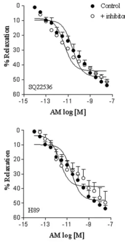
n=6) did not alter the relaxation induced by AM (Figure 4). Neither H89 (Emax: 49.7±7.7%; pD2: 11.1±0.4, n=5) nor
SQ22536 (Emax: 51.6±1.8%; pD2: 11.4±0.2, n=5) altered
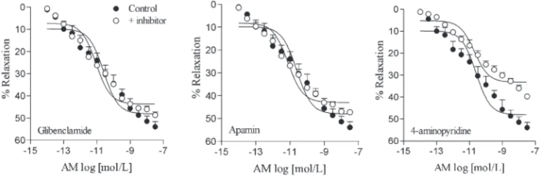
AM-induced relaxation (Figure 5). L-NAME, ODQ, Rp-8-Br-PET-cGMPS, and SC560 reduced AM-induced relaxation to a similar extent (Figure 6, Table 1). The combination of L-NAME and SC560 showed further suppression of AM relaxation than that observed with either L-NAME or SC560 alone. However, even when combined, these compounds were not able to abolish AM-induced relaxation. Sildenafil induced a leftward displacement in the concentration-response curve for AM. Conversely, 7-nitroindazole and wortmannin did not alter the relaxation induced by AM (Figure 6, Table 1). 4-Aminopyridine, but not apamin or glibenclamide, reduced the relaxation induced by AM in rat CSM (Figure 7, Table 1).
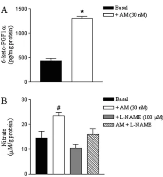
Nitrate and 6-keto-PGF1ameasurements
AM significantly increased 6-keto-PGF1a (a stable
product of PGI2) in rat CSM compared with tissues that
were not stimulated with the peptide (Figure 8A). AM significantly increased nitrate generation in rat CSM compared with tissues that were not stimulated with the peptide (Figure 8B). AM-induced nitrate generation was significantly inhibited by L-NAME, which had no effectper seon basal nitrate levels.
Discussion
In the present study, protein and mRNA expression of AM, CRLR, and RAMP1, -2, and -3 were detected in rat CSM. Immunohistochemical assays showed that AM and CRLR are expressed in the cavernous tissue. AM acts as a circulating hormone and locally in an autocrine/ paracrine fashion. Because AM is expressed in rat CSM, it may play a role in the autocrine/paracrine regulation of penile erection due to its vasodilator action. AM is considered an important regulatory peptide that helps to regulate cardiovascular homeostasis. AM levels in cardiovascular tissues are elevated to compensate for changes induced by cardiovascular diseases such as atherosclerosis and hypertension (24). Thus, increased AM expression in CSM could exert a protective action against ED. In fact, it has been suggested that combina-tion therapy using PGE1and proerection agents such as
AM may be beneficial in the treatment of ED (25). A pharmacological characterization of the mechan-isms mediating the relaxant effect of AM in rat CSM was attempted with functional assays, using standard muscle bath procedures. AM induced CSM relaxation in a concentration-dependent manner. AM was similar in potency to CGRP, and both were more potent than acetylcholine, which is in accordance with previous findings in rat aorta (26), rat mesenteric arterial bed (27), and cat CSM (6). Relaxation induced by AM has
Figure 6.Relaxation responses induced by adrenomedullin (AM) on rat cavernosal smooth muscle strips pre-contracted with phenylephrine. The concentration-response curves were obtained in the absence (control) or after incubation for 30 min with the following drugs: 100mM L-NAME, 100mM 7-nitroinda-zole, 1mM ODQ, 3mM Rp-8-Br-PET-cGMPS, 10mM sildenafil,
1mM wortmannin, 10mM SC560, or the combination of L-NAME
been previously described in isolated rabbit CSM in a concentration range different from that employed in the present study (11). A possible explanation for such discrepancy is that the mechanism by which AM induces vasorelaxation or erection varies with species, vascular bed studied, and experimental procedure employed (5-7,11,28).
The AM receptor is composed of the CRLR and specific RAMP (9,10). RAMPs are a class of type I transmembrane proteins that interact with and modulate the activities of G protein-coupled receptors. Cell surface RAMP2-CRLR and RAMP3-CRLR complexes are AM receptors, while the RAMP1-CRLR complex forms the CGRP receptor (9,10). RAMP interaction with its asso-ciated receptor can lead to three potential consequences: trafficking of receptor protein from an intracellular com-partment to the cell surface, alteration in the terminal glycosylation of the receptor, and alteration of receptor phenotype, presumably through a direct or indirect effect
on the ligand-binding site (29). Although the antagonist AM22-52 has been shown to selectively inhibit AM
receptors, CGRP8-37is a CGRP receptor antagonist that
has been shown to be able to block some, but not all, of the actions of AM in the vasculature (30). This observation indicates that the vasorelaxation induced by AM may occur due to its interaction with both AM or CGRP receptors. The present findings show that AM-induced CSM relaxation was attenuated by AM22-52, but not
CGRP8-37. Our study provides the first functional
evi-dence that relaxation induced by AM in rat CSM is solely mediated by AM receptors.
Activation of adenylate cyclase with consequent increase in cAMP and cAMP-dependent protein kinase activation has been implicated in the vascular relaxation induced by AM (31,32). In our study, neither SQ22536 nor H89 altered AM-induced relaxation, which is not consis-tent with the participation of adenylate cyclase and protein kinase in this response.
Table 1. Effect of L-NAME, 7-nitroindazole, ODQ (1H-(1,2,4)oxadiazolo[4,3-a]quinoxalin-1-one), wortmannin, Rp-8-Br-PET-cGMPS, sildenafil, and SC560 on the Emaxand pD2values for adrenomedullin in the isolated rat cavernosal smooth muscle.
Inhibitor Emax(% relaxation) pD2
Absent 53.9 ± 2.5 10.9 ± 0.3 (6)
L-NAME (100mM) 38.6 ± 2.8* 11.6 ± 0.2 (6)
7-nitroindazole (100mM) 48.2 ± 4.1 11.4 ± 0.4 (6)
ODQ (1mM) 29.8 ± 3.4* 10.5 ± 0.4 (5)
Rp-8-Br-PET-cGMPS (3mM) 24.9 ± 4.3* 10.6 ± 0.5 (5)
Sildenafil (10mM) 59.9 ± 2.6 12.1 ± 0.2* (6)
Wortmannin (1mM) 45.1 ± 4.7 10.5 ± 0.3 (5)
SC560 (10mM) 35.5 ± 1.5* 10.2 ± 0.1 (5)
L-NAME ++ SC560 23.0 ± 0.8*# 11.1 ± 0.3 (5)
Glibenclamide (3mM) 48.6 ± 1.3 11.2 ± 0.1 (6)
Apamin (1mM) 47.3 ± 1.2 11.3 ± 0.2 (5)
4-aminopiridine (1 mM) 39.7 ± 0.7* 10.6 ± 0.2 (6)
Data are reported as means±SE. Number between parentheses indicates the number of animals. * P,0.05, compared to control; #P
,0.05, compared to L-NAME and SC560 (ANOVA followed by the Bonferroni multiple comparison test).
Figure 7. Relaxation responses induced by adrenomedullin (AM) on rat cavernosal smooth muscle strips pre-contracted with phenylephrine. The concentration-response curves were obtained in the absence (control) or after incubation for 30 min with 3mM
In some vascular tissues, AM induces relaxation via production of NO, with consequent increases in cGMP levels (33,34). NO is formed from L-arginine by the catalytic action of the enzyme NOS. The latter has three isoforms: nNOS (or NOS type I), inducible NOS (or NOS type II), and endothelial NOS (eNOS or NOS type III). nNOS and eNOS are the main isoforms involved in penile erection and are present in the nerves and endothelium of the penis, respectively (35). Our data show that L-NAME partially, but significantly, reduced AM-mediated relaxa-tion. In addition, AM increased nitrate levels in rat CSM, and this response was inhibited by L-NAME, further implicating NOS in this process. Taken together, these results show that activation of NOS with consequent NO generation play a role in AM-mediated relaxation. 7-Nitroindazole, a selective nNOS inhibitor, had no effect on AM-induced relaxation, suggesting that this NOS isoform could not account for the AM-mediated relaxation in rat CSM. The selective inhibitor of guanylyl cyclase enzyme, ODQ, reduced the relaxant action of AM, confirming the involvement of the NO-cGMP pathway in this response as previously observed in cat and rat CSM (5-7). cGMP-stimulated protein kinase (PKG) acts downstream to reduce Ca2++ concentration and/or the sensitivity of the
contractile proteins to Ca2++, thus leading to smooth muscle relaxation. Our findings show that PKG activation plays a role in AM-induced relaxation because Rp-8-Br-PET-cGMPS reduced this response. Phosphodiesterase type 5 (PDE5) is widely expressed in CSM, where it catalyzes cGMP hydrolysis (36). Sildenafil, a PDE5 inhibitor, induced a leftward displacement of the con-centration-response curves for AM, further suggesting that PDE5 negatively modulates the relaxation induced by AM.
AM has also been shown to elicit phosphatidylinositol 3-kinase (PI3K) activation and Akt phosphorylation, resulting in the stimulation of eNOS (37). Our findings with wortmannin discard the participation of the PI3K/Akt pathway on AM-induced relaxation. It is important to note that blockade of the NO-cGMP pathway only partially attenuated the relaxant response induced by AM, indicat-ing that mediators unrelated to the production of NO also participate in this response. In fact, we observed that the relaxation evoked by AM was partially blunted by SC560, an inhibitor of cyclooxygenase-1, suggesting the involve-ment of vasodilator prostanoids in the relaxing effect of AM. Additionally, AM increased 6-keto-PGF1a, a stable
product of PGI2. These results agree with a previous
finding showing the participation of prostanoids in AM-induced relaxation in porcine ciliary arteries (38). When L-NAME and SC560 were simultaneously added to the organ bath, an additional inhibitory effect on AM-induced relaxation was observed, indicating that both NO and vasodilator prostanoids participate in this response.
Activation of K+channels is an important mechanism in vascular smooth muscle hyperpolarization and relaxa-tion, and cGMP can modulate the activity of K+channels to elicit vasodilatation. A role for K+ channels in AM-mediated relaxation has already been described for vasculature (27,38). We found that 4-aminopyridine, but not glibenclamide or apamin, reduced AM-induced relaxa-tion, indicating that the activation of voltage-sensitive K+ channels plays a role in such responses.
The major new finding of the present study is that AM receptors mediate CSM relaxation via the NO-cGMP pathway, vasodilator prostanoids (probably PGI2), and the
opening of K+ channels. Studies of the expression and function of AM receptors in CSM may provide valuable information on the contribution of AM to CSM tone, since this tissue is a useful model for the study of penile erectile responses and ED.
Acknowledgments
Research supported by FAPESP (#2006/60076-7 and #2011/12911-2). L.N. Leite was supported by a master fellowship from CAPES.
Figure 8. 6-keto-PGF1a and nitrate levels in rat cavernosal
smooth muscle strips stimulated with adrenomedullin (AM). Data are reported as means±SE of 5 to 6 independent preparations. *P,0.05, compared to basal (Student t-test); #P
References
1. Andersson KE, Wagner G. Physiology of penile erection. Physiol Rev1995; 75: 191-236.
2. Saenz de Tejada I, Goldstein I, Krane RJ. Local control of penile erection. Nerves, smooth muscle, and endothelium. Urol Clin North Am1988; 15: 9-15.
3. Burnett AL, Lowenstein CJ, Bredt DS, Chang TS, Snyder SH. Nitric oxide: a physiologic mediator of penile erection. Science1992; 257: 401-403, doi: 10.1126/science.1378650. 4. Lue TF. Erectile dysfunction. N Engl J Med 2000; 342:
1802-1813, doi: 10.1056/NEJM200006153422407. 5. Champion HC, Wang R, Shenassa BB, Murphy WA, Coy
DH, Hellstrom WJ, et al. Adrenomedullin induces penile erection in the cat.Eur J Pharmacol1997; 319: 71-75, doi: 10.1016/S0014-2999(96)00924-7.
6. Champion HC, Wang R, Santiago JA, Murphy WA, Coy DH, Kadowitz PJ, et al. Comparison of responses to adrenome-dullin and calcitonin gene-related peptide in the feline erection model.J Androl1997; 18: 513-521.
7. Nishimatsu H, Hirata Y, Hayakawa H, Nagata D, Satonaka H, Suzuki E, et al. Effects of intracavernous administration of adrenomedullin on erectile function in rats. Peptides 2001; 22: 1913-1918, doi: 10.1016/S0196-9781(01)00521-6.
8. Kitamura K, Kangawa K, Kawamoto M, Ichiki Y, Nakamura S, Matsuo H, et al. Adrenomedullin: a novel hypotensive peptide isolated from human pheochromocytoma.Biochem Biophys Res Commun1993; 192: 553-560, doi: 10.1006/ bbrc.1993.1451.
9. McLatchie LM, Fraser NJ, Main MJ, Wise A, Brown J, Thompson N, et al. RAMPs regulate the transport and ligand specificity of the calcitonin-receptor-like receptor. Nature1998; 393: 333-339, doi: 10.1038/30666.
10. Poyner DR, Sexton PM, Marshall I, Smith DM, Quirion R, Born W, et al. International Union of Pharmacology. XXXII. The mammalian calcitonin gene-related peptides, adreno-medullin, amylin, and calcitonin receptors.Pharmacol Rev 2002; 54: 233-246, doi: 10.1124/pr.54.2.233.
11. Gokce G, Bagcivan I, Kilicarslan H, Yildirim S, Gultekin YE, Sarioglu Y. Relaxation effects of adrenomedullin in isolated rabbit corpus cavernosum smooth muscle.BJU Int 2004; 93: 859-862, doi: 10.1111/j.1464-410X.2003.04728.x. 12. Marinoni E, di Iorio R, Villaccio B, Vellucci O, Di Netta T,
Sessa M, et al. Adrenomedullin in human male reproductive system.Eur J Obstet Gynecol Reprod Biol2005; 122: 195-198, doi: 10.1016/j.ejogrb.2005.03.021.
13. Hinson JP, Kapas S, Smith DM. Adrenomedullin, a multi-functional regulatory peptide. Endocr Rev2000; 21: 138-167.
14. Kloner RA. Sex and the patient with cardiovascular risk factors: focus on sildenafil.Am J Med2000; 109 (Suppl 9A): 13S-21S, doi: 10.1016/S0002-9343(00)00656-2.
15. Carneiro FS, Carneiro ZN, Giachini FR, Lima VV, Nogueira E, Rainey WE, et al. Murine and rat cavernosal responses to endothelin-1 and urotensin-II Vasoactive Peptide Symposium. J Am Soc Hypertens2008; 2: 439-447, doi: 10.1016/j.jash.2008.07.001.
16. Lizarte FS, Morgueti M, Tirapelli CR, Claudino MA, Evora PR, Novais PC, et al. Chronic alcoholism associated with
diabetes impairs erectile function in rats.BJU Int2010; 105: 1592-1597, doi: 10.1111/j.1464-410X.2009.09084.x. 17. Lizarte FS, Claudino MA, Tirapelli CR, Morgueti M, Tirapelli
DP, Batalhao ME, et al. Chronic ethanol consumption induces cavernosal smooth muscle dysfunction in rats. Urology2009; 74: 1250-1256, doi: 10.1016/j.urology.2009. 04.043.
18. Lin RJ, Wu BN, Lo YC, Shen KP, Lin YT, Huang CH, et al. KMUP-1 relaxes rabbit corpus cavernosum smooth muscle in vitroandin vivo: involvement of cyclic GMP and K(+) channels. Br J Pharmacol 2002; 135: 1159-1166, doi: 10.1038/sj.bjp.0704554.
19. Dalaklioglu S, Ozbey G. Role of different types of potassium channels in the relaxation of corpus cavernosum induced by resveratrol. Pharmacogn Mag 2014; 10: 47-52, doi: 10. 4103/0973-1296.126658.
20. Williams BA, Liu C, Deyoung L, Brock GB, Sims SM. Regulation of intracellular Ca2++release in corpus caverno-sum smooth muscle: synergism between nitric oxide and cGMP.Am J Physiol Cell Physiol2005; 288: C650-C658, doi: 10.1152/ajpcell.00475.2004.
21. Simplicio JA, Pernomian L, Simao MR, Carnio EC, Batalhao ME, Ambrosio SR, et al. Mechanisms underlying the vascular and hypotensive actions of the labdane ent-3-acetoxy-labda-8(17),13-dien-15-oic acid. Eur J Pharmacol 2014; 726C: 66-76, doi: 10.1016/j.ejphar.2014.01.018. 22. Yang L, Tang Q, Hu BR, Xiang JZ. [Relaxant effect of
ethanol on isolated rabbit corpus cavernosum and its mechanism].Zhonghua Nan Ke Xue2007; 13: 910-914. 23. Yogi A, Callera GE, Hipolito UV, Silva CR, Touyz RM,
Tirapelli CR. Ethanol-induced vasoconstriction is mediated via redox-sensitive cyclo-oxygenase-dependent mechan-isms.Clin Sci2010; 118: 657-668, doi: 10.1042/CS20090 352.
24. Yanagawa B, Nagaya N. Adrenomedullin: molecular mechanisms and its role in cardiac disease.Amino Acids 2007; 32: 157-164, doi: 10.1007/s00726-005-0279-5. 25. Lau DH, Kommu S, Mumtaz FH, Morgan RJ, Thompson
CS, Mikhailidis DP. The management of phosphodiester-ase-5 (PDE5) inhibitor failure.Curr Vasc Pharmacol2006; 4: 89-93, doi: 10.2174/157016106776359871.
26. Hipolito UV, Rocha JT, Martins-Oliveira A, Tirapelli DP, Jacob-Ferreira A, Batalhao ME, et al. Chronic ethanol consumption reduces adrenomedullin-induced relaxation in the isolated rat aorta.Alcohol 2011; 45: 805-814, doi: 10.1016/j.alcohol.2011.06.005.
27. Rocha JT, Hipolito UV, Martins-Oliveira A, Tirapelli DP, Batalhao ME, Carnio EC, et al. Ethanol consumption alters the expression and reactivity of adrenomedullin in the rat mesenteric arterial bed.Alcohol Alcohol2012; 47: 9-17, doi: 10.1093/alcalc/agr141.
28. Ishimitsu T, Ono H, Minami J, Matsuoka H. Pathophy-siologic and therapeutic implications of adrenomedullin in cardiovascular disorders.Pharmacol Ther2006; 111: 909-927, doi: 10.1016/j.pharmthera.2006.02.004.
30. Hay DL, Conner AC, Howitt SG, Smith DM, Poyner DR. The pharmacology of adrenomedullin receptors and their relationship to CGRP receptors.J Mol Neurosci2004; 22: 105-113, doi: 10.1385/JMN:22:1-2:105.
31. Okamura T, Ayajiki K, Kangawa K, Toda N. Mechanism of adrenomedullin-induced relaxation in isolated canine retinal arteries.Invest Ophthalmol Vis Sci1997; 38: 56-61. 32. Takata F, Dohgu S, Nishioku T, Takahashi H, Harada E,
Makino I, et al. Adrenomedullin-induced relaxation of rat brain pericytes is related to the reduced phosphorylation of myosin light chain through the cAMP/PKA signaling pathway.Neurosci Lett2009; 449: 71-75, doi: 10.1016/j.neulet.2008.10.082. 33. Hirata Y, Hayakawa H, Suzuki Y, Suzuki E, Ikenouchi H,
Kohmoto O, et al. Mechanisms of adrenomedullin-induced vasodilation in the rat kidney.Hypertension1995; 25: 790-795, doi: 10.1161/01.HYP.25.4.790.
34. Hayakawa H, Hirata Y, Kakoki M, Suzuki Y, Nishimatsu H, Nagata D, et al. Role of nitric oxide-cGMP pathway in
adrenomedullin-induced vasodilation in the rat.Hypertension 1999; 33: 689-693, doi: 10.1161/01.HYP.33.2.689. 35. Prieto D. Physiological regulation of penile arteries and
veins. Int J Impot Res 2008; 20: 17-29, doi: 10.1038/sj. ijir.3901581.
36. Rybalkin SD, Yan C, Bornfeldt KE, Beavo JA. Cyclic GMP phosphodiesterases and regulation of smooth muscle function. Circ Res 2003; 93: 280-291, doi: 10.1161/01. RES.0000087541.15600.2B.
37. Nishimatsu H, Suzuki E, Nagata D, Moriyama N, Satonaka H, Walsh K, et al. Adrenomedullin induces endothelium-dependent vasorelaxation via the phosphatidylinositol 3-kinase/Akt-dependent pathway in rat aorta.Circ Res2001; 89: 63-70, doi: 10.1161/hh1301.092498.