AUTOCHTHONOUS PHYTASE-PRODUCING BACTERIA ISOLATED FROM THE GASTROINTESTINAL TRACTS
OF FOUR INDIAN FRESHWATER TELEOSTS: CHARACTERIZATION AND IDENTIFICATION
Suhas Kumar Dan, Goutam Banerjee, Ankita Nandi, Pinki Ghosh, Arun Kumar Ray*
Address(es): Prof. Arun Kumar Ray
Fisheries Laboratory, Department of Zoology, Visva-Bharati University, Santiniketan-731 235, West Bengal, India.
*Corresponding author: [email protected] / [email protected]
ABSTRACT
Keywords: Fish GI tract, bacteria, phytase, 16S rDNA, identification
INTRODUCTION
Phytases (myo-inositol hexaphosphohydrolases; E.C. 3.1.3.8) are the enzymes which catalyze the hydrolysis of phytic acid to myo-inositol and inorganic phosphates. Phytic acid or phytate (myo-inositol-1,2,3,4,5,6-hexakisphosphates) is abundantly present in plant materials, such as edible legumes, cereals, oilseeds, pollen and nuts (Singh et al., 2011) and serves as the major storage form of phosphorous (about 60-90% of total phosphorous). Phytate is generally regarded as an anti-nutrient in animal feed and for human consumption, as it effectively chelates metal ions such as Ca2+, Mg2+, Zn2+and Fe 2+ and may results in mineral deficiency (Noureddini and Dang, 2008). It also forms complexes with lipid, starch and proteins and thus affects their digestion and may also inhibit digestive enzymes like α-amylase, trypsin, acid phosphatase and tyrosinase (Cosgrove, 1966; Harland and Morris, 1995; Caipang et al., 2011; Khan and Ghosh, 2013). Intestinal phytase activity has been detected in some fish species, but it is insufficient to degrade the phytate content of the feed (Debnath et al., 2005), which passes undigested in feces, accumulates in the environment and causes eutrophication (Baruah et al., 2004). In addition, to meet the nutritional demand, inorganic P has to be supplied that increase the associated cost of aquaculture farming. The prospect of utilization of plant protein sources replacing fish meal in aquaculture is of considerable interest. However, the plant proteins sources are generally rich in anti-nutritional factors like phytic acid which restrict their inclusion in animal diet.One way to reduce the phytate content is treating them with microbial phytase which is considered as a better option than chemical digestion (Roy et al., 2009). Dietary supplementation of phytase has been shown to decrease the phytate content of phytate rich diets and also resulted in improved P availability, protein digestibility and growth in different fish species with a simultaneous decrease in fecal P concentration (Vielma et al., 2002; Debnath et al., 2005; Roy et al., 2013). Besides diminishing the anti-nutritional effect of phytic acid, it also reduces the requirement of supplemental inorganic phosphate. The use of microbial phytases has several advantages over plant phytases as they are more stable at high temperatures and low pH and also have higher specific activity which makes it more suitable for application in fish feed (Cao et al., 2007). However, the application of phytase in aquaculture is limited because of the lack of phytases with appropriate properties, insufficient information about feeding experiment, and manufacturing difficulties (Huang et al., 2009).
It has been proved in different studies that fish harbor their own resident microbial flora which may help the host in digestion by secreting a variety of enzymes (for review, please see Ray et al., 2012). Autochthonous phytase-producing bacteria have been isolated from the gastrointestinal (GI) tract of different fish species and a number of studies have also been done on phytase producing resident gut bacteria in the Indian major carps (Roy et al., 2009; Khan
et al., 2011, 2012; Das and Ghosh, 2013; Dan and Ray, 2014). In spite of some
limited information on phytase-producing fish gut bacteria, more research is needed to get clear ideas about the phytase producing bacteria that are able to colonize within the GI tract of freshwater teleosts. In the present study, the bacterial strains isolated from the GI tract of three Indian major carps, rohu (Labeo rohita), catla (Catla catla) and mrigal (Cirrhinus mrigala) and one minor carp, bata (Labeo bata) were screened for phytase activity, both qualitatively and quantitatively. The most promising strains, two from the proximal intestine (PI) and two from the distal intestine (DI) were characterized and identified by 16S rDNA partial sequence analysis..
MATERIALS AND METHODS
Fishes examined
In the present study, four species of adult healthy freshwater fish namely, rohu (L. rohita), catla (C. catla), mrigal (C. mrigala) and bata (L. bata) were examined. The fish were obtained from a local fish farm near Santiniketan, West Bengal, India (23o41 30 N; 87o41 20 E). For acclimatization, the fish were kept in large tanks and fed a diet containing a mixture of fish meal and mustard oil cake (1:1). The water temperature ranged from 30-35 oC. The fish were starved for 24 hours before sacrifice to empty their alimentary tract. The average weight, average length, gut weight and feeding habits of the fishes examined are given in Table 1.
Phytase producing autochthonous bacteria have been isolated from the proximal intestine (PI) and distal intestine (DI) of three Indian major carps, rohu (Labeo rohita), catla (Catla catla) and mrigal (Cirrhinus mrigala) and one minor carp, bata (Labeo bata). In modified phytase screening medium (MPSM), phytase-producing strains were recorded at higher densities in the PI of rohu and minimum in the PI of bata. Out of 45 isolates, 4 bacterial strains were selected as potent phytase producers according to quantitative enzyme assay. Maximum phytase activity was detected in LRF5 isolated from the PI of L. rohita followed by CCF2 isolated from the PI of C. catla. Among the selected isolates, three (LRF5, LRH2 and CMH1) were Gram positive rods, whereas CCF2 was Gram positive coccus. All the four isolates could tolerate a wide range of temperature (25–42 °C) and pH (6.0-9.0). The isolate LRH2 was most thermostable as it was able to survive up to 55 oC. On the basis of 16SrDNA partial sequence analysis, isolates LRF5 and CCF2 were identified as Bacillus
cereus (GenBank Accession no. KC894957.1) and Staphylococcus caprae (Accession no. KC894956.1), respectively. Whereas, the
isolates LRH2 and CMH1 were most closely related to Bacillus licheniformis (Accession no. KF011267.1) and Lysinibacillus fusiformis (Accession No. KF011266.1), respectively.
ARTICLE INFO
Received 2. 5. 2014 Revised 3. 12. 2014 Accepted 10. 12. 2014 Published 1. 2. 2015
Regular article
Table 1 Food habits and average weight of gastrointestinal tract of the fishes examined
Fish species Feeding habits* Average weight (g)1 Average length (cm)1 Average gut weight (g)1
Labeo rohita Catla catla Cirrhinus mrigala Labeo bata
Omnivorous, mostly plant matter Zooplanktophagous Detritivorous Herbivorous 121.6 (2.6) 132.5 (3.1) 96.2 (2.1) 73.2 (1.5) 20.3 (1.5) 18.5(1.2) 19.3 (1.4) 19.5 (1.2) 5.3 (0.6) 4.5 (0.48) 3.3 (0.39) 1.5 (0.16) 1Standard deviations are given within the brackets. * Jhingran (1997).
Postmortem Examination and Microbial culture
The fish were killed with a sharp blow on the head and the ventral surfaces were carefully scrubbed with 1% iodine solution (Trust and Sparrow, 1974). The fish were then dissected aseptically on ice to remove the alimentary tracts. The alimentary tracts were longitudinally slit and repeatedly (5 times) washed with chilled sterilized physiological saline (pH 7.2) solution (Mondal et al., 2010). The alimentary tracts were divided into proximal (PI) and distal (DI) parts and homogenized in sterilized chilled 0.9% sodium chloride solution (Das and Tripathi, 1991) as separate samples to isolate bacteria from both PI and DI regions. The gut segment homogenates were subjected to serial dilution up to 10-8 with sterilized distilled water (Beveridge et al., 1991). Diluted samples (100 µl) were used as inoculums for microbial culture on sterilized tryptone soya agar (TSA, Himedia Laboratories, Mumbai, India) plates in duplicate. The plates were incubated at 37 oC for 48 h. To determine phytase-producing bacterial load, gut samples were inoculated seperately to modified phytase screening medium (MPSM) containing plates. The composition and preparation of the MPSM medium has been described elsewhere (Roy et al., 2009). After inoculation, the MPSM plates were incubated at 37oC for 72 h. Since most of the bacterial cells grow optimally at 35-37 oC, the aforementioned temperature was chosen for incubation. Moreover, the bacteria were grown in vitro. The bacterial colonies appeared on TSA and MPSM plates were enumerated and phytase producing bacterial population per g of intestinal tissue were calculated. Bacterial colonies of different morphological appearance (colony, color, configuration, surface margin and opacity) were cultured in fresh MPSM plates until purity is achieved.
Screening of the isolatesfor phytase production
The bacterial isolates were first qualitatively screened (106 cells/ml of inoculum) following the method described by Yanke et al. (1998). The diameter of the clear zones appeared on the MPSM plates, surrounding the bacterial growth regions were measured and were used to determine extracellular phytase producing ability of the bacterial isolates. After discarding some isolates based on the results of qualitative screening, quantitative phytase assay was performed with the remaining bacterial isolates. The quantitative assay was carried out according to the methods of Engelen et al. (1994). Briefly, the bacterial isolates were cultured in TSA broth for 24 h. 0.5 ml of this seed culture was used to inoculate 25 mL of liquid MPSM production media in conical flasks. The culture flasks were incubated with vigorous shaking (180-200 rpm) at 37 oC for 72 h. After incubation, the medium was centrifuged at 10,000 g for 10 min at 4 oC. The supernatant was collected and used for enzyme assay using sodium phytate as substrate. The colour developed after addition of the colour reagent was measured spectrophotometrically at 415 nm. The enzymatic assay for each bacterial strain was carried out in triplicate. One unit (U) of phytase activity corresponds to the amount of enzyme that released 1µg of inorganic phosphorous per min per ml of culture filtrate under the assay conditions (pH 5.5, 37 oC). Statistical analysis (ANOVA) of the data was performed using MICROSOFT EXCEL software which was followed by Duncan’s multiple range test (Duncan, 1955).
Phenotypic characterization and 16S rDNA identification of the bacterial isolates
For phenotypic characterization, the selected promising isolates were cultured in MPSM broth (pH 5-10) and incubated at different temperatures ranging from 10-55 oC. Salt tolerance was tested by growing at different NaCl concentrations (2.5-11.5 %). Colony morphology of the bacterial isolates was studied visually and endospore forming ability was tested by subjecting them to nutrient deprivation.The promising bacterial isolates were identified by 16S rDNA gene sequence analysis following the method described by Roy et al. (2009). Sequenced data were aligned with National Center for Biotechnology Information (NCBI) GenBank nucleotide database and analyzed for finding the closest homolog of the isolates. The partial 16S rDNA sequences of the four selected isolates were deposited in the NCBI GenBank database to obtain their accession numbers.
RESULTS
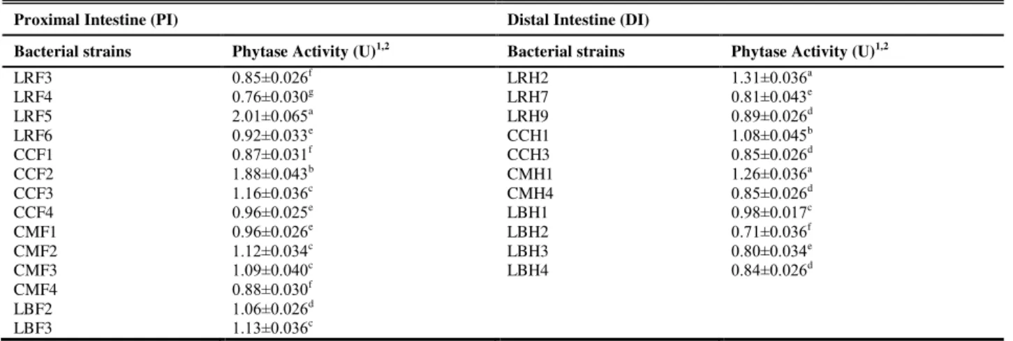
The cultivable aerobic bacterial count and phytase-producing bacterial count of the fish GI tract were determined from the colony forming unit (CFU). Heterotrophic bacterial population was maximum in the PI of L. rohita (log viable count=6.05 per g intestinal tissue), while in the DI, it was maximum in C. mrigala (log =5.91 per g intestinal tissue). Phytase-producing bacterial count was also maximum in the PI of L. rohita (log =4.36 per g intestinal tissue) and in DI it was maximum in L. bata (log =4.01 per g intestinal tissue). It was noticed that in the four fish species, the phytase-producing bacterial population of the gut comprised only a fraction of the total bacterial population. A total of 45 bacterial strains were isolated from the GI tracts of the four fish species and qualitative screening was carried out. Out of 45 strains, 25 isolates were tested positive (7 isolates from L. rohita, 6 isolates each from C. catla, C. mrigala and L. bata) for phytase activity. These 25 bacterial strains produced transparent zones around their colonies in MPSM plates indicating phytase activity. The strain LRF5, isolated from the PI of L. rohita and CCF2, isolated from the PI of C. catla produced halo zones of maximum diameter (16-18 mm), indicating high extracellular phytase activity, while most other strains produced clear zones of shorter radius. Other 20 strains did not produce any visible halo on MPSM plates in qualitative assay and were discarded. All the selected 25 strains were subjected to quantitative phytase assay for quantification of their phytase activity. The results of the quantitative phytase activity are presented in Table 2. The strain LRF5 showed highest phytase activity among all the strains, followed by CCF2, while the strain LRH2, isolated from the DI of L. rohita showed highest activity among the strains isolated from distal intestine, followed by CMH1, isolated from the DI of C. mrigala. Among other strains, CCF3 and CCH1 (isolated from the PI and DI of C. catla, respectively), CMF2 and CMF3 (isolated from the PI of
C. mrigala) and LBF2 and LBF3 (isolated from the PI of L. bata) were found to
have phytase activity in excess of 1.00. Four highest phytase producing strains, 2 from the PI (LRF5 and CCF2) and 2 from the DI, (LRH2 and CMH1) were selected as promising strains and were further studied.
Table 2 Quantitative phytase activity of the bacterial strains isolated from the four fishes
Proximal Intestine (PI) Distal Intestine (DI)
Bacterial strains Phytase Activity (U)1,2 Bacterial strains Phytase Activity (U)1,2
LRF3 LRF4 LRF5 LRF6 CCF1 CCF2 CCF3 CCF4 CMF1 CMF2 CMF3 CMF4 LBF2 LBF3 0.85±0.026f 0.76±0.030g 2.01±0.065a 0.92±0.033e 0.87±0.031f 1.88±0.043b 1.16±0.036c 0.96±0.025e 0.96±0.026e 1.12±0.034c 1.09±0.040c 0.88±0.030f 1.06±0.026d 1.13±0.036c LRH2 LRH7 LRH9 CCH1 CCH3 CMH1 CMH4 LBH1 LBH2 LBH3 LBH4 1.31±0.036a 0.81±0.043e 0.89±0.026d 1.08±0.045b 0.85±0.026d 1.26±0.036a 0.85±0.026d 0.98±0.017c 0.71±0.036f 0.80±0.034e 0.84±0.026d
1Data are means ± SE of the three determinations. Means in the same column with different lower case letters are significantly different (P<0.05).
The bacterial strains, LRF5, LRH2 and CMH1 were Gram positive rods, while CCF2 was a coccus and the isolates LRF5 and LRH2 were capable of forming endospores under unfavorable conditions. The strain CCF2 was found to occur in grapes like clusters while LRF5 occurred in tangled chains and LRH2 and CMH1 as single. All the four strains were able to grow in the temperature range of 25 to 42oC. LRH2 was most thermally resistant as it was capable of surviving up to the temperature of 55oC. The strain LRF5 had the widest pH range of 6.0-9.0 and all the strains had their pH optima around 7.0. The strain CCF2 was very much tolerant to salinity as it was able to grow at the NaCl concentration of 10.5%, while the other three strains were able to grow up to 7.5% of NaCl. The four strains were capable to utilize catalase, citrate and gelatin, whereas only CCF2 was able to utilize urease.
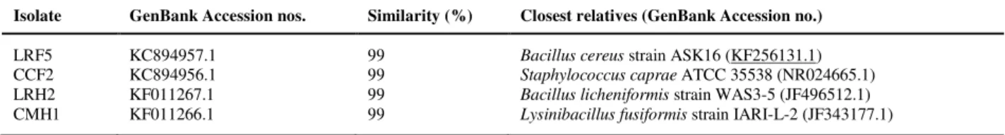
On the basis of phenotypic characteristics, the strains were tentatively identified up to the genus level following Bergey’s Manual of Systematic Bacteriology (Williams et al., 1986). To gain more information about their identity, 16S rDNA sequencing of the bacterial isolates were carried out. The isolate LRF5 (GenBank Accession no. KC894957.1) showed maximum similarity with Bacillus cereus strain ASK16 (Accession no. KF256131.1), whereas,the bacterial isolates CCF2, LRH2 and CMH1 (Accession nos. KC894956.1, KF011267.1 and KF011266.1, respectively) were identified as Staphylococcus caprae, Bacillus licheniformis and Lysinibacillus fusiformis, respectively (Table 3).
Table 3 16S rDNA identification of the four selected bacterial strains
Isolate GenBank Accession nos. Similarity (%) Closest relatives (GenBank Accession no.)
LRF5 CCF2 LRH2 CMH1
KC894957.1 KC894956.1 KF011267.1 KF011266.1
99 99 99 99
Bacillus cereus strain ASK16 (KF256131.1)
Staphylococcus caprae ATCC 35538 (NR024665.1)
Bacillus licheniformis strain WAS3-5 (JF496512.1)
Lysinibacillus fusiformis strain IARI-L-2 (JF343177.1)
DISCUSSION
Bacteria are known to colonize within the GI tract of most terrestrial and aquatic animals and many of them have been proved to be beneficial to the host organism. Fishes are also no exception to this. The microbial gut symbiotic populations of higher animals are mostly anaerobic, whereas monogastric or agastric animals like fish generally harbor microbial population that are mostly aerobic or facultative anaerobic in nature (Mondal et al., 2010). To the authors’ knowledge, a few studies have focused on phytase-producing autochthonous gut bacteria of Indian freshwater fishes (Roy et al., 2009; Khan et al., 2011; Khan and Ghosh, 2012; Das and Ghosh, 2013; Dan and Ray, 2014). Earlier, Li et al. (2008) isolated several marine yeast strains from the gut of sea cucumber
(Holothuria scabra) and marine fish (Hexagrammes otakii and Synecogobius
hasts), which can produce substantial amounts of extra-cellular phytase, and opined that such marine yeasts can play an important role in degradation of phytate in the guts of marine animals. They found that one of the yeast strains, Kodamea ohmeri BG3, isolated from the gut of H. otakii, could produce more phytase than any other marine yeast strains tested. Roy et al. (2009) investigated the presence of phytase producing bacteria in the GI tract of 9 Indian freshwater teleosts. In their study, a total of 25 bacterial isolates were reported to show phytase activity in the quantitative phytase assay of which two promising strains, LF1 and LH1, isolated from PI and DI of L. rohita respectively were identified as two strains of B. licheniformis. In another study, Khan and Ghosh (2012) studied gut-associated phytase producing bacteria in 14 freshwater fish. Several phytase-producing strains were isolated of which two were characterized and identified as Bacillus subtilis and Bacillus atropheus. Askarian et al. (2012) reported phytase activity in Bacillus sp., B. subtilis, B. thurigiensis, B. cereus and
Acinetobacter sp. isolated from the GI tract of Atlantic salmon, Salmo salar, fed
with or without a chitin-supplemented diet. In addition, two bacterial strains,
Brochothrix sp. and B. thermosphacta isolated from the GI tract of Atlantic cod,
Gadus morhua have also been described as phytase producers (Askarian et al., 2013). However, in neither of the studies, did the authors quantify the phytase activity of the bacterial strains.The autochthonous gut microbial population of fish is very much dependent on the environment the fish living in. Recently, Dan and Ray (2014) isolated and enumerated phytase-producing bacteria in the proximal intestine (PI) and distal intestine(DI) of four freshwater teleosts, Nile tilapia (Oreochromis niloticus), murrel (Channa punctatus), climbing perch (Anabas testudineus), and stinging catfish (Heteropneustes fossilis). Out of 32 isolates, 20 phytase-producing strains (9 from the DI and 11 from the PI) were primarily selected on the basis of qualitative assay on MPSM plates. Among these isolates, 3 strains (2 from the PI and 1 from the DI) were selected as potent phytase producers according to quantitative enzyme assay and were identified as different strains of B. licheniformis on the basis of 16S rDNA sequence analysis. The bacteria present in the water and ingested by the fish with food, may colonize and form symbiotic relationship with the fish in their GI tract. It appears that the gut bacterial population may vary in the same fish species and to acquire sufficient knowledge about the phytase-producing bacterial diversity in their GI tract, more independent investigations are necessary. In the present study, a common culture-based technique has been used to evaluate the autochthonous bacterial population of the fish gut. It has been debated that conventional culture-based methods are unable to represent the correct picture of the bacterial load of the fish gut (Ray et al., 2012), and also that these methods are time consuming and lack accuracy (Asfie et al., 2003). Use of more reliable culture-independent methods, like denaturing gradient gel electrophoresis (DGGE) and 16S rDNA clone library based identification are more suitable for studying bacterial diversity in the fish GI tract than the traditional culture based methods (Ray et al., 2012). However, our main target was to isolate and identify culturable autochthonous phytase-producing gut bacteria colonizing in the gut of three
Indian major carps L. rohita, C. catla and C. mrigala and one Indian minor carp L. bata. All the four fishes studied seem to have a large heterotropic aerobic bacterial population in their gut. Four promising phytase-producing strains (LRF5, CCF2, LRH2 and CMH1) were selected on the basis of quantitative phytase assay and were characterized and identified. Characterization of the isolated strains revealed that they could grow within a wide range of temperatures
(25–42 °C). The strain LRH2 was capable of surviving up to a temperature of 55
oC. Similar ranges of temperature and pH tolerance have been reported in other strains of bacilli isolated from fish gut (Ghosh et al., 2002; Saha et al., 2006; Mondal et al., 2010; Dan and Ray, 2014). Although the primary identifications of bacteria are based on their biochemical reaction with different substrates, most of the bacteria within the same genera share common biochemical properties and it is difficult to differentiate them based on biochemical reactions (Akolkar et al., 2006). In the present investigation, since the most promising phytase-producing strains could not be identified to species level by phenotypic criteria, they were placed by 16S rDNA sequence analysis. Based on 16S rDNA sequence analysis,
S. caprae strain ATCC 35538 (Genbank Accession no. NR 024665.1) and L.
fusiformis strain IARI-L-2 (Genbank Accession no. JF343177.1) were identified
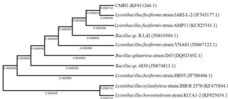
as the closest homologues of the isolates CCF2 and CMH1 (GenBank Accession nos.KC894956.1 and KF011266.1 respectively). Phytase-producing S. caprae (CCF2) and L. fusiformis (CMH1) have not been reported previously in the GI tract of freshwater fishes and hence, the phylogenetic relationships with their close homologs are presented in dendograms in Figures 1 and 2.
Figure 1 Dendogram showing phylogenetic relationship of the strain CCF2 with other close homologs; the horizontal bars represent the branch length; similarity and homology of the neighbouring sequences are indicated by the bootstrap values.
The isolate LRH2 was found to have maximum (99%) similarity with B.
licheniformis strain WAS3-5 (Accession no. JF496512.1). A number of strains of
B. licheniformis with high phytase activity were previously isolated from the GI
Figure 2 Dendogram showing phylogenetic relationship of the strain CMH1 with other close homologs; the horizontal bars represent the branch length; similarity and homology of the neighbouring sequences are indicated by the bootstrap values.
In some recent studies, it has been demonstrated that phytase producing bacteria, colonizing fish GI tract are able to effectively reduce phytate content in plant derived phytate rich diets. Roy et al. (2014) used two bacterial strains LF1 and LH1 (both identified as B. licheniformis) isolated from GI tract of L. rohita to ferment phytate rich sesame (Sesamum indicum) oilseed meal which significantly reduced its phytic acid content and increased the nutritional value whereas, Khan and Ghosh (2013) reported reduction in the contents of the anti-nutritional factors like, tannins, phytic acid, and trypsin inhibitor in the leaf meal of the aquatic weed water spinach (Ipomea aquatica) after fermentation with B. subtilis isolated from the gut of bata, L. bata. Still, there is need for isolation of novel phytase-producing bacteria as any single phytase may never be able to meet all the needs from commercial, environmental or physiological point of view of the animal and in this context the resident microbiota of the host may be of special importance (Lazado et al. 2010). These phytase producing bacteria are better suited to the gut environment and can be used as feed supplement for carps at different life cycle stages which may help to modify the resident gut bacterial flora, increase secretion of phytase and may also competitively reduce pathogenic bacterial population.
In the present study, two new phytase-producing bacterial strains, S. caprae and L. fusiformis (CCF2 and CMH1) not previously known to colonize the fish GI tract, were detected in the GI tract of Indian carps along with two other bacterial strains of B. cereus and B. licheniformis (LRF5 and LRH2, respectively).Their efficacy in improving the nutritional quality of phytate rich plant ingredients are subject to further research. It has to be evaluated through feeding trials if their inclusion in the diet as live bacterial supplement or fermentation of the phytate rich plant ingredients before their inclusion in the fish feed could improve fish health, inorganic P availability and growth rate of Indian major carps.
Acknowledgments: We are grateful to the Council of Scientific and Industrial Research (CSIR), New Delhi [Project No. 37(1415)/10/EMR-II] for financial support.
REFERENCES
AKOLKAR, D., SAMANTA, M., MOHANTY, S., MUKHOPADHYAY, P.K., MAITI, N.K. 2006. Molecular characterization of cellulolytic bacteria from two freshwater cyprinids by immunoblotting and RAPD-PCR. Journal of Aquaculture in the Tropics, 21, 133–147.
ASFIE, M., YOSHIJIMA, T., SUGITA, H. 2003. Characterization of the goldfish fecal microflora by the fluorescent in situ hybridization method. Fisheries
Science, 69, 21–26. http://dx.doi.org/10.1046/j.1444-2906.2003.00583.x
ASKARIAN, F., ZHOU, Z., OLSEN, R.E., SPERSTAD, S., RINGØ, E. 2012.
Culturable autochthonous gut bacteria in Atlantic salmon (SalmosalarL.) fed diets with or without chitin. Characterization by 16S rRNA gene sequencing, ability to produce enzymes and in vitro growth inhibition of four fish pathogens. Aquaculture, 3 26–329, 1–8. http://dx.doi.org/10.1016/j.aquaculture.2011.10.016 ASKARIAN, F., SPERSTAD, S., MERRIFIELD, D.L., RAY, A.K., RINGØ, E.2013. The effect of different feeding regimes on enzyme activity of gut microbiota in Atlantic cod (Gadus morhua L.). Aquaculture Research, 44, 841-846. http://dx.doi.org/10.1111/j.1365-2109.2011.03079.x
BANERJEE, G., RAY, A.K., ASKARIAN, F., RINGØ, E. 2013.Characterisation
and identification of enzyme-producing autochthonous bacteria from the gastrointestinal tract of two Indian air-breathing fish. Beneficial Microbes, 4, 277-284. http://dx.doi.org/10.3920/BM2012.0051
BARUAH, K., SAHU, N.P., PAL, A.K., DEBNATH, D.2004. Dietary phytase: an ideal approach for a cost effective and low-polluting aquafeed. NAGA, World
fish Center Quarterly, 27, 15-19.
BEVERIDGE, M.C.M.,SIKDAR, P.K., FRERICHS, G.N., MILLAR, S.1991. The ingestion of bacteria in suspension by the common carp Cyprinus carpio L.
Journal of Fish Biology, 39, 825-831.
http://dx.doi.org/10.1111/j.1095-8649.1991.tb04412.x
CAIPANG, C.M.A., DECHAVEZ, R.B., APINES-AMAR, M.J.S. 2011. Potential application of microbial phytase in aquaculture. ELBA Bioflux, 3, 55– 66.
CAO, L., WANG, W., YANG, C., YANG, Y., DIANA, J., YAKUPITIYAGE, A., LUO, Z., LI, D. 2007. Application of microbial phytase in fish feed. Enzyme
and Microbial Technology, 40, 497–507.
http://dx.doi.org/10.1016/j.enzmictec.2007.01.007
COSGROVE, D.J. 1966. The chemistry and biochemistry of inositol polyphosphates. Reviews of pure and applied chemistry, 16, 209–224.
DAN, S.K., RAY, A.K. 2014. Characterization and identification of phytase producing bacteria isolated from the gastrointestinal tract of four freshwater teleosts. Annals of Microbiology, 64, 297-306. http://dx.doi.org/10.1007/s13213-013-0664-3
DAS, P., GHOSH, K. 2013. Evaluation of Phytase-producing ability by a fish gut bacterium, Bacillus subtilis sub sp. subtilis. Journal of Biological Sciences, 13, 691-700. http://dx.doi.org/10.3923/jbs.2013.691.700
DAS, K.M., TRIPATHI, S.D. 1991. Studies on the digestive enzymes of grass carp, Ctenopharyngodon idella (Val.). Aquaculture, 92, 21-32. http://dx.doi.org/10.1016/0044-8486(91)90005-R
DEBNATH, D., PAL, A.K., SAHU, N.P. 2005. Effect of dietary microbial phytase supplementation on growth and nutrient digestibility of Pangasius
pangasius (Hamilton) fingerlings. Aquaculture Research, 36, 180–187.
http://dx.doi.org/10.1111/j.1365-2109.2004.01203.x
DUNCAN, D.B. 1955. Multiple range and multiple F-tests. Biometrics, 11, 1-42. ENGELEN, A.J., VAN DER HEEFT, F.C., RANDSDORF, P.H.G., SMIT, E.L.C.1994. Simple and rapid determination of phytase activity. Journal of
AOAC International, 7, 760-764.
GHOSH, K., SEN, S.K., RAY, A.K. 2002. Characterization of bacilli isolated from gut of rohu, Labeo rohita, fingerlings and its significance in digestion.
Journal of Applied Aquaculture, 12, 33-42.
http://dx.doi.org/10.1300/J028v12n03_04
HARLAND, B.F., MORRIS, E.R. 1995. Phytate: A good or a bad food component. Nutrition Research, 15, 733-754. http://dx.doi.org/10.1016/0271-5317(95)00040-P
HUANG, H., SHAO, N., WANG, Y., LUO, H., YANG, P., ZHOU, Z., ZHAN, Z., YAO, B. 2009. A novel beta-propeller phytase from Pedobacter nyackensis MJ11 CGMCC 2503 with potential as an aquatic feed additive. Applied
Microbiology and Biotechnology, 83, 249–259.
http://dx.doi.org/10.1007/s00253-008-1835-1
JHINGRAN, V.G. 1997. Fish and Fisheries of India. 3rd edition. Hindustan Publishing Corporation, Delhi, India, 335-337.
KHAN, A., MANDAL, S., SAMANTA, D., CHATTERJEE, S., GHOSH, K. 2011. Phytase-producing Rhodococcus sp. (MTCC 9508) from fish gut: a preliminary study. Proceedings of the Zoological Society, 64, 29–34. http://dx.doi.org/10.1007/s12595-011-0004-1
KHAN, A., GHOSH, K. 2012. Characterization and identification of gut-associated phytase-producing bacteria in some freshwater fish cultured in ponds.
Acta Ichthyologica et Piscatoria, 42, 37-45.
http://dx.doi.org/10.3750/AIP2011.42.1.05
KHAN, A., GHOSH, K. 2013. Evaluation of phytase production by fish gut bacterium, Bacillus subtilis for processing of Ipomea aquatica leaves as probable aquafeed ingredient. Journal of Aquatic Food Product Technology, 22, 508-519. http://dx.doi.org/10.1080/10498850.2012.669032
LAZADO, C.C., CAIPANG, C.M.A., GALLAGE, S., BRINCHMANN, M.F., KIRON, V. 2010. Responses of Atlantic cod Gadus morhua head kidney leukocytes to phytase produced by gastrointestinal-derived bacteria. Fish
Physiology and Biochemistry, 36, 883–891.
http://dx.doi.org/10.1007/s10695-009-9364-0
LI, X., CHI, Z., LIU, Z., YAN, K., LI, H. 2008. Phytase production by a marine yeast Kodamea ohmeri BG3. Applied Biochemistry and Biotechnology, 149, 183– 193. http://dx.doi.org/10.1007/s12010-007-8099-6
MONDAL, S., ROY, T., RAY, A.K.2010. Characterization and identification of enzyme producing bacteria isolated from the digestive tract of bata, Labeo bata.
Journal of the World Aquaculture Society, 41, 369-377.
http://dx.doi.org/10.1111/j.1749-7345.2010.00378.x
NOUREDDINI, H., DANG, J. 2008. Degradation of phytate in distillers’ grains and corn gluten feed by Aspergillus niger phytase. Applied Biochemistry and
Biotechnology, 159, 11–23. http://dx.doi.org/10.1007/s12010-008-8365-2
RAY, A.K., GHOSH, K., RINGO, E. 2012. Enzyme-producing bacteria isolated from fish gut: a review. Aquaculture Nutrition, 18, 465-492. http://dx.doi.org/10.1111/j.1365-2095.2012.00943.x
ROY, T., MONDAL, S., RAY, A.K. 2009. Phytase-producing bacteria in the digestive tracts of some freshwater fish. Aquaculture Research, 40, 344–353. http://dx.doi.org/10.1111/j.1365-2109.2008.02100.x
SAHA, S., ROY, R.N., SEN, S.K., RAY, A.K. 2006. Characterization of cellulase-producing bacteria from the digestive tract of tilapia, Oreochromis
mossambica (Peters) and grass carp, Ctenopharyngodon idella (Valenciennes).
Aquaculture Research, 37, 380–388.
http://dx.doi.org/10.1111/j.1365-2109.2006.01442.x
SINGH, B., KUNZE, G., SATYANARAYANA, T. 2011. Developments in biochemical aspects and biotechnological applications of microbial phytases.
Biotechnology and Molecular Biology Reviews, 6, 69–87.
TRUST, T.J., SPARROW, R.A.H. 1974. The bacterial flora in the alimentary tract of freshwater salmonid fishes. Canadian Journal of Microbiology, 20, 1219-1228. http://dx.doi.org/10.1139/m74-188
VIELMA, J., RUOHONEN, K., PEISKER, M. 2002. Dephytinization of two soy proteins increases phosphorus and protein utilization by rainbow trout,
Oncorhynchus mykiss. Aquaculture, 204, 145-156.
http://dx.doi.org/10.1016/S0044-8486(01)00653-6
WILLIAMS, S.T., SHARPE, M.E., HOLT, G. (eds.), 1986. Bergey’s Manual of Systematic Bacteriology, vol.1.Williams & Wilkins, Baltimore.